1. Abstract
Standard orthopedic surgical procedures fail for reasons that are
currently not understood. Joint replacements become unexpect-
edly stiff, well-fixed implants become loose and fusions fail to
heal. These failures are costly to the patient and the healthcare
system, especially if they result in substantial number of these
‘aseptic’ failures may be due to pernicious infection with relatively
low virulence organisms, such as Propionibacteria, that do not
stimulate the body’s usual immune response or usual clinical evi-
dence of infection.
Propionibacterium acnes is aerotolerant anaerobic, slow-grow-
ing, and found in the skin’s sebaceous gland as commensal mi-
crobial flora. The Bumgarner group has sequenced a large
number of P. acnes isolated from prosthetic implants.
There is a strong interest in understanding the genes responsible
for variation in biofilm forming capacity. My project within the
Bumgarner group involves measuring biofilm production capacity
across several strains of P. acnes and a related species, P. hu-
merusii.
The goal is to catalog and associate the phenotypes with the
genotypes associated with pathogenic strains. This will contribute
to the understanding of surgical pathology, and lead to improved
aseptic practice in future operations.
Methods
1. Incubate isolates in BHI remel tubes anaerobically at 37c for 7 days.
2. Take 200 uL of new BHI solution, and 2uL of incubated isolate, and
innoculate into multiwell plates, with several redundancies. Incubate an-
aerobically at 37c for another 7 days.
3. After incubation period, dispose of BHI media (and loose cells) in
multiwell plates by inverting and gently shaking, but avoid tapping
(which would dislodge the biofilm).
4. Dispose of further loose cells by submerging multiwell plates in de-
ionized water. Repeat once.
5. Let multiwell plates dry for 15 minutes.
6. Inject each multiwell with 250 uL of 0.1% crystal violet dye solution.
7. Close multiwell cover and let the solution incubate for 15 minutes.
8. After incubation period, dispose of crystal violet solution by inverting
and gently shaking, but avoid tapping.
9. Dispose of further excess crystal violet solution by submerging multi-
well plates in deionized water. Repeat once.
10. Inject each multiwell with 250 uL of 95% ethanol.
11. Close multiwell cover and let the solution incubate for 15 minutes.
12. Measure OD of each sample with nanodrop device.
Note: plastic, and titanium multiwells were also used.
- 20x to 40x of 10mL BHI Remel Tubes
- Deionized Water
- Crystal Violet
- 95% Ethanol
- Multiwell Plates
- Bacteria Strains
- Nanodrop device
Roger Bumgarner, Ph.D; Susan Butler-Wu, Ph.D; Michael Watling; Rob Hall
Skin P. acnes strains tested: 16
Deep P. acnes strains tested: 9
P. Humerusii strains tested: 7
Points of data generated per run: 96-384
Redundancies per strain per run: 3
One run duration: 3 weeks
Runs: 6
Contrary to original prediction, P. Humerusii isolated from surgical samples did not show significant biofilm for-
mation compared to skin isolates, supposing that their virulence factor may not be associated with biofilm for-
mation.
Deep isolates show significantly higher biofilm production than those found on skin, this is consistent with the
prediction that strains infecting deep tissue in surgery is promoted for biofilm generation. Surgical procedures
involving sectioning of the skin causes flora from the sebaceous gland to leak into the wound, the strains of P.
acnes isolated from the infected deep tissue exhibit extra biofilm production.
Titanium multiwell exhibit slightly more biofilm growth than plastic multiwell. Perhaps this is due to the rougher,
grainier surface of the titanium plating, allowing easier attachment of cells onto surface.
The highly variable nature of biofilm production in the lab is influenced by several factors: BHI media quality, ini-
tial cell innoculation with beads, different growth rates of different strains, and probably variable gene expres-
sion of the innoculated cells.
Though the different groups of isolates (skin vs deep vs humerusii) have shown a consistent trend in biofilm
production, their respective difference falls within one standard deviation to each other. The p-value, however, is
consistently small (<0.05). Further testing is recommended.
Due to their association with surgical infections, thorough UV treatment of surgical procedures involving skin in-
cisions is recommended.
With this generated biofilm data, genetic sequencing of available strains of P. acnes and P. humerusii may asso-
ciate the group of genes involved with biofilm production. This dataset may be useful in future experiments in-
volving biofilm gene expression.
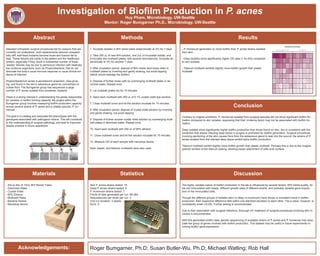
19B219B1 19B1 19B1
20B
19B2 19B2
25G 25G 25G 25G 25G 25G
21G 21G 21G 21G 21G 21G
31N 31N 31N 18B 18B 18B
27N 27N 27N 20B 20B
20B20B 20B 32N 32N 32N
28N1 28N1 28N1 30N 30N 30N
27N 27N 27N 32N 32N 32N
518520 520 520
522
518 518
512 512 512 525 525 525
516 516 516 526 526 526
519 519 519 521 521 521
513 513 513 522 522
514514 514 524 524 524
510 510 510 527 527 527
523 523 523 515 515 515
Multiwell 1: Deep Isolates and P. Humerusii Multiwell 2: Skin Isolates
Multiwell OD Colormaps
- P. Humerusii generates no more biofilm than P. acnes strains isolated
from skin.
- Deep isolates show significantly higher OD data (1.5x-20x) compared
to skin isolates.
- Titanium multiwell exhibits slightly more biofilm growth than plastic
multiwell.
Results
Conclusion
DiscussionStatisticsMaterials
Acknowledgements:
Investigation of Biofilm Production in P. acnes
Huy Pham, Microbiology, UW-Seattle
Mentor: Roger Bumgarner Ph.D., Microbiology, UW-Seattle