
Recommended
More Related Content
Similar to Respiratory_System-10.ppt
Similar to Respiratory_System-10.ppt (20)
Recently uploaded
Recently uploaded (20)
Respiratory_System-10.ppt
- 2. What is It Respiration is the process of exchanging gases between the atmosphere and body cells Non-Respiratory Air Movements: coughing, sneezing, laughing, crying, hiccuping, yawning, speech
- 3. Respiration Pulmonary ventilation (breathing): movement of air into and out of the lungs External respiration: O2 and CO2 exchange between the lungs and the blood Transport: O2 and CO2 in the blood Internal respiration: O2 and CO2 exchange between systemic blood vessels and tissues Respiratory system Circulatory system
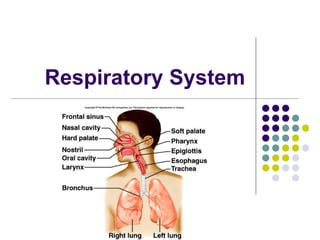
- 4. Organs Nose Pharynx Larynx Trachea Bronchi Lungs Bronchioles Alveoli
- 5. The Nose only externally visible part of the respiratory system The interior of the nose consists of a nasal cavity divided by a nasal septum Nose is the 1st line of defense against airborne antigens so it is also part of our immune system. Functions: Moisten, warm, filter, olfaction, resonance
- 7. Nasal Cavity Olfactory receptors are located in the mucosa on the superior surface cavity is lined with respiratory mucosa and cilia Lateral walls have projections called conchae
- 8. Cont…. The nasal cavity is separated from the oral cavity by the palate Anterior hard palate (bone) Posterior soft palate (muscle Cavities within bones surrounding the nasal cavity are called sinuses Function of the sinuses Lighten the skull Act as resonance chambers for speech Produce mucus that drains into the nasal cavity
- 9. Pharynx (Throat) Three regions of the pharynx Nasopharynx Oropharynx Laryngopharynx
- 10. Figure 22.3b Pharynx Nasopharynx Oropharynx Laryngopharynx (b) Regions of the pharynx
- 11. Larynx or Voice box Routes air and food into proper channels Plays a role in speech Made of eight rigid hyaline cartilages and a spoon- shaped flap of elastic cartilage (epiglottis) Thyroid cartilage (Adam’s apple) Epiglottis Routes food to the larynx and air toward the trachea
- 12. Figure 22.4b Epiglottis Body of hyoid bone Thyrohyoid membrane Vestibular fold (false vocal cord) Vocal fold (true vocal cord) Cricothyroid ligament Cricotracheal ligament Fatty pad Thyroid cartilage Cuneiform cartilage Corniculate cartilage Arytenoid cartilage Cricoid cartilage Tracheal cartilages Arytenoid muscles (b) Sagittal view; anterior surface to the right Thyrohyoid membrane
- 13. Figure 22.5 (a) Vocal folds in closed position; closed glottis (b) Vocal folds in open position; open glottis Base of tongue Epiglottis Vestibular fold (false vocal cord) Vocal fold (true vocal cord) Glottis Inner lining of trachea Cuneiform cartilage Corniculate cartilage
- 14. Voice Production Speech: intermittent release of expired air while opening and closing the glottis Pitch is determined by the length and tension of the vocal cords Loudness depends upon the force of air Chambers of pharynx, oral, nasal, and sinus cavities amplify and enhance sound quality Sound is “shaped” into language by muscles of the pharynx, tongue, soft palate, and lips
- 15. Trachea (Windpipe) Lined with pseudo stratified ciliated mucosa Goblet cells Walls are reinforced with C- shaped hyaline cartilage
- 16. Figure 22.6a (a) Cross section of the trachea and esophagus Hyaline cartilage Submucosa Mucosa Seromucous gland in submucosa Posterior Lumen of trachea Anterior Esophagus Trachealis muscle Adventitia
- 17. Figure 22.6b (b) Photomicrograph of the tracheal wall (320x) Hyaline cartilage • Lamina propria (connective tissue) Submucosa Mucosa Seromucous gland in submucosa • Pseudostratified ciliated columnar epithelium
- 18. Figure 22.7 Trachea Superior lobe of right lung Middle lobe of right lung Inferior lobe of right lung Superior lobe of left lung Left main (primary) bronchus Lobar (secondary) bronchus Segmental (tertiary) bronchus Inferior lobe of left lung
- 19. Conducting Zone Structures Trachea branches into brochi that have 23 orders of branching Bronchioles are less than 1 mm in diameter Terminal bronchioles are the smallest, less than 0.5 mm diameter No cartilage on bronchioles
- 20. Lungs Occupy most of the thoracic cavity Apex is near the clavicle (superior portion) Base rests on the diaphragm (inferior portion) Left lung – two lobes Right lung – three lobes Coverings: (visceral) pleura Parietal pleura
- 22. Site of Gas Exchange Gas exchange takes place within the alveoli 300 million + Pulmonary capillaries cover external surfaces of alveoli and basement membranes connect .5 um thick Total surface area = 40 times your skin
- 23. Figure 22.9a Elastic fibers (a) Diagrammatic view of capillary-alveoli relationships Smooth muscle Alveolus Capillaries Terminal bronchiole Respiratory bronchiole
- 24. Figure 22.9b
- 25. Figure 22.9c Capillary Type II (surfactant- secreting) cell Type I cell of alveolar wall Endothelial cell nucleus Macrophage Alveoli (gas-filled air spaces) Red blood cell in capillary Alveolar pores Capillary endothelium Fused basement membranes of the alveolar epithelium and the capillary endothelium Alveolar epithelium Respiratory membrane Red blood cell O2 Alveolus CO2 Capillary Alveolus Nucleus of type I (squamous epithelial) cell (c) Detailed anatomy of the respiratory membrane
- 26. Alveoli Pores for gas exchange between alveoli
- 27. Respiration Events Pulmonary ventilation External respiration Respiratory gas transport Internal respiration
- 28. Pulmonary Ventilation mechanical process Depends on volume changes in the thoracic cavity It is all about pressure outside, inside, and around the lungs Two phases Inspiration Expiration
- 29. Inspiration
- 30. Figure 22.13 (1 of 2) Sequence of events Changes in anterior- posterior and superior- inferior dimensions Changes in lateral dimensions (superior view) Ribs are elevated and sternum flares as external intercostals contract. Diaphragm moves inferiorly during contraction. External intercostals contract. Inspiratory muscles contract (diaphragm descends; rib cage rises). 2 1 Thoracic cavity volume increases. 3 Lungs are stretched; intrapulmonary volume increases. 4 Intrapulmonary pressure drops (to –1 mm Hg). 5 Air (gases) flows into lungs down its pressure gradient until intrapulmonary pressure is 0 (equal to atmospheric pressure).
- 31. Pressure Difference in Thoracic Cavity Differences in lung and pleural space pressures keep lungs from collapsing Atelectasis (lung collapse) is due to Plugged bronchioles collapse of alveoli Wound that admits air into pleural cavity (pneumothorax)
- 32. Figure 22.16b Respiratory volumes Tidal volume (TV) Amount of air inhaled or exhaled with each breath under resting conditions 3100 ml Inspiratory reserve volume (IRV) Expiratory reserve volume (ERV) Residual volume (RV) Amount of air remaining in the lungs after a forced exhalation 500 ml Amount of air that can be forcefully inhaled after a nor- mal tidal volume inhalation Amount of air that can be forcefully exhaled after a nor- mal tidal volume exhalation 1200 ml 1200 ml Measurement Description Adult male average value 1900 ml 500 ml 700 ml 1100 ml Adult female average value Respiratory Volumes
- 33. Figure 22.16b Respiratory capacities (b) Summary of respiratory volumes and capacities for males and females Functional residual capacity (FRC) Volume of air remaining in the lungs after a normal tidal volume expiration: FRC = ERV + RV Maximum amount of air contained in lungs after a maximum inspiratory effort: TLC = TV + IRV + ERV + RV Maximum amount of air that can be expired after a maxi- mum inspiratory effort: VC = TV + IRV + ERV Maximum amount of air that can be inspired after a normal expiration: IC = TV + IRV Total lung capacity (TLC) Vital capacity (VC) Inspiratory capacity (IC) 6000 ml 4800 ml 3600 ml 2400 ml 4200 ml 3100 ml 2400 ml 1800 ml
- 34. Figure 22.16a Inspiratory reserve volume 3100 ml Tidal volume 500 ml (a) Spirographic record for a male Expiratory reserve volume 1200 ml Residual volume 1200 ml Functional residual capacity 2400 ml Inspiratory capacity 3600 ml Vital capacity 4800 ml Total lung capacity 6000 ml
- 35. Alveolar Ventilation Alveolar ventilation rate (AVR): flow of gases into and out of the alveoli during a particular time Dead space is normally constant Rapid, shallow breathing decreases AVR AVR = frequency X (TV – dead space) (ml/min) (breaths/min) (ml/breath)
- 36. Table 22.2
- 37. External Respiration Oxygen movement into the blood Carbon dioxide movement out of the blood Blood leaving the lungs is oxygen-rich and carbon dioxide-poor
- 38. Gas Transport in Blood Oxygen transport in the blood attached to hemoglobin (oxyhemoglobin [HbO2]) A small amount (1.5%) is carried dissolved in the plasma Carbon dioxide transport in the blood transported in the plasma as bicarbonate ion (HCO3–) (70%) 10% free in plasma A small amount is carried inside red blood cells on hemoglobin, but at different binding sites than those of oxygen (20%)
- 40. Figure 22.18 Time in the pulmonary capillary (s) P 104 mm Hg O2 End of capillary Start of capillary
- 41. Figure 22.17 Inspired air: P 160 mm Hg P 0.3 mm Hg Blood leaving lungs and entering tissue capillaries: P 100 mm Hg P 40 mm Hg Alveoli of lungs: P 104 mm Hg P 40 mm Hg O2 Heart Blood leaving tissues and entering lungs: P 40 mm Hg P 45 mm Hg Systemic veins Systemic arteries Tissues: P less than 40 mm Hg P greater than 45 mm Hg Internal respiration External respiration Pulmonary veins (P 100 mm Hg) Pulmonary arteries CO2 O2 CO2 O2 CO2 O2 CO2 O2 CO2 O2
- 42. Factors that Increase Release of O2 by Hemoglobin As cells metabolize glucose Pco2 and H+ increase in concentration in capillary blood Declining pH weakens the hemoglobin-O2 bond (Bohr effect) Heat production increases Increasing temperature directly and indirectly decreases Hb affinity for O2
- 43. Internal respiration Exchange of gases between blood and body cells An opposite reaction to what occurs in the lungs Carbon dioxide diffuses out of tissue to blood Oxygen diffuses from blood into tissue
- 45. Figure 22.23 Pons Pons Ventral respiratory group (VRG) contains rhythm generators whose output drives respiration. Pontine respiratory centers interact with the medullary respiratory centers to smooth the respiratory pattern. Medulla Medulla To inspiratory muscles External intercostal muscles Diaphragm Dorsal respiratory group (DRG) integrates peripheral sensory input and modifies the rhythms generated by the VRG.
- 46. Factors affecting Breathing Physical factors: Increased body temperature, Exercise, Talking, Coughing Volition (conscious control) Emotional factors- fight or flight Chemical factors Carbon dioxide levels Level of carbon dioxide in the blood is the main regulatory chemical for respiration Increased carbon dioxide increases respiration Changes in carbon dioxide act directly on the medulla oblongata
- 47. Disorders COPD
- 48. Lung Cancer SIDS Asthma Etc. Lung Cancer: Accounts for 1/3 of all cancer deaths in the United States Increased incidence associated with smoking (90%) TB- bacterial infection…1 year of antibiotics SIDS Some cases are thought to be a problem of the neural respiratory control center One third of cases appear to be due to heart rhythm abnormalities Asthma- Chronic inflamed hypersensitive bronchiole passages
- 49. That’s Life reflect accumulation of environmental influences reflect the effects of aging in other organ systems cilia less active mucous thickens swallowing, gagging, and coughing reflexes slow macrophages in lungs lose efficiency increased susceptibility to respiratory infections “barrel chest” may develop bronchial walls thin and collapse dead space increases
- 50. The Death Stick Cigarette affects cilia disappear excess mucus produced lung congestion increases lung infections lining of bronchioles thicken bronchioles lose elasticity emphysema fifteen times more common lung cancer more common about 90% smoke other usually have jobs where air in full of impurities. much damage repaired when smoking stops And those who smoke say they need this!!!!
- 51. Cont… About half of all Americans who keep smoking will die because of the habit. Each year about 443,600 people in the United States die from illnesses related to tobacco use. 1 of 5 deaths in US. Smoking cigarettes kills more Americans than alcohol, car accidents, suicide, AIDS, homicide, and illegal drugs combined smokers are at increased risk for cancer of the larynx, oral cavity, esophagus, bladder, kidney, and pancreas. About 48 million people in the United States smoke an estimated total of 430 billion cigarettes each year the average cigarette contains around 4,000 chemicals, some of which are highly toxic and at least 60 of which cause cancer
- 52. Smoking causes a fivefold increase in the risk of dying from chronic bronchitis and emphysema, and a twofold increase in deaths from diseases of the heart and coronary arteries. Smoking also increases the risk of stroke by 50 percent—40 percent among men and 60 percent among women. Other research has shown that mothers who smoke give birth more frequently to premature or underweight babies
- 53. Why Smoke Recent findings may explain why cigarettes are addictive. An unknown component of tobacco smoke appears to destroy an important brain enzyme, monoamine oxidase B (MAO B). The enzyme is vital for breaking down excess amounts of dopamine, a neurotransmitter that triggers pleasure-seeking behavior. Smokers have decreased levels of MAO B and abnormally high levels of dopamine, which may encourage the smoker to seek the pleasure of more tobacco smoke.
- 54. Innocent Bystander the effect of tobacco smoke on nonsmokers who must share the same environment with a smoker. The United States Environmental Protection Agency (EPA) estimates that exposure to ETS, which contains all the toxic agents inhaled by a smoker, causes 3,400 cancer deaths and an estimated 46,000 deaths from heart disease per year in nonsmokers. Secondhand smoke can aggravate asthma, pneumonia, bronchitis, and impaired blood circulation.