SlideShare a Scribd company logo
1 of 79
ANATOMY
OF KIDNEYS, URETERS,
BLADDER AND URETHRA
By
AREMU ABDULMUJEEB BABATUNDE
KIDNEYS
 Its main function is to excrete
most of the waste products of
metabolism
 It control the water and electrolyte
balance of the body
 It maintain acid-base balance of
the blood
 The waste products leave the
kidneys as urine, which passes
down the ureters to the urinary
bladder
 The urine leaves the body
through the urethra
KIDNEYS
 Kidneys are reddish brown
in color
 Lie behind the peritoneum of
the posterior abdominal wall
 It lie high up on either side
of the vertebral column
 Are largely under cover of
the costal margin
 The right kidney lies slightly
lower than the left due to the
large size of right lobe of the
liver
KIDNEYS
 With contraction of the
diaphragm during respiration,
both kidneys move downward
in a vertical direction as much
as 2.5 cm
 On the medial border of each
kidney is a vertical slit called
the hilum
 The hilum extends into a
large cavity called the renal
sinus
 The hilum transmits the renal
vein, two branches of renal
artery, ureter, and the third
branch of renal artery from
the front backward (V.A.U.A.)
COVERINGS
 1- Fibrous capsule:
 It surrounds the kidney
 2- Perirenal fat:
 It covers the fibrous capsule
 3- Renal fascia:
 it is a condensation of
connective tissue that
encloses the kidneys and
suprarenal glands,
 it is continuous laterally with
the fascia transversalis
 4- Pararenal fat:
 it lies external to the renal
fascia, and forms part of the
retroperitoneal fat
THE KIDNEYS
 Each Kidney Is Protected and Stabilized
 By three concentric layers of connective
tissue
1. Fibrous capsule
• A layer of collagen fibers
• Covers outer surface of entire organ
2. Perinephric fat capsule
• A thick layer of adipose tissue
• Surrounds renal capsule
3. Renal fascia
• A dense, fibrous outer layer
• Anchors kidney to surrounding structures
A superior view of a transverse
section at the level indicated in part (a)
Connective
tissue layers
Fibrous capsule
Perinephric fat
Renal fascia
Left
kidney
External
oblique
Ureter
Quadratus
lumborum
Psoas
major
Inferior
vena cava
Parietal
peritoneumStomach Aorta
Renal
vein
Renal
artery
Spleen
Pancreas
Vertebra
A posterior view of the trunk
Abdominal
aorta
Urinary
bladder
Urethra
Iliac crest
Inferior
vena cava
Ureter
Lumbar
(L1) vertebra
Right
kidney
Renal artery
and vein
11th and
12th ribs
Adrenal
gland
Diaphragm Left kidney
Anterior view
Urinary bladder
Peritoneum (cut)
Hilum
Right kidney
Diaphragm
Celiac trunk
Inferior vena
cava
Right adrenal
gland
Quadratus
lumborum
muscle
Iliacus muscle
Psoas major
muscle
Rectum (cut)
Gonadal artery
and vein
Abdominal aorta
Left common iliac
artery
Superior mesenteric
artery
Left adrenal gland
Esophagus (cut)
Left kidney
Left renal artery
Left renal vein
Left ureter
THE KIDNEYS
 Typical Adult Kidney
 Is about 10 cm long, 50.5 cm wide, and 3
cm thick (4 in.  2.2 in.  1.2 in.)
 Weighs about 150 g (5.25 oz)
 Hilum
 Point of entry for renal artery and renal
nerves
 Point of exit for renal vein and ureter
THE KIDNEYS
 Sectional Anatomy of the Kidneys
 Renal sinus
 Internal cavity within kidney
 Lined by fibrous renal capsule
 Bound to outer surfaces of structures in renal sinus
 Stabilizes positions of ureter, renal blood vessels, and
nerves
THE KIDNEYS
 Renal Cortex
 Superficial portion of kidney in contact with
renal capsule
 Reddish brown and granular
 Renal Pyramids
 6 to 18 distinct conical or triangular
structures in renal medulla
 Base abuts cortex
 Tip (renal papilla) projects into renal sinus
THE KIDNEYS
 Renal Columns
 Bands of cortical tissue separate adjacent
renal pyramids
 Extend into medulla
 Have distinct granular texture
THE KIDNEYS
 Renal Lobe
 Consists of:
 Renal pyramid
 Overlying area of renal cortex
 Adjacent tissues of renal columns
 Produces urine
THE KIDNEYS
 Renal Papilla
 Ducts discharge urine into minor calyx, a
cup-shaped drain
 Major Calyx
 Formed by four or five minor calyces
THE KIDNEYS
 Renal Pelvis
 Large, funnel-shaped chamber
 Consists of two or three major calyces
 Fills most of renal sinus
 Connected to ureter, which drains kidney
A diagrammatic view of a frontal
section through the left kidney
Ureter
Renal papilla
Hilum
Renal pelvis
Adipose tissue
in renal sinus
Renal sinus
Inner layer of
fibrous capsule
Fibrous capsule
Renal columns
Renal lobe
Major calyx
Minor calyx
Connection to
minor calyx
Renal pyramid
Renal medulla
Renal cortex
A frontal section of the left
kidney
Renal columns
Renal medulla
Renal cortex
Ureter
Hilum
Renal
pyramids
Renal sinus
Renal pelvis
Major calyx
Minor calyx
Renal papilla
Renal lobe
Fibrous
capsule
THE KIDNEYS
 Blood Supply to the Kidneys
 Kidneys receive 20%–25% of total cardiac
output
 1200 mL of blood flows through kidneys each
minute
 Kidney receives blood through renal artery
THE KIDNEYS
 Segmental Arteries
 Receive blood from renal artery
 Divide into interlobar arteries
 Which radiate outward through renal columns
between renal pyramids
 Supply blood to arcuate arteries
 Which arch along boundary between cortex and
medulla of kidney
THE KIDNEYS
 Afferent Arterioles
 Branch from each cortical radiate artery
(also called interlobular artery)
 Deliver blood to capillaries supplying
individual nephrons
THE KIDNEYS
 Cortical Radiate Veins
 Also called interlobular veins
 Deliver blood to arcuate veins
 Empty into interlobar veins
 Which drain directly into renal vein
THE KIDNEYS
 Renal Nerves
 Innervate kidneys and ureters
 Enter each kidney at hilum
 Follow tributaries of renal arteries to
individual nephrons
THE KIDNEYS
 Sympathetic Innervation
 Adjusts rate of urine formation
 By changing blood flow and blood pressure at
nephron
 Stimulates release of renin
 Which restricts losses of water and salt in urine
 By stimulating reabsorption at nephron
A sectional view, showing major
arteries and veins
Interlobar
veins
Renal
vein
Renal
artery
Adrenal
artery
Segmental
artery
Interlobar
arteries
Cortical
radiate
arteries
Cortical
radiate
veins
Cortex
Medulla
Arcuate
veins
Arcuate
arteries
Circulation in a single renal lobe
Cortical radiate vein
Cortical radiate artery
Arcuate artery
Arcuate vein
Renal
pyramid
Glomerulus
Afferent
arterioles
Cortical
nephron
Juxtamedullary
nephron
Interlobar vein
Interlobar artery
Minor calyx
A flowchart of renal circulation
Renal vein Renal artery
Segmental arteries
Interlobar arteries
Arcuate arteries
Cortical radiate arteries
Afferent arterioles
Glomerulus
Efferent
arteriole
Peritubular
capillaries
Interlobar veins
Arcuate veins
Cortical radiate veins
Venules
NEPHRONS
THE KIDNEYS
 The Nephron
 Consists of renal tubule and renal corpuscle
 Renal tubule
 Long tubular passageway
 Begins at renal corpuscle
THE KIDNEYS
 The Nephron
 Renal corpuscle
 Spherical structure consisting of:
 Glomerular capsule (Bowman’s capsule)
 Cup-shaped chamber
 Capillary network (glomerulus)
RENAL STRUCTURE
 Each kidney has a dark
brown outer cortex and a
light brown inner medulla
 Medulla is composed of
about a dozen renal
pyramids
 Each pyramid having its
base directed laterally
toward the cortex, and its
apex, (the renal papilla),
projecting medially
 The cortex extends into
the medulla between
adjacent pyramids as the
renal column
RENAL STRUCTURE
 Extending from the bases
of the renal pyramids into
the cortex are striations
known as medullary rays
 The renal sinus within the
hilum, contains the upper
expanded end of the
ureter, the renal pelvis
 Renal pelvis divides into
two or three major
calyces, which divides
into two or three minor
calyces
ANTERIOR RELATIONS OF
THE KIDNEYS
 RIGHT KIDNEY
 Suprarenal gland
 Liver,
 Second part of the
duodenum,
 Right colic flexure
 Coils of small intestine
 LEFT KIDNEY
 Suprarenal gland,
 Spleen,
 Stomach,
 Pancreas (body),
 Left colic flexure,
 Descending colon
 Coils of jejunum
RIGHT KIDNEY
Diaphragm,
Costodiaphragmatic recess of the
pleura,
Twelfth rib & last intercostal
space,
Psoas major
Quadratus lumborum,
Transversus abdominis muscle
Subcostal nerve (T12)
Iliohypogastric nerve (L1)
Ilioinguinal nerve (L1)
LEFT KIDNEY
Same as the right except it
is higher it reaches up to the
11th rib.
Posterior Relations of the
Kidneys
BLOOD SUPPLY
 The renal artery arises from
the aorta at the level of the
second lumbar vertebra
 Each renal artery divides
into five segmental arteries
that enter the hilum of the
kidney, four infront and one
behind the renal pelvis
 Lobar artery arise from each
segmental artery, one for
each renal pyramid
BLOOD SUPPLY
 Each lobar artery gives two
or three interlobar arteries
 The interlobar arteries run
toward the cortex on each
side of the renal pyramid
 Interlobar arteries give the
arcuate arteries at the
junction of the cortex and
medulla
 The arcuate arteries give
several interlobular arteries
 Afferent glomerular
arterioles arise as branches
of interlobular arteries
VEINS, LYMPH,
NERVE SUPPLY
 Renal vein emerges from the
hilum in front of the renal
artery and drains into the IVC
 Left renal vein is longer than
the right renal vein
 Lateral aortic lymph nodes lie
around the origin of the renal
artery
 Renal sympathetic plexus:
afferent fibers that travel
through the renal plexus
enter the spinal cord in the
10th, 11th, and 12th thoracic
nerves
URETERS
 The two ureters are muscular
tubes that extend from the
kidneys to the posterior
surface of the urinary bladder
 The urine is propelled along
the ureter by peristaltic
contractions
 Each ureter measures about
25 cm long
 Each ureter has three
constrictions along its course,
 1- Where the renal pelvis
joins the ureter,
 2- As it crosses the pelvic
brim,
 3- Where it pierces the
bladder wall ( intra-mural
URETER
 The renal pelvis is the
funnel-shaped expanded
upper end of the ureter
 It lies within the hilum of the
kidney and receives the
major calyces
 It enters the pelvis by
crossing the bifurcation of
the common iliac artery in
front of the sacroiliac joint
 It runs downward & forward
on the lateral wall of the
pelvis to enter the lateral
angle of the bladder
RELATIONS, RIGHT URETER
 Anterior:
 Duodenum,
 Terminal part of the ileum,
 Right colic vessels,
 Iliocolic vessels,
 Right testicular or ovarian
vessels, and the
 Root of the mesentery.
 Posterior:
 Right psoas muscle,
 Bifurcation of the right
common iliac artery
RELATIONS, LEFT URETER
 Anterior:
 Sigmoid colon,
 Sigmoid mesocolon,
 Left colic vessels,
 Left testicular or
ovarian vessels
 Posterior:
 Left psoas muscle,
 Bifurcation of left
common iliac artery
BLOOD SUPPLY
 Upper end is supplied
by the renal artery
 Middle portion by
(gonadal) testicular or
ovarian artery
 In the pelvis is supplied
by the superior vesical
artery
 The lymph drains to the
lateral aortic nodes and
the iliac nodes
NERVE SUPPLY
 Renal, testicular (or
ovarian), and hypogastric
plexus (in the pelvis)
 Afferent fibers travel with
the sympathetic nerves
and enter the spinal cord
in the first and second
lumbar segments
URINARY BLADDER
SHAPE:
 It has the shape of three-sided pyramid
placed on one of its angles, with the apex
of pyramid is directed forward & its base
is directed backward
SITE:
 It lies behind the body of pubis & is
separated from it by the retropubic space
Upper Surface
Inferolateral surface
Apex
Neck
MALE PELVIS
MALE PELVIS
BASE OF MALE BLADDER
FEMALE PELVIS
FEMALE PELVIS
URINARY BLADDER
RETROPUBIC SPACE:
 It is a space filled with extraperitoneal
fatty tissue continuous with that of lower
part of anterior abdominal wall
 It accomodates distention of urinary
bladder
 In case of rupture of urinary bladder,
urine may escape upward into the
anterior abdominal wall
URINARY BLADDER
APEX:
 Is directed forward
 Is related to upper border of symphysis
pubis
 Is connected to umbilicus by the median
umbilical ligament (obliterated part of
urachus)
URINARY BLADDER
BASE (POSTERIOR SURFACE):
 Is directed backward
 Its superolateral angles receive the ureters
 In male:
1. Its upper part is covered by peritoneum
2. It is related to vasa deferentia & seminal
vesicles separating it from rectum
 In female:
1. It has no peritoneal covering
2. It is related to vagina
URINARY BLADDER
SUPERIOR SURFACE:
 Is covered by peritoneum in both sexes
 In male: it is related to sigmoid colon &
loops of ileum
 In female: it is related to the uterus
separating it from sigmoid colon & loops
of ileum
URINARY BLADDER
INFEROLATERAL SURFACES:
 Are related to retropubic fat separating
them from:
1. Body of pubis
2. Levator ani
3. Obturator internus
URINARY BLADDER
NECK:
 Is the lowest & most fixed part
 Lies behind symphysis pubis
 Is continuous with urethra
 In male:
1. It rests on upper surface of prostate
2. Anteriorly: it is attached to puboprostatic ligament
3. Posteriorly: it is related to beginning of ejaculatory
ducts
 In female:
1. Anteriorly: it is attached to pubovesical ligament
2. Posteriorly: it is related to anterior wall of vagina
URINARY BLADDER
LIGAMENTS:
1. Median umbilical ligament
2. Puboprostatic (pubovesical) ligament:
 Forms the floor of retropubic space
 In male: is called “puboprostatic” &
extends from body of pubis to prostatic
fascia & neck of bladder
 In female: is called “pubovesical” &
extends from body of pubis to neck of
bladder
INTERIOR OF URINARY BLADDER
 The mucous membrane forms folds (rugae)
that disappear when the bladder is
distended
 TRIGONE:
1. A triangular area in the base of bladder,
bounded by the 2 ureteric orifices & the
internal urethral orifice
2. Its mucous membrane is elastic, more
vascular & more sensitive
 UVULA VESICA: is an elevation
immediately behind internal urethral orifice
produced by the underlying median lobe of
prostate
URINARY BLADDER
Distended Empty
URINARY BLADDER
CAPACITY:
 Is about 300 ml with a maximum capacity
of 500 ml
 Distended bladder:
 Is circular in shape
 Bulges upward into abdominal cavity
 Removes peritoneum form lower part of
anterior abdominal wall & becomes into
direct contact with it
URINARY BLADDER IN CHILD
 It is an abdominal
organ even when
empty
 It begins to enter the
enlarging pelvis at
six years of age
 It is not entirely a
pelvic organ till after
puberty
Median sagittal section of
a new-born female child
URINARY BLADDER
ARTERIAL SUPPLY:
 Superior & inferior vesical arteries
VENOUS DRAINAGE:
 Veins from the vesical venous plexus that
drain into the internal iliac vein
LYMPHATIC DRAINAGE:
 Into internal & external iliac lymph nodes
URINARY BLADDER
 NERVE SUPPLY: by the inferior
hypogastric plexuses
1. Parasympathetic fibers: from S2,3,4
motor to detrusor muscle (muscle
coat of bladder) & inhibitory to
internal urethral sphincter, produce
micturation
2. Sympathetic fibers: from first &
second lumbar ganglia
3. Ascending sensory fibers: carry
sensation of fullness (distention) &
pain sensation
PELVIC PART OF URETER
 Crosses the front of
bifurcation of
common iliac artery
to reach the pelvis
 Descends downward
& backward, along
the lower border of
internal iliac artery,
crossing (from
above downward):
1. External iliac artery
& vein
2. Obturator nerve,
artery & vein
 Curves forward &
medially
IN MALE
 It is crossed
anteriorly by
vas deferens
IN FEMALE
 It passes below
the root of
broad
ligament,
lateral to lateral
fornix of
vagina & is
crossed
superiorly by
the uterine
artery
PELVIC PART OF URETER
TERMINATION:
 It reaches the posterosuperior angle of
bladder
 It runs an oblique course of about 2 cm
through the wall of bladder before it
opens into its lumen (intramural part of
ureter). This part forms a valve-like
mechanism that prevents reflux of urine
into the ureter when bladder is distended
MALE URETHRA
 About 8 inches (20cm)
long
 Extends from the neck
of bladder to the
external urinary meatus
on the tip of the glans
penis
 Divided into three
parts:
 Prostatic
 Membranous
 Penile
Prostatic urethra
 Length=3 cm
 Widest & most dilatable
 Extends from neck of
bladder inside prostate
gland
 Structures openings into
prostatic urethra:
 Ejaculatory ducts
 Ducts of prostate gland
Membranous urethra
 Length=1 cm
 Surrounded by external
urethral sphincter
 Penile (spongy) urethra
 Length=16 cm
 narrowest part of whole urethra
 Extends inside penis & opens
externally through external
urethral orifice
FEMALE URETHRA
 Extends from neck
of urinary bladder
to open externally
through the
external urethral
orifice (anterior to
the vaginal
opening)
 Has only urinary
function
DIURETICS
 Diuretics are medicines that increase the
amount of urine that is produced.
 People who have high blood pressure might be
prescribed diuretics to decrease the blood
volume.
 Alcohol is a diuretic and this is what
contributes to the symptoms of a hangover.
The best way to prevent a hangover after
drinking is to consume a lot of water before
you go to bed.
 Caffeine is also a diuretic, so coffee, energy
drinks, and regular Coca-cola are diuretics.
 You should drink one cup of water for every
cup of those beverages to prevent
dehydration.
73
PROBLEMS
 URETHRITIS = infection and inflammation of the
urethra
 PYELITIS= infection of the renal calyxes
 CYSTITIS = infection of the urinary bladder.
 CHOLEOCYTITIS = infection of the gall bladder
 Nephritis: inflammation of the nephrons.
 Hydronephritis: excess fluid in the nephron.
74
PROBLEMS
 KIDNEY STONES
 Develop in the renal pelvis
 Stones are made out of a variety of things: uric
acid, calcium, cystine (an amino acid), or
cholesterol.
 They keep growing.
75
KIDNEY STONES
 They can block the ureter, causing the kidney to
enlarge. As the kidney stretches, the capsule
stretches, causing excruciating pain in cycles of
hours. As pressure builds up from fluid
accumulating around the stone, urine can pass,
and the kidney stone moves down the urethra
slowly.
 Symptomatic kidney stones may be pea sized or
larger (up to 1 ½ inches).
 They get stuck in three places:
 Renal pelvis
 In the ureter as it bends over the common iliac artery
 In the urinary bladder at the trigone.
76
KIDNEY FAILURE
 Things can happen to the kidney that can lead to
kidney failure: infection, excess proteins, pH
change, and blood pressure drops.
 Treatment for kidney failure is DIALYSIS, which
removes blood, send it through a filter, and returns
it without the wastes. It is done three times a week,
six hours a day. Ideally, they need a kidney
transplant because the kidney has other functions
as well.
 The brain, heart, and kidney are the only three
organs in the body that have to get oxygen to
sustain life.
77
WHO IS MOST LIKELY TO GET RENAL
FAILURE?
 Kidney or Liver Disease
 Diabetes
 High Blood Pressure
 Heart Failure
 Obesity
 Bone Marrow Transplant
 Heart or Belly Surgery
78
KIDNEY LECTURE 2017.ppt

More Related Content

Similar to KIDNEY LECTURE 2017.ppt

Anatomy of Urinary system
Anatomy of Urinary systemAnatomy of Urinary system
Anatomy of Urinary systemdrsukriti1
 
the urinary system
the urinary systemthe urinary system
the urinary systemDeb Smu
 
Radiological anatomy of kidney, ureter & bladder
Radiological anatomy of kidney, ureter & bladderRadiological anatomy of kidney, ureter & bladder
Radiological anatomy of kidney, ureter & bladderrajss007
 
urinary system(urinary tract)
urinary system(urinary tract)urinary system(urinary tract)
urinary system(urinary tract)Muhammad Bilal
 
Anatomy of urinary system
Anatomy of urinary systemAnatomy of urinary system
Anatomy of urinary systemJohny Wilbert
 
The Anatomy of the Systemic Circulation.ppt
The Anatomy of the Systemic Circulation.pptThe Anatomy of the Systemic Circulation.ppt
The Anatomy of the Systemic Circulation.pptMiguelJohnson8
 
Final biology project work
Final biology project workFinal biology project work
Final biology project workOliviaobligado
 
Urinary system-presentationyorkville-college-1203376567476185-2
Urinary system-presentationyorkville-college-1203376567476185-2Urinary system-presentationyorkville-college-1203376567476185-2
Urinary system-presentationyorkville-college-1203376567476185-2Ksenja Ljashenko
 
Urinary System.pdf
Urinary System.pdfUrinary System.pdf
Urinary System.pdfDr Musadiq
 
Anatomy of Upper & Lower Urinary Tract.pptx
Anatomy  of Upper & Lower  Urinary Tract.pptxAnatomy  of Upper & Lower  Urinary Tract.pptx
Anatomy of Upper & Lower Urinary Tract.pptxDr Ndayisaba Corneille
 
Portal vein and porta caval anastomoses.pptx
Portal vein and porta caval anastomoses.pptxPortal vein and porta caval anastomoses.pptx
Portal vein and porta caval anastomoses.pptxSundip Charmode
 
Urinary System Presentation-Yorkville College
Urinary System Presentation-Yorkville CollegeUrinary System Presentation-Yorkville College
Urinary System Presentation-Yorkville Collegeguestae94d9
 
cardiovascular-system-1216560905497953-8 (1).pptx
cardiovascular-system-1216560905497953-8 (1).pptxcardiovascular-system-1216560905497953-8 (1).pptx
cardiovascular-system-1216560905497953-8 (1).pptxsafuraqazi
 
circulatory system mammals.ppt
circulatory system mammals.pptcirculatory system mammals.ppt
circulatory system mammals.pptssuser880f82
 
Urinary System Presentationyorkville College 1203376567476185 2
Urinary System Presentationyorkville College 1203376567476185 2Urinary System Presentationyorkville College 1203376567476185 2
Urinary System Presentationyorkville College 1203376567476185 2guest8b2ebd
 

Similar to KIDNEY LECTURE 2017.ppt (20)

Anatomy of Urinary system
Anatomy of Urinary systemAnatomy of Urinary system
Anatomy of Urinary system
 
the urinary system
the urinary systemthe urinary system
the urinary system
 
Uninary System
Uninary SystemUninary System
Uninary System
 
Radiological anatomy of kidney, ureter & bladder
Radiological anatomy of kidney, ureter & bladderRadiological anatomy of kidney, ureter & bladder
Radiological anatomy of kidney, ureter & bladder
 
urinary system(urinary tract)
urinary system(urinary tract)urinary system(urinary tract)
urinary system(urinary tract)
 
Anatomy of urinary system
Anatomy of urinary systemAnatomy of urinary system
Anatomy of urinary system
 
Urinary System
Urinary SystemUrinary System
Urinary System
 
The Anatomy of the Systemic Circulation.ppt
The Anatomy of the Systemic Circulation.pptThe Anatomy of the Systemic Circulation.ppt
The Anatomy of the Systemic Circulation.ppt
 
Final biology project work
Final biology project workFinal biology project work
Final biology project work
 
Urinary system-presentationyorkville-college-1203376567476185-2
Urinary system-presentationyorkville-college-1203376567476185-2Urinary system-presentationyorkville-college-1203376567476185-2
Urinary system-presentationyorkville-college-1203376567476185-2
 
Urinary System.pdf
Urinary System.pdfUrinary System.pdf
Urinary System.pdf
 
Anatomy of Upper & Lower Urinary Tract.pptx
Anatomy  of Upper & Lower  Urinary Tract.pptxAnatomy  of Upper & Lower  Urinary Tract.pptx
Anatomy of Upper & Lower Urinary Tract.pptx
 
Kidney
KidneyKidney
Kidney
 
Portal vein and porta caval anastomoses.pptx
Portal vein and porta caval anastomoses.pptxPortal vein and porta caval anastomoses.pptx
Portal vein and porta caval anastomoses.pptx
 
Urinary System Presentation-Yorkville College
Urinary System Presentation-Yorkville CollegeUrinary System Presentation-Yorkville College
Urinary System Presentation-Yorkville College
 
cardiovascular-system-1216560905497953-8 (1).pptx
cardiovascular-system-1216560905497953-8 (1).pptxcardiovascular-system-1216560905497953-8 (1).pptx
cardiovascular-system-1216560905497953-8 (1).pptx
 
circulatory system mammals.ppt
circulatory system mammals.pptcirculatory system mammals.ppt
circulatory system mammals.ppt
 
Urinary system.pptx
Urinary system.pptxUrinary system.pptx
Urinary system.pptx
 
Urinary System Presentationyorkville College 1203376567476185 2
Urinary System Presentationyorkville College 1203376567476185 2Urinary System Presentationyorkville College 1203376567476185 2
Urinary System Presentationyorkville College 1203376567476185 2
 
Excretory products n their elimination by BNP.pdf
Excretory products n their elimination by BNP.pdfExcretory products n their elimination by BNP.pdf
Excretory products n their elimination by BNP.pdf
 

More from HarunMohamed7

RegulatoryIssues In Drug management cycle
RegulatoryIssues In Drug management cycleRegulatoryIssues In Drug management cycle
RegulatoryIssues In Drug management cycleHarunMohamed7
 
Endocrine lecture HYPOTHALAMUS AND PITUITARY HORMONES 2023.pptx
Endocrine lecture  HYPOTHALAMUS AND PITUITARY HORMONES 2023.pptxEndocrine lecture  HYPOTHALAMUS AND PITUITARY HORMONES 2023.pptx
Endocrine lecture HYPOTHALAMUS AND PITUITARY HORMONES 2023.pptxHarunMohamed7
 
THE THYROID GLAND AND DRUGS USED IN THYROID.pdf
THE THYROID GLAND AND DRUGS USED IN THYROID.pdfTHE THYROID GLAND AND DRUGS USED IN THYROID.pdf
THE THYROID GLAND AND DRUGS USED IN THYROID.pdfHarunMohamed7
 
1_ Sample size determination.pptx
1_ Sample size determination.pptx1_ Sample size determination.pptx
1_ Sample size determination.pptxHarunMohamed7
 
basic fracture management JUNE.pptx
basic fracture management JUNE.pptxbasic fracture management JUNE.pptx
basic fracture management JUNE.pptxHarunMohamed7
 
24-Scrotal_Swelling.pptx
24-Scrotal_Swelling.pptx24-Scrotal_Swelling.pptx
24-Scrotal_Swelling.pptxHarunMohamed7
 
trypanosoma-171119074846.pdf
trypanosoma-171119074846.pdftrypanosoma-171119074846.pdf
trypanosoma-171119074846.pdfHarunMohamed7
 
principlesinfracturesmanagement-131009203955-phpapp02.pdf
principlesinfracturesmanagement-131009203955-phpapp02.pdfprinciplesinfracturesmanagement-131009203955-phpapp02.pdf
principlesinfracturesmanagement-131009203955-phpapp02.pdfHarunMohamed7
 
Cryptococcal Meningitis.pptx
Cryptococcal Meningitis.pptxCryptococcal Meningitis.pptx
Cryptococcal Meningitis.pptxHarunMohamed7
 
abdominalwallherniae-130518052821-phpapp02.pdf
abdominalwallherniae-130518052821-phpapp02.pdfabdominalwallherniae-130518052821-phpapp02.pdf
abdominalwallherniae-130518052821-phpapp02.pdfHarunMohamed7
 
Clinical Anatomy of The Upper Limb 2017 NEW.ppt
Clinical Anatomy of The Upper Limb 2017 NEW.pptClinical Anatomy of The Upper Limb 2017 NEW.ppt
Clinical Anatomy of The Upper Limb 2017 NEW.pptHarunMohamed7
 
Wounds and Wound healing..pptx
Wounds and Wound healing..pptxWounds and Wound healing..pptx
Wounds and Wound healing..pptxHarunMohamed7
 
INDUCTION & AUGUMENTATION OF LABOUR.ppt
INDUCTION & AUGUMENTATION OF LABOUR.pptINDUCTION & AUGUMENTATION OF LABOUR.ppt
INDUCTION & AUGUMENTATION OF LABOUR.pptHarunMohamed7
 

More from HarunMohamed7 (20)

RegulatoryIssues In Drug management cycle
RegulatoryIssues In Drug management cycleRegulatoryIssues In Drug management cycle
RegulatoryIssues In Drug management cycle
 
Endocrine lecture HYPOTHALAMUS AND PITUITARY HORMONES 2023.pptx
Endocrine lecture  HYPOTHALAMUS AND PITUITARY HORMONES 2023.pptxEndocrine lecture  HYPOTHALAMUS AND PITUITARY HORMONES 2023.pptx
Endocrine lecture HYPOTHALAMUS AND PITUITARY HORMONES 2023.pptx
 
THE THYROID GLAND AND DRUGS USED IN THYROID.pdf
THE THYROID GLAND AND DRUGS USED IN THYROID.pdfTHE THYROID GLAND AND DRUGS USED IN THYROID.pdf
THE THYROID GLAND AND DRUGS USED IN THYROID.pdf
 
1_ Sample size determination.pptx
1_ Sample size determination.pptx1_ Sample size determination.pptx
1_ Sample size determination.pptx
 
basic fracture management JUNE.pptx
basic fracture management JUNE.pptxbasic fracture management JUNE.pptx
basic fracture management JUNE.pptx
 
APPENDIcitis.pptx
APPENDIcitis.pptxAPPENDIcitis.pptx
APPENDIcitis.pptx
 
ACUTE ABDOMEN.pptx
ACUTE ABDOMEN.pptxACUTE ABDOMEN.pptx
ACUTE ABDOMEN.pptx
 
24-Scrotal_Swelling.pptx
24-Scrotal_Swelling.pptx24-Scrotal_Swelling.pptx
24-Scrotal_Swelling.pptx
 
12.Peritonitis.pdf
12.Peritonitis.pdf12.Peritonitis.pdf
12.Peritonitis.pdf
 
trypanosoma-171119074846.pdf
trypanosoma-171119074846.pdftrypanosoma-171119074846.pdf
trypanosoma-171119074846.pdf
 
principlesinfracturesmanagement-131009203955-phpapp02.pdf
principlesinfracturesmanagement-131009203955-phpapp02.pdfprinciplesinfracturesmanagement-131009203955-phpapp02.pdf
principlesinfracturesmanagement-131009203955-phpapp02.pdf
 
Headache
HeadacheHeadache
Headache
 
Cryptococcal Meningitis.pptx
Cryptococcal Meningitis.pptxCryptococcal Meningitis.pptx
Cryptococcal Meningitis.pptx
 
abdominalwallherniae-130518052821-phpapp02.pdf
abdominalwallherniae-130518052821-phpapp02.pdfabdominalwallherniae-130518052821-phpapp02.pdf
abdominalwallherniae-130518052821-phpapp02.pdf
 
Clinical Anatomy of The Upper Limb 2017 NEW.ppt
Clinical Anatomy of The Upper Limb 2017 NEW.pptClinical Anatomy of The Upper Limb 2017 NEW.ppt
Clinical Anatomy of The Upper Limb 2017 NEW.ppt
 
THE ARM.pptx
THE ARM.pptxTHE ARM.pptx
THE ARM.pptx
 
ca stomach.ppt
ca stomach.pptca stomach.ppt
ca stomach.ppt
 
Wounds and Wound healing..pptx
Wounds and Wound healing..pptxWounds and Wound healing..pptx
Wounds and Wound healing..pptx
 
chest trauma.pdf
chest trauma.pdfchest trauma.pdf
chest trauma.pdf
 
INDUCTION & AUGUMENTATION OF LABOUR.ppt
INDUCTION & AUGUMENTATION OF LABOUR.pptINDUCTION & AUGUMENTATION OF LABOUR.ppt
INDUCTION & AUGUMENTATION OF LABOUR.ppt
 

Recently uploaded

Mumbai Call Girls Service 9910780858 Real Russian Girls Looking Models
Mumbai Call Girls Service 9910780858 Real Russian Girls Looking ModelsMumbai Call Girls Service 9910780858 Real Russian Girls Looking Models
Mumbai Call Girls Service 9910780858 Real Russian Girls Looking Modelssonalikaur4
 
call girls in green park DELHI 🔝 >༒9540349809 🔝 genuine Escort Service 🔝✔️✔️
call girls in green park  DELHI 🔝 >༒9540349809 🔝 genuine Escort Service 🔝✔️✔️call girls in green park  DELHI 🔝 >༒9540349809 🔝 genuine Escort Service 🔝✔️✔️
call girls in green park DELHI 🔝 >༒9540349809 🔝 genuine Escort Service 🔝✔️✔️saminamagar
 
Call Girls Jayanagar Just Call 7001305949 Top Class Call Girl Service Available
Call Girls Jayanagar Just Call 7001305949 Top Class Call Girl Service AvailableCall Girls Jayanagar Just Call 7001305949 Top Class Call Girl Service Available
Call Girls Jayanagar Just Call 7001305949 Top Class Call Girl Service Availablenarwatsonia7
 
Housewife Call Girls Bangalore - Call 7001305949 Rs-3500 with A/C Room Cash o...
Housewife Call Girls Bangalore - Call 7001305949 Rs-3500 with A/C Room Cash o...Housewife Call Girls Bangalore - Call 7001305949 Rs-3500 with A/C Room Cash o...
Housewife Call Girls Bangalore - Call 7001305949 Rs-3500 with A/C Room Cash o...narwatsonia7
 
College Call Girls Vyasarpadi Whatsapp 7001305949 Independent Escort Service
College Call Girls Vyasarpadi Whatsapp 7001305949 Independent Escort ServiceCollege Call Girls Vyasarpadi Whatsapp 7001305949 Independent Escort Service
College Call Girls Vyasarpadi Whatsapp 7001305949 Independent Escort ServiceNehru place Escorts
 
See the 2,456 pharmacies on the National E-Pharmacy Platform
See the 2,456 pharmacies on the National E-Pharmacy PlatformSee the 2,456 pharmacies on the National E-Pharmacy Platform
See the 2,456 pharmacies on the National E-Pharmacy PlatformKweku Zurek
 
Kolkata Call Girls Services 9907093804 @24x7 High Class Babes Here Call Now
Kolkata Call Girls Services 9907093804 @24x7 High Class Babes Here Call NowKolkata Call Girls Services 9907093804 @24x7 High Class Babes Here Call Now
Kolkata Call Girls Services 9907093804 @24x7 High Class Babes Here Call NowNehru place Escorts
 
VIP Call Girls Pune Vrinda 9907093804 Short 1500 Night 6000 Best call girls S...
VIP Call Girls Pune Vrinda 9907093804 Short 1500 Night 6000 Best call girls S...VIP Call Girls Pune Vrinda 9907093804 Short 1500 Night 6000 Best call girls S...
VIP Call Girls Pune Vrinda 9907093804 Short 1500 Night 6000 Best call girls S...Miss joya
 
VIP Call Girls Lucknow Nandini 7001305949 Independent Escort Service Lucknow
VIP Call Girls Lucknow Nandini 7001305949 Independent Escort Service LucknowVIP Call Girls Lucknow Nandini 7001305949 Independent Escort Service Lucknow
VIP Call Girls Lucknow Nandini 7001305949 Independent Escort Service Lucknownarwatsonia7
 
Call Girl Bangalore Nandini 7001305949 Independent Escort Service Bangalore
Call Girl Bangalore Nandini 7001305949 Independent Escort Service BangaloreCall Girl Bangalore Nandini 7001305949 Independent Escort Service Bangalore
Call Girl Bangalore Nandini 7001305949 Independent Escort Service Bangalorenarwatsonia7
 
Glomerular Filtration and determinants of glomerular filtration .pptx
Glomerular Filtration and  determinants of glomerular filtration .pptxGlomerular Filtration and  determinants of glomerular filtration .pptx
Glomerular Filtration and determinants of glomerular filtration .pptxDr.Nusrat Tariq
 
Ahmedabad Call Girls CG Road 🔝9907093804 Short 1500 💋 Night 6000
Ahmedabad Call Girls CG Road 🔝9907093804  Short 1500  💋 Night 6000Ahmedabad Call Girls CG Road 🔝9907093804  Short 1500  💋 Night 6000
Ahmedabad Call Girls CG Road 🔝9907093804 Short 1500 💋 Night 6000aliya bhat
 
Call Girl Service Bidadi - For 7001305949 Cheap & Best with original Photos
Call Girl Service Bidadi - For 7001305949 Cheap & Best with original PhotosCall Girl Service Bidadi - For 7001305949 Cheap & Best with original Photos
Call Girl Service Bidadi - For 7001305949 Cheap & Best with original Photosnarwatsonia7
 
Call Girl Koramangala | 7001305949 At Low Cost Cash Payment Booking
Call Girl Koramangala | 7001305949 At Low Cost Cash Payment BookingCall Girl Koramangala | 7001305949 At Low Cost Cash Payment Booking
Call Girl Koramangala | 7001305949 At Low Cost Cash Payment Bookingnarwatsonia7
 
Asthma Review - GINA guidelines summary 2024
Asthma Review - GINA guidelines summary 2024Asthma Review - GINA guidelines summary 2024
Asthma Review - GINA guidelines summary 2024Gabriel Guevara MD
 
Hemostasis Physiology and Clinical correlations by Dr Faiza.pdf
Hemostasis Physiology and Clinical correlations by Dr Faiza.pdfHemostasis Physiology and Clinical correlations by Dr Faiza.pdf
Hemostasis Physiology and Clinical correlations by Dr Faiza.pdfMedicoseAcademics
 
VIP Call Girls Mumbai Arpita 9910780858 Independent Escort Service Mumbai
VIP Call Girls Mumbai Arpita 9910780858 Independent Escort Service MumbaiVIP Call Girls Mumbai Arpita 9910780858 Independent Escort Service Mumbai
VIP Call Girls Mumbai Arpita 9910780858 Independent Escort Service Mumbaisonalikaur4
 
call girls in Connaught Place DELHI 🔝 >༒9540349809 🔝 genuine Escort Service ...
call girls in Connaught Place  DELHI 🔝 >༒9540349809 🔝 genuine Escort Service ...call girls in Connaught Place  DELHI 🔝 >༒9540349809 🔝 genuine Escort Service ...
call girls in Connaught Place DELHI 🔝 >༒9540349809 🔝 genuine Escort Service ...saminamagar
 

Recently uploaded (20)

Mumbai Call Girls Service 9910780858 Real Russian Girls Looking Models
Mumbai Call Girls Service 9910780858 Real Russian Girls Looking ModelsMumbai Call Girls Service 9910780858 Real Russian Girls Looking Models
Mumbai Call Girls Service 9910780858 Real Russian Girls Looking Models
 
call girls in green park DELHI 🔝 >༒9540349809 🔝 genuine Escort Service 🔝✔️✔️
call girls in green park  DELHI 🔝 >༒9540349809 🔝 genuine Escort Service 🔝✔️✔️call girls in green park  DELHI 🔝 >༒9540349809 🔝 genuine Escort Service 🔝✔️✔️
call girls in green park DELHI 🔝 >༒9540349809 🔝 genuine Escort Service 🔝✔️✔️
 
Call Girls Jayanagar Just Call 7001305949 Top Class Call Girl Service Available
Call Girls Jayanagar Just Call 7001305949 Top Class Call Girl Service AvailableCall Girls Jayanagar Just Call 7001305949 Top Class Call Girl Service Available
Call Girls Jayanagar Just Call 7001305949 Top Class Call Girl Service Available
 
Housewife Call Girls Bangalore - Call 7001305949 Rs-3500 with A/C Room Cash o...
Housewife Call Girls Bangalore - Call 7001305949 Rs-3500 with A/C Room Cash o...Housewife Call Girls Bangalore - Call 7001305949 Rs-3500 with A/C Room Cash o...
Housewife Call Girls Bangalore - Call 7001305949 Rs-3500 with A/C Room Cash o...
 
College Call Girls Vyasarpadi Whatsapp 7001305949 Independent Escort Service
College Call Girls Vyasarpadi Whatsapp 7001305949 Independent Escort ServiceCollege Call Girls Vyasarpadi Whatsapp 7001305949 Independent Escort Service
College Call Girls Vyasarpadi Whatsapp 7001305949 Independent Escort Service
 
See the 2,456 pharmacies on the National E-Pharmacy Platform
See the 2,456 pharmacies on the National E-Pharmacy PlatformSee the 2,456 pharmacies on the National E-Pharmacy Platform
See the 2,456 pharmacies on the National E-Pharmacy Platform
 
Kolkata Call Girls Services 9907093804 @24x7 High Class Babes Here Call Now
Kolkata Call Girls Services 9907093804 @24x7 High Class Babes Here Call NowKolkata Call Girls Services 9907093804 @24x7 High Class Babes Here Call Now
Kolkata Call Girls Services 9907093804 @24x7 High Class Babes Here Call Now
 
VIP Call Girls Pune Vrinda 9907093804 Short 1500 Night 6000 Best call girls S...
VIP Call Girls Pune Vrinda 9907093804 Short 1500 Night 6000 Best call girls S...VIP Call Girls Pune Vrinda 9907093804 Short 1500 Night 6000 Best call girls S...
VIP Call Girls Pune Vrinda 9907093804 Short 1500 Night 6000 Best call girls S...
 
VIP Call Girls Lucknow Nandini 7001305949 Independent Escort Service Lucknow
VIP Call Girls Lucknow Nandini 7001305949 Independent Escort Service LucknowVIP Call Girls Lucknow Nandini 7001305949 Independent Escort Service Lucknow
VIP Call Girls Lucknow Nandini 7001305949 Independent Escort Service Lucknow
 
sauth delhi call girls in Bhajanpura 🔝 9953056974 🔝 escort Service
sauth delhi call girls in Bhajanpura 🔝 9953056974 🔝 escort Servicesauth delhi call girls in Bhajanpura 🔝 9953056974 🔝 escort Service
sauth delhi call girls in Bhajanpura 🔝 9953056974 🔝 escort Service
 
Call Girl Bangalore Nandini 7001305949 Independent Escort Service Bangalore
Call Girl Bangalore Nandini 7001305949 Independent Escort Service BangaloreCall Girl Bangalore Nandini 7001305949 Independent Escort Service Bangalore
Call Girl Bangalore Nandini 7001305949 Independent Escort Service Bangalore
 
Glomerular Filtration and determinants of glomerular filtration .pptx
Glomerular Filtration and  determinants of glomerular filtration .pptxGlomerular Filtration and  determinants of glomerular filtration .pptx
Glomerular Filtration and determinants of glomerular filtration .pptx
 
Ahmedabad Call Girls CG Road 🔝9907093804 Short 1500 💋 Night 6000
Ahmedabad Call Girls CG Road 🔝9907093804  Short 1500  💋 Night 6000Ahmedabad Call Girls CG Road 🔝9907093804  Short 1500  💋 Night 6000
Ahmedabad Call Girls CG Road 🔝9907093804 Short 1500 💋 Night 6000
 
Call Girl Service Bidadi - For 7001305949 Cheap & Best with original Photos
Call Girl Service Bidadi - For 7001305949 Cheap & Best with original PhotosCall Girl Service Bidadi - For 7001305949 Cheap & Best with original Photos
Call Girl Service Bidadi - For 7001305949 Cheap & Best with original Photos
 
Escort Service Call Girls In Sarita Vihar,, 99530°56974 Delhi NCR
Escort Service Call Girls In Sarita Vihar,, 99530°56974 Delhi NCREscort Service Call Girls In Sarita Vihar,, 99530°56974 Delhi NCR
Escort Service Call Girls In Sarita Vihar,, 99530°56974 Delhi NCR
 
Call Girl Koramangala | 7001305949 At Low Cost Cash Payment Booking
Call Girl Koramangala | 7001305949 At Low Cost Cash Payment BookingCall Girl Koramangala | 7001305949 At Low Cost Cash Payment Booking
Call Girl Koramangala | 7001305949 At Low Cost Cash Payment Booking
 
Asthma Review - GINA guidelines summary 2024
Asthma Review - GINA guidelines summary 2024Asthma Review - GINA guidelines summary 2024
Asthma Review - GINA guidelines summary 2024
 
Hemostasis Physiology and Clinical correlations by Dr Faiza.pdf
Hemostasis Physiology and Clinical correlations by Dr Faiza.pdfHemostasis Physiology and Clinical correlations by Dr Faiza.pdf
Hemostasis Physiology and Clinical correlations by Dr Faiza.pdf
 
VIP Call Girls Mumbai Arpita 9910780858 Independent Escort Service Mumbai
VIP Call Girls Mumbai Arpita 9910780858 Independent Escort Service MumbaiVIP Call Girls Mumbai Arpita 9910780858 Independent Escort Service Mumbai
VIP Call Girls Mumbai Arpita 9910780858 Independent Escort Service Mumbai
 
call girls in Connaught Place DELHI 🔝 >༒9540349809 🔝 genuine Escort Service ...
call girls in Connaught Place  DELHI 🔝 >༒9540349809 🔝 genuine Escort Service ...call girls in Connaught Place  DELHI 🔝 >༒9540349809 🔝 genuine Escort Service ...
call girls in Connaught Place DELHI 🔝 >༒9540349809 🔝 genuine Escort Service ...
 

KIDNEY LECTURE 2017.ppt

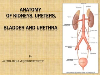
  • 1. ANATOMY OF KIDNEYS, URETERS, BLADDER AND URETHRA By AREMU ABDULMUJEEB BABATUNDE
  • 2. KIDNEYS  Its main function is to excrete most of the waste products of metabolism  It control the water and electrolyte balance of the body  It maintain acid-base balance of the blood  The waste products leave the kidneys as urine, which passes down the ureters to the urinary bladder  The urine leaves the body through the urethra
  • 3. KIDNEYS  Kidneys are reddish brown in color  Lie behind the peritoneum of the posterior abdominal wall  It lie high up on either side of the vertebral column  Are largely under cover of the costal margin  The right kidney lies slightly lower than the left due to the large size of right lobe of the liver
  • 4. KIDNEYS  With contraction of the diaphragm during respiration, both kidneys move downward in a vertical direction as much as 2.5 cm  On the medial border of each kidney is a vertical slit called the hilum  The hilum extends into a large cavity called the renal sinus  The hilum transmits the renal vein, two branches of renal artery, ureter, and the third branch of renal artery from the front backward (V.A.U.A.)
  • 5. COVERINGS  1- Fibrous capsule:  It surrounds the kidney  2- Perirenal fat:  It covers the fibrous capsule  3- Renal fascia:  it is a condensation of connective tissue that encloses the kidneys and suprarenal glands,  it is continuous laterally with the fascia transversalis  4- Pararenal fat:  it lies external to the renal fascia, and forms part of the retroperitoneal fat
  • 6. THE KIDNEYS  Each Kidney Is Protected and Stabilized  By three concentric layers of connective tissue 1. Fibrous capsule • A layer of collagen fibers • Covers outer surface of entire organ 2. Perinephric fat capsule • A thick layer of adipose tissue • Surrounds renal capsule 3. Renal fascia • A dense, fibrous outer layer • Anchors kidney to surrounding structures
  • 7. A superior view of a transverse section at the level indicated in part (a) Connective tissue layers Fibrous capsule Perinephric fat Renal fascia Left kidney External oblique Ureter Quadratus lumborum Psoas major Inferior vena cava Parietal peritoneumStomach Aorta Renal vein Renal artery Spleen Pancreas Vertebra
  • 8. A posterior view of the trunk Abdominal aorta Urinary bladder Urethra Iliac crest Inferior vena cava Ureter Lumbar (L1) vertebra Right kidney Renal artery and vein 11th and 12th ribs Adrenal gland Diaphragm Left kidney
  • 9. Anterior view Urinary bladder Peritoneum (cut) Hilum Right kidney Diaphragm Celiac trunk Inferior vena cava Right adrenal gland Quadratus lumborum muscle Iliacus muscle Psoas major muscle Rectum (cut) Gonadal artery and vein Abdominal aorta Left common iliac artery Superior mesenteric artery Left adrenal gland Esophagus (cut) Left kidney Left renal artery Left renal vein Left ureter
  • 10. THE KIDNEYS  Typical Adult Kidney  Is about 10 cm long, 50.5 cm wide, and 3 cm thick (4 in.  2.2 in.  1.2 in.)  Weighs about 150 g (5.25 oz)  Hilum  Point of entry for renal artery and renal nerves  Point of exit for renal vein and ureter
  • 11. THE KIDNEYS  Sectional Anatomy of the Kidneys  Renal sinus  Internal cavity within kidney  Lined by fibrous renal capsule  Bound to outer surfaces of structures in renal sinus  Stabilizes positions of ureter, renal blood vessels, and nerves
  • 12. THE KIDNEYS  Renal Cortex  Superficial portion of kidney in contact with renal capsule  Reddish brown and granular  Renal Pyramids  6 to 18 distinct conical or triangular structures in renal medulla  Base abuts cortex  Tip (renal papilla) projects into renal sinus
  • 13. THE KIDNEYS  Renal Columns  Bands of cortical tissue separate adjacent renal pyramids  Extend into medulla  Have distinct granular texture
  • 14. THE KIDNEYS  Renal Lobe  Consists of:  Renal pyramid  Overlying area of renal cortex  Adjacent tissues of renal columns  Produces urine
  • 15. THE KIDNEYS  Renal Papilla  Ducts discharge urine into minor calyx, a cup-shaped drain  Major Calyx  Formed by four or five minor calyces
  • 16. THE KIDNEYS  Renal Pelvis  Large, funnel-shaped chamber  Consists of two or three major calyces  Fills most of renal sinus  Connected to ureter, which drains kidney
  • 17. A diagrammatic view of a frontal section through the left kidney Ureter Renal papilla Hilum Renal pelvis Adipose tissue in renal sinus Renal sinus Inner layer of fibrous capsule Fibrous capsule Renal columns Renal lobe Major calyx Minor calyx Connection to minor calyx Renal pyramid Renal medulla Renal cortex
  • 18. A frontal section of the left kidney Renal columns Renal medulla Renal cortex Ureter Hilum Renal pyramids Renal sinus Renal pelvis Major calyx Minor calyx Renal papilla Renal lobe Fibrous capsule
  • 19. THE KIDNEYS  Blood Supply to the Kidneys  Kidneys receive 20%–25% of total cardiac output  1200 mL of blood flows through kidneys each minute  Kidney receives blood through renal artery
  • 20. THE KIDNEYS  Segmental Arteries  Receive blood from renal artery  Divide into interlobar arteries  Which radiate outward through renal columns between renal pyramids  Supply blood to arcuate arteries  Which arch along boundary between cortex and medulla of kidney
  • 21. THE KIDNEYS  Afferent Arterioles  Branch from each cortical radiate artery (also called interlobular artery)  Deliver blood to capillaries supplying individual nephrons
  • 22. THE KIDNEYS  Cortical Radiate Veins  Also called interlobular veins  Deliver blood to arcuate veins  Empty into interlobar veins  Which drain directly into renal vein
  • 23. THE KIDNEYS  Renal Nerves  Innervate kidneys and ureters  Enter each kidney at hilum  Follow tributaries of renal arteries to individual nephrons
  • 24. THE KIDNEYS  Sympathetic Innervation  Adjusts rate of urine formation  By changing blood flow and blood pressure at nephron  Stimulates release of renin  Which restricts losses of water and salt in urine  By stimulating reabsorption at nephron
  • 25. A sectional view, showing major arteries and veins Interlobar veins Renal vein Renal artery Adrenal artery Segmental artery Interlobar arteries Cortical radiate arteries Cortical radiate veins Cortex Medulla Arcuate veins Arcuate arteries
  • 26. Circulation in a single renal lobe Cortical radiate vein Cortical radiate artery Arcuate artery Arcuate vein Renal pyramid Glomerulus Afferent arterioles Cortical nephron Juxtamedullary nephron Interlobar vein Interlobar artery Minor calyx
  • 27. A flowchart of renal circulation Renal vein Renal artery Segmental arteries Interlobar arteries Arcuate arteries Cortical radiate arteries Afferent arterioles Glomerulus Efferent arteriole Peritubular capillaries Interlobar veins Arcuate veins Cortical radiate veins Venules NEPHRONS
  • 28. THE KIDNEYS  The Nephron  Consists of renal tubule and renal corpuscle  Renal tubule  Long tubular passageway  Begins at renal corpuscle
  • 29. THE KIDNEYS  The Nephron  Renal corpuscle  Spherical structure consisting of:  Glomerular capsule (Bowman’s capsule)  Cup-shaped chamber  Capillary network (glomerulus)
  • 30. RENAL STRUCTURE  Each kidney has a dark brown outer cortex and a light brown inner medulla  Medulla is composed of about a dozen renal pyramids  Each pyramid having its base directed laterally toward the cortex, and its apex, (the renal papilla), projecting medially  The cortex extends into the medulla between adjacent pyramids as the renal column
  • 31. RENAL STRUCTURE  Extending from the bases of the renal pyramids into the cortex are striations known as medullary rays  The renal sinus within the hilum, contains the upper expanded end of the ureter, the renal pelvis  Renal pelvis divides into two or three major calyces, which divides into two or three minor calyces
  • 32. ANTERIOR RELATIONS OF THE KIDNEYS  RIGHT KIDNEY  Suprarenal gland  Liver,  Second part of the duodenum,  Right colic flexure  Coils of small intestine  LEFT KIDNEY  Suprarenal gland,  Spleen,  Stomach,  Pancreas (body),  Left colic flexure,  Descending colon  Coils of jejunum
  • 33.
  • 34. RIGHT KIDNEY Diaphragm, Costodiaphragmatic recess of the pleura, Twelfth rib & last intercostal space, Psoas major Quadratus lumborum, Transversus abdominis muscle Subcostal nerve (T12) Iliohypogastric nerve (L1) Ilioinguinal nerve (L1) LEFT KIDNEY Same as the right except it is higher it reaches up to the 11th rib. Posterior Relations of the Kidneys
  • 35. BLOOD SUPPLY  The renal artery arises from the aorta at the level of the second lumbar vertebra  Each renal artery divides into five segmental arteries that enter the hilum of the kidney, four infront and one behind the renal pelvis  Lobar artery arise from each segmental artery, one for each renal pyramid
  • 36. BLOOD SUPPLY  Each lobar artery gives two or three interlobar arteries  The interlobar arteries run toward the cortex on each side of the renal pyramid  Interlobar arteries give the arcuate arteries at the junction of the cortex and medulla  The arcuate arteries give several interlobular arteries  Afferent glomerular arterioles arise as branches of interlobular arteries
  • 37. VEINS, LYMPH, NERVE SUPPLY  Renal vein emerges from the hilum in front of the renal artery and drains into the IVC  Left renal vein is longer than the right renal vein  Lateral aortic lymph nodes lie around the origin of the renal artery  Renal sympathetic plexus: afferent fibers that travel through the renal plexus enter the spinal cord in the 10th, 11th, and 12th thoracic nerves
  • 38. URETERS  The two ureters are muscular tubes that extend from the kidneys to the posterior surface of the urinary bladder  The urine is propelled along the ureter by peristaltic contractions  Each ureter measures about 25 cm long  Each ureter has three constrictions along its course,  1- Where the renal pelvis joins the ureter,  2- As it crosses the pelvic brim,  3- Where it pierces the bladder wall ( intra-mural
  • 39. URETER  The renal pelvis is the funnel-shaped expanded upper end of the ureter  It lies within the hilum of the kidney and receives the major calyces  It enters the pelvis by crossing the bifurcation of the common iliac artery in front of the sacroiliac joint  It runs downward & forward on the lateral wall of the pelvis to enter the lateral angle of the bladder
  • 40. RELATIONS, RIGHT URETER  Anterior:  Duodenum,  Terminal part of the ileum,  Right colic vessels,  Iliocolic vessels,  Right testicular or ovarian vessels, and the  Root of the mesentery.  Posterior:  Right psoas muscle,  Bifurcation of the right common iliac artery
  • 41. RELATIONS, LEFT URETER  Anterior:  Sigmoid colon,  Sigmoid mesocolon,  Left colic vessels,  Left testicular or ovarian vessels  Posterior:  Left psoas muscle,  Bifurcation of left common iliac artery
  • 42. BLOOD SUPPLY  Upper end is supplied by the renal artery  Middle portion by (gonadal) testicular or ovarian artery  In the pelvis is supplied by the superior vesical artery  The lymph drains to the lateral aortic nodes and the iliac nodes
  • 43. NERVE SUPPLY  Renal, testicular (or ovarian), and hypogastric plexus (in the pelvis)  Afferent fibers travel with the sympathetic nerves and enter the spinal cord in the first and second lumbar segments
  • 44. URINARY BLADDER SHAPE:  It has the shape of three-sided pyramid placed on one of its angles, with the apex of pyramid is directed forward & its base is directed backward SITE:  It lies behind the body of pubis & is separated from it by the retropubic space
  • 48. BASE OF MALE BLADDER
  • 51. URINARY BLADDER RETROPUBIC SPACE:  It is a space filled with extraperitoneal fatty tissue continuous with that of lower part of anterior abdominal wall  It accomodates distention of urinary bladder  In case of rupture of urinary bladder, urine may escape upward into the anterior abdominal wall
  • 52. URINARY BLADDER APEX:  Is directed forward  Is related to upper border of symphysis pubis  Is connected to umbilicus by the median umbilical ligament (obliterated part of urachus)
  • 53. URINARY BLADDER BASE (POSTERIOR SURFACE):  Is directed backward  Its superolateral angles receive the ureters  In male: 1. Its upper part is covered by peritoneum 2. It is related to vasa deferentia & seminal vesicles separating it from rectum  In female: 1. It has no peritoneal covering 2. It is related to vagina
  • 54. URINARY BLADDER SUPERIOR SURFACE:  Is covered by peritoneum in both sexes  In male: it is related to sigmoid colon & loops of ileum  In female: it is related to the uterus separating it from sigmoid colon & loops of ileum
  • 55. URINARY BLADDER INFEROLATERAL SURFACES:  Are related to retropubic fat separating them from: 1. Body of pubis 2. Levator ani 3. Obturator internus
  • 56. URINARY BLADDER NECK:  Is the lowest & most fixed part  Lies behind symphysis pubis  Is continuous with urethra  In male: 1. It rests on upper surface of prostate 2. Anteriorly: it is attached to puboprostatic ligament 3. Posteriorly: it is related to beginning of ejaculatory ducts  In female: 1. Anteriorly: it is attached to pubovesical ligament 2. Posteriorly: it is related to anterior wall of vagina
  • 57. URINARY BLADDER LIGAMENTS: 1. Median umbilical ligament 2. Puboprostatic (pubovesical) ligament:  Forms the floor of retropubic space  In male: is called “puboprostatic” & extends from body of pubis to prostatic fascia & neck of bladder  In female: is called “pubovesical” & extends from body of pubis to neck of bladder
  • 59. INTERIOR OF URINARY BLADDER  The mucous membrane forms folds (rugae) that disappear when the bladder is distended  TRIGONE: 1. A triangular area in the base of bladder, bounded by the 2 ureteric orifices & the internal urethral orifice 2. Its mucous membrane is elastic, more vascular & more sensitive  UVULA VESICA: is an elevation immediately behind internal urethral orifice produced by the underlying median lobe of prostate
  • 61. URINARY BLADDER CAPACITY:  Is about 300 ml with a maximum capacity of 500 ml  Distended bladder:  Is circular in shape  Bulges upward into abdominal cavity  Removes peritoneum form lower part of anterior abdominal wall & becomes into direct contact with it
  • 62. URINARY BLADDER IN CHILD  It is an abdominal organ even when empty  It begins to enter the enlarging pelvis at six years of age  It is not entirely a pelvic organ till after puberty Median sagittal section of a new-born female child
  • 63. URINARY BLADDER ARTERIAL SUPPLY:  Superior & inferior vesical arteries VENOUS DRAINAGE:  Veins from the vesical venous plexus that drain into the internal iliac vein LYMPHATIC DRAINAGE:  Into internal & external iliac lymph nodes
  • 64. URINARY BLADDER  NERVE SUPPLY: by the inferior hypogastric plexuses 1. Parasympathetic fibers: from S2,3,4 motor to detrusor muscle (muscle coat of bladder) & inhibitory to internal urethral sphincter, produce micturation 2. Sympathetic fibers: from first & second lumbar ganglia 3. Ascending sensory fibers: carry sensation of fullness (distention) & pain sensation
  • 65. PELVIC PART OF URETER  Crosses the front of bifurcation of common iliac artery to reach the pelvis  Descends downward & backward, along the lower border of internal iliac artery, crossing (from above downward): 1. External iliac artery & vein 2. Obturator nerve, artery & vein  Curves forward & medially
  • 66. IN MALE  It is crossed anteriorly by vas deferens
  • 67. IN FEMALE  It passes below the root of broad ligament, lateral to lateral fornix of vagina & is crossed superiorly by the uterine artery
  • 68. PELVIC PART OF URETER TERMINATION:  It reaches the posterosuperior angle of bladder  It runs an oblique course of about 2 cm through the wall of bladder before it opens into its lumen (intramural part of ureter). This part forms a valve-like mechanism that prevents reflux of urine into the ureter when bladder is distended
  • 69. MALE URETHRA  About 8 inches (20cm) long  Extends from the neck of bladder to the external urinary meatus on the tip of the glans penis  Divided into three parts:  Prostatic  Membranous  Penile
  • 70.
  • 71. Prostatic urethra  Length=3 cm  Widest & most dilatable  Extends from neck of bladder inside prostate gland  Structures openings into prostatic urethra:  Ejaculatory ducts  Ducts of prostate gland Membranous urethra  Length=1 cm  Surrounded by external urethral sphincter  Penile (spongy) urethra  Length=16 cm  narrowest part of whole urethra  Extends inside penis & opens externally through external urethral orifice
  • 72. FEMALE URETHRA  Extends from neck of urinary bladder to open externally through the external urethral orifice (anterior to the vaginal opening)  Has only urinary function
  • 73. DIURETICS  Diuretics are medicines that increase the amount of urine that is produced.  People who have high blood pressure might be prescribed diuretics to decrease the blood volume.  Alcohol is a diuretic and this is what contributes to the symptoms of a hangover. The best way to prevent a hangover after drinking is to consume a lot of water before you go to bed.  Caffeine is also a diuretic, so coffee, energy drinks, and regular Coca-cola are diuretics.  You should drink one cup of water for every cup of those beverages to prevent dehydration. 73
  • 74. PROBLEMS  URETHRITIS = infection and inflammation of the urethra  PYELITIS= infection of the renal calyxes  CYSTITIS = infection of the urinary bladder.  CHOLEOCYTITIS = infection of the gall bladder  Nephritis: inflammation of the nephrons.  Hydronephritis: excess fluid in the nephron. 74
  • 75. PROBLEMS  KIDNEY STONES  Develop in the renal pelvis  Stones are made out of a variety of things: uric acid, calcium, cystine (an amino acid), or cholesterol.  They keep growing. 75
  • 76. KIDNEY STONES  They can block the ureter, causing the kidney to enlarge. As the kidney stretches, the capsule stretches, causing excruciating pain in cycles of hours. As pressure builds up from fluid accumulating around the stone, urine can pass, and the kidney stone moves down the urethra slowly.  Symptomatic kidney stones may be pea sized or larger (up to 1 ½ inches).  They get stuck in three places:  Renal pelvis  In the ureter as it bends over the common iliac artery  In the urinary bladder at the trigone. 76
  • 77. KIDNEY FAILURE  Things can happen to the kidney that can lead to kidney failure: infection, excess proteins, pH change, and blood pressure drops.  Treatment for kidney failure is DIALYSIS, which removes blood, send it through a filter, and returns it without the wastes. It is done three times a week, six hours a day. Ideally, they need a kidney transplant because the kidney has other functions as well.  The brain, heart, and kidney are the only three organs in the body that have to get oxygen to sustain life. 77
  • 78. WHO IS MOST LIKELY TO GET RENAL FAILURE?  Kidney or Liver Disease  Diabetes  High Blood Pressure  Heart Failure  Obesity  Bone Marrow Transplant  Heart or Belly Surgery 78