SlideShare a Scribd company logo
1 of 35
Download to read offline
Sequence Instability and Functional Inactivation
of Murine Y Chromosomes Can Occur on a
Specific Genetic Background ’
Ferez S. Nallaseth
Department of Cell and Developmental Biology, Roche Research Center
When the Y chromosome from Mus. poschiavinus (Y ‘OS)is backcrossed onto the
C57BL/6J laboratory strain, testicular dysmnction occurs at high frequencies. When
five different multicopy probes from the recombinationally suppressed region of
the Y chromosome were used, genomic DNAs from sibling female progeny of
C57BL/6J Y ‘OSmales were found to contain Y ““-specific sequences ranging from
trace levels to levels consistent with an intact Y chromosome. Females with a high
copy number of Y ‘OS-specific sequences had a karyotype of XY ‘OSand were sterile.
Females with trace levels of these sequences were XX and fertile. Repeated sequences
in the testis-determining-region (Sxr) of inactive Y ‘OSchromosomes were unstable
relative to sequences in non-Sxr regions. In contrast, the Y ‘OSchromosome was
stable and functioned normally in other inbred laboratory strains such as 129/Sv.
The frequency and extent of Y ‘OSchromosome instability increased with successive
backcrosses from stable ( 129/Sv) to unstable (C57BL/6J) genetic backgrounds.
Traces of Y ‘Or-specific sequences were first detected in N2 female offspring of Fl
males. Therefore, sequences were deleted from Y ‘OSchromosomes in the Fl male
germ line and were transmitted to N2 females; inactive Y ‘OSchromosomes (XY ‘OS
females) were first detected in the N3 generation. The mouse line being derived
by backcrossing the Y ‘Orchromosome onto C57BL/6J inbred strains ended in the
N7 generation, since all XY ‘OSoffspring were sterile. Even stable repeated sequences
from the non-Sxr regions of their inactive Y ‘OSchromosomes were precisely rear-
ranged in these N7 offspring at high frequencies. These data are consistent with
hybrid dysgenesis in mammals.
Introduction
Haldane first observed that, when hybrids of evolutionarily diverged species or
races are either sterile or inviable, they belong to the heterogametic (XY or ZW) sex,
rather than to the homogametic (XX or ZZ) sex (Haldane 1922). It was subsequently
shown that both genie and chromosomal incompatibilities were responsible for invi-
ability and infertility in hybrids of Drosophila species (Sturtevant 1939; Dobzhan-
sky 1951).
Furthermore, it is recognized that the heterogametic sex- and genetic-background-
dependent induction of specific genotypic and phenotypic traits constitutes hybrid
dysgenesis in Drosophila (Hiraizumi 1971; Kidwell et al. 1977; Bregliano et al. 1980;
Bingham et al. 1982). A major genotypic trait characterizing hybrid dysgenesis is the
1. Key words: Y chromosome instability, XY females, murine hybrid dysgenesis.
Address for correspondence and reprints: Ferez S. Nallasetb, Department of Cell and Developmental
Biology, Roche Institute of Molecular Biology, Roche Research Center, 340 Kingsland Avenue, Nutley,
New Jersey 07110-I 199.
Mol. Bid. Ed. 9(2):331-365. 1992.
0 1992 by The University of Chicago. All rights reserved.
0737-4038/92/0902-0012$02.00
331
332 Nallaseth
generalized induction of recombination ( Hiraizumi 197 1). This loss of control results
from the availability of breakpoints at sites of excision of transposons but also includes
suppression of recombination, transmission-ratio distortion, translocations, aneuploi-
dies, and high frequencies of mutations (Hiraizumi 197 1;Kidwell et al. 1977; Hinton
1979; Bregliano et al. 1980; Bingham et al. 1982; Yannopoulos et al. 1987 ) . Mutations
of gonadal functions are also detected frequently in hybrid genomes undergoing random
recombination because of ( 1) their nonlethal effects, (2) the large numbers of encoding
target loci, and ( 3) the simplicity of monitoring mutant phenotypes (e.g., by sex ratios,
external genitalia, fertility, and karyotypic analysis) (Kidwell et al. 1977; Bingham et
al, 1982; Yannopoulos et al. 1987).
Similar genotypic and phenotypic traits have been reported in hybrids or strains
derived from hybrids of several murine species. Chromosomal aneuploidy and em-
bryonal inviability (Gropp et al. 1972; Cattanach and Moseley 1973; Capanna 1982),
sterility (Gropp et al. 1972; Cattanach and Moseley 1973; Forejt and Ivanyi 1975;
Capanna 1982; Handel 1987 ), and sex reversal (Either and Washburn 1986) occur
at high frequencies. Testicular dysfunction in these hybrids/strains, however, has not
been molecularly correlated to mutations resulting from induction of sequence insta-
bility. The presence of major testicular determinants on the murine Y chromosome
has now been established by the canonical loss-of-function/gain-of-function analyses.
This locus is referred to as testis-determining Y-linked, or Tdy (Either and Washburn
1986), and is contained within the Sxr region of the murine Y chromosome, which
on translocation causes XX maleness (Cattanach et al. 1982; Evans and Burtenshaw
1982; McLaren and Monk 1982; Singh and Jones 1982; McLaren et al. 1988 and
Roberts et al. 1988). The necessity for sequestering the Y-linked copies of the testis-
determining locus from the female genome is illustrated by the dominant maleness
of the sterile translocation mutant XX’“’ (Cattanach et al. 1982; McLaren and Monk
1982; Singh and Jones 1982 ). This sequestration is thought to be effected by the male-
limited haploidy and by the suppression of interchromosomal X-Y recombination
over the predominant length of the Y chromosome (Tres 1977; Chandley et al. 1984).
High frequencies of sex reversal (XY females) result when the Y chromosome
from the feral species Mus poschiavinus (Y ‘OSchromosome) is backcrossed onto the
C57BL/6J genetic background (Either et al. 1982). When this same Y ‘OSchromosome
is backcrossed onto the DBA / 25 or 129/ Sv genetic backgrounds, testicular function
is unaffected and normal ratios of male and female progeny are obtained (Either et
al. 1982; Nallaseth 1987 ). Testicular dysfunction may result from improper reception
by (C57BL/6J) autosomal loci of trans-activating testis-determining signals from the
Tdy-locus of Y ‘OSchromosomes (Either et al. 1982 ). However, some of the molecular
analyses of genomic DNAs from female offspring of C57BL/6J Y ‘OSmales provide
an alternative explanation. It is shown here that random mutations due to a generalized
loss of control of recombination apparently result in the observed testicular dysfunction
in C57BL/6J Y ‘OSmice. Most genotypic and phenotypic traits characteristic of hybrid
dysgenesis in other species (Kidwell et al. 1977; Hinton 1979; Bregliano et al. 1980;
Bingham et al. 1982; Yannopoulos et al. 1987) are also observed in (M. musculus
X A4. poschiavinus) Fl hybrids and in various lines derived from them (Gropp et al.
1972; Cattanach and Moseley 1973; Capanna 1982; Either et al. 1982; Magnuson et
al. 1985; Nallaseth 1987). These data collectively argue that the C57BL/6J Y ‘OSmouse
represents the first case of mammalian hybrid dysgenesis recognized at the level of
molecular resolution.
Instability and Inactivation of Murine Y Chromosomes 333
Material and Methods
Mice
XX’“’ males (C57BL/6J genetic backgrounds) and pure strain C57BL/6J and
129/ Sv were obtained from Jackson Laboratories. 129/ Sv Y ‘OSand C57BL/ 65 Y ‘OS
males were initially transferred from the colonies of J. Barry Whitney III to the colonies
at the University of South Carolina. Mus poschiavinus (Valende) and M. domesticus
(Centreville Light) were provided by Michael J. Potter.
Southern and Densitometric Analysis
Genomic DNAs were extracted from livers by using a procedure modified from
published methods (Nallaseth and Dewey 1986; Nallaseth 1987). DNAs were digested
as per instructions, with restriction endonucleases that are commercially available.
Digested DNAs were heat inactivated in 10 mM ethylene diaminetetraacetate (EDTA )
and were ethanol precipitated, electrophoresed through (0.8%) agarose, 40 mM Tris,
20 mM sodium acetate, 1 mM EDTA pH 8.0, and 1 pg ethidium bromide/ml and
were transferred either to nylon (N-Bond, Amersham) or, occasionally, to nitrocellulose
(BA45; Schleicher and Schuell) filters. Hybridization and washing protocols are as
described elsewhere (Nallaseth and Dewey 1986; Nallaseth 1987 ) . Nomenclature of
genomic clones and of the various Y-specific fragments subcloned from them are as
described elsewhere (Nallaseth and Dewey 1986; Nallaseth 1987). EcoRI inserts of
the Y-specific plasmid subclones were labeled either by replacement synthesis with
T4 DNA polymerase or by nick-translation. (GATA)S oligonucleotides were end-
labeled with T4 polynucleotide kinase. Some single-stranded probes of Y-specific se-
quences cloned into M 13 were probes of choice. Details of choices of labeling protocols
are as described elsewhere (Nallaseth 1987). Multiple exposures of autoradiographs
were necessary for identifying and quantifying sequences that either (a) were IO-fold
different in copy numbers or (b) because of nucleotide sequence mismatches annealed
poorly with the probe. High- and low-level exposures of autoradiographs were attained
by varying the following conditions: times and temperatures (-70°C or ambient) of
exposure, use of intensifier screens, and preflashed X-ray films. XAR-5 (Kodak) X-
ray films were developed in X-O-MAT (Kodak) processors. Restriction fragments of
known and comparable copy numbers were routinely included as internal standards
in densitometric quantifications. Densitometric analysis of X-ray films was performed
in a Helena Laboratories densitometer, and the signal was analyzed with an Apple
computer and Chromatochart programs (Nallaseth 1987 ).
Results
Locations of Y-linked Sequences Used as Probes, and Their Advantages
for Detecting Aberrant Recombination Products
Because a generalized loss of genetic control of recombination affects multiple
loci, noncoding repeated sequences are the probes of choice for identifying the resultant
genotypic effects. An important and frequent technical complication of repeated se-
quence probes is the inability to detect minor aberrant recombination products in the
presence of a vast excess of normal copies. Y-linked repeated sequences are sensitive
probes because, owing to their male-limited haploidy and their location in the recom-
binationally suppressed chromosomal region, they allow the circumvention of this
complication. Minor aberrant interchromosomal recombination products from the
Y chromosome can be unambiguously detected in genomic DNAs of XX females
334 Nallaseth
probed with Y-linked sequences. In contrast, major aberrant recombination products
can be unambiguously detected in XY individuals only when most copies of sequences
hybridizing to these probes are either deleted or undergo mobility shifts. It is shown
here that the probes used in the present study represent multiple non-pseudoautosomal
loci; i.e., they are Y linked.
Several Y-specific EcoRI fragments are present at 90-170 copies/male genome
(Nallaseth et al. 1983; Nallaseth and Dewey 1986; Nallaseth 1987). Sequences hy-
bridizing to cloned copies of these fragments used as probes collectively represent a
minimum of 15% of the murine Y chromosome (fig. 1A). This was determined by
the product of the sizes of the inserts of genomic (lambda) clones and the copy numbers
of the Y-specific probes subcloned from them. The sizes and the copy numbers of the
major restriction fragments hybridizing to each of these Y-specific probes are listed
in figure 1A. Each of these Y-specific EcoRI fragments cross-hybridized with l-5
copies/ haploid genome of autosomal or X-chromosomal EcoRI fragments (Nallaseth
and Dewey 1986; Nallaseth 1987).
Three of these Y-specific probes-A, B, and C-failed to detect differences in
restriction-endonuclease fragments between Y ‘OSchromosomes in feral Mus poschia-
vinus ( Valende ) males and 129/ Sv Y ‘OS(backcrossed: N 18) males (Nallaseth 1987 ) .
This confirmed that the Y chromosomes in laboratory strains 129/Sv Y ‘OSand C57BL/
65 Y ‘OSmice were derived from M. poschiavinus (Valende). These probes identified
three distinct Y chromosomes in M. poschiavinus, M. domesticus (Centreville Light),
and M. musculus (C57BL/6J) (Nallaseth 1987).
The Sxr region and the pseudoautosomal region are located, respectively, on the
short arm near the centromere and on the long arm in the telomere of the murine Y
chromosome (McLaren et al. 1988; Roberts et al. 1988) (fig. 1A). These two regions
permit the cytogenetic localization of our probes. EcoRI-digested genomic DNAs from
XX’“’ males were hybridized with probes A-C. They failed to detect Y-specific EcoRI
fragments (data not shown). The expected cross-hybridizing X-linked fragments of
probe A (Nallaseth and Dewey 1986; Nallaseth 1987) were clearly detected. These
three Y-specific sequences were absent from the Sxr region. A fourth probe, (GATA)5,
identifies HaeIII or AluI fragments of -23 kb and 2 100 kb that are concentrated in
the Sxr region and absent in XX females (Singh et al. 1984). Therefore, I monitored
the structural integrity of the repetitive sequence fraction of the Sxr region by using
the (GATA)s oligonucleotide (fig. 1A).
The frequency of X-Y exchange in the pseudoautosomal region is reflected as
the frequency of females inheriting Y-chromosomal restriction fragments (Cooke and
Smith 1986; Goodfellow et al. 1986; Harbers et al. 1986; Rouyer et al. 1986; Simmler
et al. 1987). Y-chromosomal sequences hybridizing to probes A-C were tested for
their frequency of transfer to X chromosomes of inbred strains. Major Y-specific
EcoRI fragments of expected size (0.6 kb) and copy number ( - 100) were present
(+++) in male genomic DNA (fig. 1B, lane 11) but absent (-) in all female genomic
DNAs (fig. lB, lanes l- 10) hybridized with probe A. Probe A also hybridized to
several minor Y-specific EcoRI fragments (fig. 1B, lane 11) . Although the background
at 0.6 kb in one female genomic DNA (fig. lB, in lane 10) was high, independent
hybridizations have confirmed the absence of Y-specific EcoRI fragments in this DNA.
Cross-hybridizing EcoRI fragments of 2.2 kb, 1.7 kb, 1.4 kb, and 0.8 kb were present
in all female DNAs (fig. 1B, lanes 1- 10). These fragments served as internal standards
for probe A. After densitometric quantitation and correction for sizes of probes, the
copy numbers of the 2.2-kb and 1.7-kb EcoRI fragments of probe A were estimated
Instability and Inactivation of Murine Y Chromosomes 335
to equal the unique-copy levels of the 12-kb EcoRI fragment of a-globin (fig. lC,
lanes 1 and 2 ) . The other cross-hybridizing fragments of probe A were present at
lower copy numbers and were detected at higher exposures.
Major Y-specific EcoRI fragments of expected sizes (4.3 kb and 3.7 kb) and copy
numbers ( - 100) were present (+++ ) in male genomic DNAs (fig. ID, lanes 12 and
13) but absent ( -) in female genomic DNAs (fig. 1D, lanes l- 11) hybridized with
probe B. Probe B also hybridized to minor Y-specific EcoRI fragments of 2.2 kb and
6.6 kb (fig. 1D, lane 13). At high exposures, a unique-copy 3.9-kb EcoRI fragment
cross-hybridizing with probe B was detected in all female DNAs (fig. 1D, lanes l-
11). This fragment served as the internal standard for probe B.
Major Y-specific EcoRI fragments of expected size (6.6 kb) and copy number
( - 100) were present (+++) in feral male genomic DNA (fig. lE, lane 10) but absent
(- ) in all female genomic DNAs (fig. lE, lanes l-9) hybridized with probe C. Probe
C also hybridized to several minor Y-specific EcoRI fragments (fig. 1E, lane 10). The
multicopy 1.8-kb EcoRI fragment of probe C was not detected, as it is an RFLP
(restriction-fragment-length polymorphism) that is specific for the inbred-strain
(C57BL/6J) Y chromosome. At high exposures, a unique-copy 3.5kb EcoRI fragment
cross-hybridizing with probe C was detected in all female genomic DNAs (fig. lE,
lanes l-9). This fragment served as the internal standard for probe C.
An additional 36 sibling females also failed to reveal Y-specific fragments hy-
bridizing to probes A-C (Nallaseth 1987). Thus, if, because of pseudoautosomal ex-
change, Y-specific sequences hybridizing to these three probes normally undergo
transfer to karyotypically XX females, they do so at frequencies of ~5% (P < 0.09).
In addition, the X-linked fragments detected by probe A were monomorphic among
C57BL/6J, in six other laboratory strains, and in M. poschiavinus and M. domesticus
(Nallaseth 1987). The likelihood that these X-linked sequences were pseudouutosomal
was further reduced when five’additional restriction endonucleases failed to generate
fragments characteristic of Y-chromosomal sequences (Nallaseth 1987 ) . At least 200
independent females failed to show differences in these X-linked sequences, illustrating
their extreme stability.
Considerable (>90%) sequence identity existed between these Y-linked fragments
and published Y-specific sequences known to be distributed along the length of the
Y chromosome (author’s unpublished data). These results showed that the most likely
location of the Y-linked repeated sequences hybridizing to probes A-C was in the
entire, recombinationally suppressed Y-chromosomal region between the Sxr region
and the pseudoautosomal region (fig. 1A). Therefore, the suitability of probes A-C
for the experiments presented below was well established.
Variable Amounts of Y ‘OS-specific Sequences in Female Sibling Progeny
of C57BL/6J Y ‘OSMales
Previous karyotypic analysis had failed to identify gross structural alterations of
the Y ‘OSchromosome in C57BL/6J Y ‘OSmice (Either et al. 1982). However, others
had reported that control of segregation and recombination of autosomes and X chro-
mosomes was disrupted in mice derived from M. musculus X M. poschiavinus hybrids
(Gropp et al. 1972; Cattanach and Moseley 1973; Capanna 1982; Magnuson et al.
1985). The possibility of structural instability of the Y ‘OSchromosome in C57BL/6J
Y ‘OSmice was, therefore, pursued with the more sensitive approach of Southern analysis
with Y-linked probes. As predicted by the instability of the Yposchromosome, genomic
DNAs from female progeny of C57BL/6J Y ‘OSmales contained variable amounts of
338 Nallaseth
Y Pos-specific sequences. They were detected by hybridization of DNAs with probes
A-D and (GATA)s.
The types of progeny resulting from the background on which the Y ‘OSchro-
mosome was either structurally and functionally unstable (C57BL/6J) or stable ( 129/
Sv) are schematically represented in figure 2. Four types of sibling C57BL/6J Y ‘OS
females were classified on the basis of the estimated copy numbers of their Y ‘OS-
specific sequence content relative to normal males. For simplification of analysis, the
major Y ‘OS-specific sequence content of normal C57BL/6J Y ‘OSand 129/Sv Y ‘OS
males hybridizing to probes A-D and (GATA)5 was rounded to 100 copies, or 100%.
The four types of C57BL/6J Y ‘OSfemales identified were High Copy Y, Low Copy
Y, Traces Y, and Normal females, which had, respectively, equal amounts, lo%, l%,
and 0% of the Y “‘“-specific sequence content of males (fig. 2). Some of the phenotypic
and karyotypic characteristics of these females are also listed (fig. 2 ) .
Detection of unique-copy levels or traces of Y Pos-specific sequences in female
DNA with any one probe established Traces Y identity for that female. Regardless of
the presence or absence of Y ‘OS-specific sequences hybridizing to the five probes in
these analyses, these progeny are likely to be distinct from inbred strains. One example
of their difference was the elevated frequency of transmission of a 4.7-kb EcoRI frag-
ment cross-hybridizing with probe B. It was transmitted at six-fold higher frequencies
to daughters of C57BL/6J Y ‘OSmales than to daughters of inbred strain C57BL/6J
males (author’s unpublished data). Molecular and genetic analysis of Traces Y and
High Copy Y females are presented in the present paper.
Levels of Y ‘OS-specific Sequences Found in Traces Y Females
Transfer of unique-copy levels of Y ‘OS-specific sequences to autosomes or X
chromosomes during meiosis is one result predicted by structural instability of the
Y ‘OSchromosome. Getiomic DNAs from sibling female progeny of C57BL/6J Y ‘OS
cnB1/8J M. poschiabinus
Y-chromosome in
female c57BlAJ baekgrolltld
M. poschiavinus
Y-chromosome in
129Sv background
No No No YeS YeS
FIG. 2.-Types of progeny from matings involving genetic backgrounds on which Y chromosome from
Musposchiavinus was unstable (C57BL/6J) and stable ( 129/Sv). X and Y chromosomes are represented,
respectively, by a hatched rectangle and a shaded rectangle with a blackened, box superimposed on it. The
blackened box represents the Sxr region containing the Tdy locus that is translocated to XX’“’ males. (N)
and X represent backcross generations and matings, respectively. (N) represents backcrossing the M. pos-
chiuvinus Y chromosome for 3-7 generations onto the C57BL/6J genetic background and onto the 129/
Sv genetic background for > 18 generations. Unblackened boxes superimposed on either shaded rectangles
or closed boxes represent extent of deletions and/or rearrangements of Y-specific sequences. Shaded boxes
superimposed on hatched boxes represent unique-copy levels of Y-specific sequences that have putatively
been transferred to either X chromosomes or autosomes. High Copy Y, Low Copy Y, Traces Y, and Normal
females and males were classified on the basis of the estimated copy numbers of their Y-specific sequence
content (see text ).
Instability and Inactivation of Murine Y Chromosomes 339
males were tested for the presence of Y ‘OS-specific sequences by hybridization with
probes A-C and (GATAh . Traces Y females and Normal females that were identified
by this analysis are represented by T and F below their respective lanes in figure 3.
XXSx’and Normal male DNAs are represented by S and M below their respective
lanes in figure 3. Estimated levels of Y ‘OS-specific restriction fragments identified in
each DNA are listed below the respective lanes in figure 3.
Y Pos-specificEcoRI fragments hybridizing to probe A were of expected size (0.6
kb) and copy number (+++ ) in male DNAs (fig. 3A, lanes 5 and 6) and absent (- )
in Normal female DNAs (fig. 3A, lanes 3 and 4). X-linked internal standard EcoRI
fragments were detected in all lanes (fig. 3A, lanes l-6). The lower levels of the
internal standards in male DNAs relative to female DNAs reflected the fivefold lower
amounts of male DNAs that were loaded on the gel. Probe A hybridized to - 1.O
copy (+; see arrow in fig. 3A, lane 1) and -0.1 copies ( l/f; see arrow in fig. 3A,
lane 2) of the Y ‘OS-specific 0.6-kb EcoRI fragment in two female DNAs and therefore
identified them as Traces Y DNAs.
Y ‘“-specific EcoRI fragments hybridizing to probe B were of expected size (4.3
kb, 3.7 kb, and 2.2 kb) and copy numbers (+++) in male DNAs (fig. 3B, lanes 5-
8) and were absent (- ) in Normal female DNAs (fig. 3B, lanes 1, 2, and 4). The
internal standard 3.9-kb EcoRI fragment for probe B was detectable in all female
DNAs (fig. 3B, lanes l-4). At the high exposure levels required to detect the 3.9-kb
EcoRI fragment, Y ‘OS-specific EcoRI fragments in male DNAs hybridized to probe B
were obscured. They were clearly visualized at lower exposure levels (fig. 3B, lanes 7
and 8). Probe B hybridized to unique-copy levels of the Y ‘“-specific 4.3-kb EcoRI
fragment in one female DNA (+; see arrow in fig. 3B, lane 3) and therefore identified
it as Traces Y DNA.
Y ‘““-specific EcoRI fragments hybridizing to probe C were of expected size (6.6
kb) and copy number (+) in mixtures of male/female DNAs ( l/250) (fig. 3C, lane
9) and were absent ( - ) in normal female DNAs (fig. 3C, lanes 7 and 8). The internal
standard 3.5-kb EcoRI fragment was detectable in all DNAs (fig. 3C, lanes 1 to 9).
Probe C hybridized to unique-copy levels of the Y ““-specific 6.6-kb EcoRI fragment
in six female DNAs (+; see thick arrow in fig. 3C, lanes l-6) and therefore identified
them as Traces Y DNAs. A minor Y ‘OS-specific 9.7-kb EcoRI fragment of probe C
was also detected in one Traces Y DNA (fig. 3C, lane 1).
HueIII-digested DNA from females (fig. 3D, lanes l-4 and 7-10)) XXS”’males
(fig. 3D, lanes 5 and 11)) and normal males (fig. 3D, lanes 6 and 12) were hybridized
with end-labeled (GATA)5 oligonucleotide. Autoradiographs of the same filter were
exposed to high (fig. 3D, lanes l-6) and low (fig. 3D, lanes 7-12) intensity levels.
Hue111 fragments hybridizing to (GATA)S and specific for the Sxr region were of
expected size ( -23 kb and r 100 kb) and total copy number (> 1,000) in XXS”’male
DNAs (fig. 3D, lanes 5 and 11) and were absent in two Normal female DNAs (fig.
3D, lanes 1, 2, 7, and 8). Significantly lower copy-number levels of the 2 100-kb and
-23-kb Hue111fragments hybridizing to (GATAk were present in Normal male DNA
(fig. 3D, lanes 6 and 12). The internal standard Hue111 fragments for (GATA)s,
ranging from -5.0 kb to -0.5 kb, were detected in all DNAs (fig. 3D, lanes l-6 and
7- 12). Relative to the total levels of 2 100-kb and -23-kb Hue111fragments in XXSxr
male DNA, the (GATAh probe hybridized to unique-copy levels of the -23-kb Hue111
fragment in two female DNAs (see arrow in fig. 3D, lanes 3 and 4) and therefore
identified them as Traces Y DNAs. At the high levels of exposure required to detect
these -23-kb Hue111 fragments in Traces Y female DNAs, comigrating fragments in
Instability and Inactivation of Murine Y Chromosomes 343
XX’“’ male and normal male (fig. 3D, lanes 5 and 6) DNAs were obscured. Both
r lOO-kb and -23-kb Hue111 fragments were clearly visualized at lower levels of ex-
posure of this autoradiograph (fig. 3D, lanes 11 and 12). Comparison of the internal
standard Hue111 fragments of (GATA)s in Normal female DNAs (fig. 3D, lanes 1, 2,
7, and 8) and in Traces Y female DNAs (fig. 3D, lanes 3, 4, 9, and 10) showed that
all restriction-endonuclease digestions were complete and that more Normal female
DNAs than Traces Y female DNAs were loaded on the gel. These results excluded
trivial technical explanations such as partial digests and underloading, for the -23-
kb H&II fragment of (GATA)S being present in Traces Y female but absent in
Normal female DNAs.
It was conceivable that, despite precautions, contamination by either plasmid or
male genomic DNAs may be trivial technical explanations for the presence of traces
of Y-specific sequences in female DNAs. Both of these potential explanations were
ruled out by the data presented here. Some, but not all, of the Y-linked probes identified
Y ‘““-specific sequences in genomic DNA from the same Traces Y female. Genomic
DNAs from the individual Traces Y females represented in lanes 1, 3, and 4 in figure
3A were, respectively, represented in lanes 1,2, and 3 in figure 3C. Both probe A (fig.
3A) and probe C (fig. 3C) identified Y ‘OS-specific sequences in one of these females
(fig. 3A, lane 1, and fig. 3C, lane 1). Although the respective internal standards of
both probes A and C were clearly visible, only probe C revealed Y ““-specific sequences
in two of these females (compare fig. 3A, lanes 3 and 4, with fig. 3C, lanes 2 and 3).
To further exclude contamination as a potential explanation, Traces Y genomic DNA
on the samejilter was successively hybridized with probes A and C. Genomic DNA
from the same individual Traces Y female hybridized with probes A, C, and (GATA)s
are represented in lanes 1,4, 7, and 10 in figure 3E. Y Pos-specificsequences (see thick
blackened arrow in fig. 3E) of,probes A (fig. 3E, lanes 1 and 3) and (GATA)S (fig.
3E, lanes 7 and 10) of expected size and copy number were detected in this Traces Y
female DNA. The filter represented in lanes 1 and 2 of figure 3E was stripped of probe,
and both it and a similarly treated filter of male genomic DNA (fig. 3E, lane 3) were
rehybridized with probe C (fig. 3E, lanes 4-6). Although the internal standards of
probe C (3.5 kb) and probe A (residual signals at 2.2 kb and 1.7 kb) were clearly
detected, the expected Y ‘OS-specific 6.6-kb EcoRI fragment hybridizing to probe C
was absent in this Traces Y female DNA (see unblackened arrow in fig. 3E, lanes 4
and 6). Furthermore, the -23-kb Hue111fragment hybridizing to (GATA)5 was present
at unique-copy levels in this Traces Y female DNA (see thick blackened arrow in fig.
3E, lane 10) and was absent in Normal female DNA (fig. 3E, lane 11). The expected
copy number of the -23-kb Hue111fragment was detected in coelectrophoresed male
DNA (fig. 3E, lanes 9 and 12). Contamination by male DNA cannot explain detection
of Y pOs-specific sequences in the same Traces Y DNA by only two of the three Y-
linked probes. Since we have no plasmids of -23-kb size, detection of -23-kb Hue111
fragments in Y ‘OS-specific sequences hybridized with the (GATA)s oligonucleotide
cannot be attributed to contamination by plasmids. In any case, most plasmid and
other DNAs would be digested to small fragments, because of the high frequency of
sites for HueIII. This result also suggested the random representation of loci from
the Y ‘OSchromosome hybridizing to probes A-C and (GATA)s in Y Pos-specific
sequences of Traces Y females. Sequences from the entire length of Y ‘OSchromosomes
were transmitted to 29%-83% of Traces Y female progeny per generation (table 1;
Nallaseth 1987 ).
Karyotypic analysis of five independent Traces Y females that had 40 chromo-
344 Nallaseth
somes failed to reveal a Y ‘OSchromosome, confirming that they were karyotypically
XX. The presence of a Y ‘OSchromosome was confirmed in two of the male siblings
of these females. In an identical mating, Either et al. ( 1982) reported 42 XX females.
Y ‘OSchromosomes from High Copy Y Females
of N4 and N5 Generations
Sterile sex-reversed, karyotypically XY ‘OS(High Copy Y) females represent high
proportions of N3 progeny of C57BL/6J Y ‘OSmales (Either et al. 1982; Nallaseth
1987 ). However, extensive karyotypic analysis failed to reveal gross structural changes
of the Y ‘OSchromosome from these High Copy Y females (Either et al. 1982; author’s
unpublished data). Mutations due to higher sequence instability in the testis-deter-
mining Sxr region, which represents only 0.05% of the Y chromosome, may explain
this discrepancy. Although the testis-determining Tdy locus has been convincingly
localized to the Sxr region of the murine Y chromosome (Cattanach et al. 1982;
McLaren and Monk 1982; Singh and Jones 1982; McLaren et al. 1988; Roberts et al.
1988), fragments hybridizing specifically to the murine Tdy locus have not been cloned
(Page et al. 1987; Koopman et al. 1989; Palmer et al. 1989). An alternative approach
tested for higher instability of repeated sequences in Sxr regions than in non-Sxr
regions of inactive Y ‘OSchromosomes. Copy numbers and ratios of restriction frag-
ments from Sxr regions and non-Sxr regions were compared by hybridizing High
Copy Y female DNAs with each of probes (GATA)5 and A-D.
EcoRI-digested genomic DNAs from High Copy Y (fig. 4A, lanes 1 and 2) and
Normal females (fig. 4A, lane 3) and from inbred strain (fig. 4A, lane 4)) 129/ Sv
Y ‘OS(fig. 4A, lane 5), and C57BL/6J Y ‘OS(fig. 4A, lane 6) males were hybridized
with probes A-C and rDNA. As expected, the rDNA-specific and Y Pas-specificEcoRI
fragments were, respectively, present and absent in Normal female DNA. The mul-
ticopy 1.8-kb EcoRI fragment hybridizing to probe C was an RFLP that was char-
acteristic of inbred strain (C57BL/6J) Y chromosomes. It was present at unique-copy
levels in DNA from males with the Y ‘OSchromosome and in DNA from High Copy
Y females. This confirmed the identity of the Y ‘OSchromosome in these females. In
these High Copy Y female and male genomic DNAs, there were no differences in the
mobilities of major EcuRI fragments hybridizing to probes A-C and rDNA (fig. 4A).
Y Pas-specificEcoRI fragments from 3 1 High Copy Y female DNAs and from one of
each of the three types of listed male DNAs were densitometrically quantitated, and
their ratios were plotted as per the legend to figure 4C. Values of hybridization ratios
of seven pairs of Y ‘OS-specific EcoRI fragments hybridizing to probes A-C (fig. 4A
and fig. 4C, lower panel) did not differ significantly (>2.5 X) between either male or
inbred-strain-male DNAs and the mean for High Copy Y female DNAs (fig. 4C, upper
panel). Furthermore, in High Copy Y female DNAs, the range and the mean of the
hybridization ratios of each of these seven pairs of Y Pos-specificEcoRI fragments also
did not differ significantly (>2.5 X) (fig. 4C, lower panel).
Probe D, however, which also hybridized to sequences from the non-Sxr region,
identified maximal sequence instability of Y ‘OSchromosomes. All - 100 copies of
Y Pos-specificEcoRI fragments (fig. SB, lanes 1 and 5). hybridizing to probe D shifted
from 169 bp (see unblackened arrow in fig. 5B, lane 2) to 225 bp (see blackened arrow
in fig. 5B, lane 2) in one High Copy Y female DNA. Only partial shifts were detected
in High Copy Y female sibling DNAs (fig. 5B, lanes 2-4), in male DNAs (fig. 5B,
lane 5) , and in 27 other High Copy Y female DNAs, also from N4 and N5 generations
Instability and Inactivation of Murine Y Chromosomes 345
(data not shown). To accentuate instability of sequences hybridizing to probe D, only
genomic DNAs displaying maximal 225-bp/ 169-bp hybridization ratios were densi-
tometrically quantitated and plotted as per the legend to figure 4C. The mean of the
hybridization ratios for probe D fragments in High Copy Y female DNAs (28.9) was
1l-22-fold higher than their hybridization ratios in male DNAs (0.4 and 0.8) (fig.
4C, upper panel ). In High Copy Y female DNAs the hybridization ratios for probe
D fragments was 0.1-26, or 260-fold, which was 29-fold higher than their mean hy-
bridization ratios ( -8.9) (fig. 4C, lower panel).
However, only one of eight pairs of Y ‘OS-specific EcoRI fragments from the non-
Sxr region displayed this high degree of instability. The remaining seven pairs of Y ‘OS-
specific EcoRI fragments were stable in 31 High Copy Y female DNAs from three
types of tissue (kidney, liver, and spleen) and from two backcross generations. Thus,
non- Sxr regions of inactive Y ‘OSchromosomes from High Copy Y females of N4 and
N5 generations displayed great stability during both meiotic and somatic cell division.
In contrast, the Sxr regions of inactive Y ‘OSchromosomes from the aforemen-
tioned High Copy Y females were significantly unstable. Genomic DNAs from these
High Copy Y females (fig. 4B, lanes 1, 2, 4, and 5), a Normal female (fig. 4B, lane
3), a Normal N3 male (fig. 4B, lane 7 ),and XX’“’males (fig. 4B, lane 6 ) were digested
with either HaeIII (fig. 4B) or AluI (data not shown) and hybridized with the end-
labeled eicosanucleotide (GATA)5. Internal standards showed that digests were com-
plete, and equal amounts of DNA were loaded. Significant variations in copy numbers
and hybridization ratios of the 2 lOO-kb/ -23-kb Hue111(fig. 4B) and AZuI (data not
shown) fragments hybridizing to (GATA)5 were detected within High Copy Y female
DNAs and between High Copy Y female, male, and pooled XX’” male DNAs (see
arrows in fig. 4B, lanes l-7). The Z-lOO-kb and -23-kb HaeIII and A/u1 fragments
hybridizing to (GATA)s were ‘densitometrically quantitated and plotted as per the
legend to figure 4C. XX’“’ males have only -0.05% of the Y chromosome including
the Tdy locus (Cattanach et al. 1982; Evans and Burtenshaw 1982; Singh and Jones
1982). However, in pooled XXsx’ male genomic DNAs, the copy number of the
2 100-kb HaeIII fragments was at least 11-fold higher than that in genomic DNAs
from High Copy Y females (fig. 4B, lanes 1, 2, 4, and 5) and males (fig. 4B, lane 7 ).
The mean of the hybridization ratios of 2 lOO-kb/ -23-kb Hue111and AluI fragments
hybridizing to (GATA)5 in High Copy Y female genomic DNAs was - 3.7 (fig. 4C,
upper panel). It did not differ significantly from the hybridization ratios of the 2 1OO-
kb/ -23-kb HaeIII fragments of (GATA)5, either in pooled XX’“’ male genomic
DNAs ( - 3.8 ) or in genomic DNAs from an N3 male ( - 1.97 ) (fig. 4C, upper panel).
In contrast, in High Copy Y female genomic DNAs, the hybridization ratios of 2 lOO-
kb/-23-kb Hue111and A/u1 fragments of (GATAk were 166.5 and 100, respectively
(fig. 4C, lower panel). These ranges were, respectively, 45-fold and 5l-fold higher
than the mean hybridization ratios of these Hue111and AZuI fragments in High Copy
Y female genomic DNAs. Phenotypic inactivity of Sxr regions of Y ‘OSchromosomes
in High Copy Y females correlates well with the molecular instability of their repeated
sequence content. It may be that the high variability and the copy-number reductions
of sequences in the Sxr regions relative to sequences in the non-Sxr regions reflect
the indirect selection for analysis of inactive Y ‘OSchromosomes, i.e., High Copy Y
females.
Instability and Inactivation of Murine Y Chromosomes 347
Hybrldi~i Ratlo
0 2 8 ?l ONPrnaD
6(4.3)/A
6(3.7)/A
8(4.3)/8(3.7)
C(6.6)/A
C(l.6)/A
C(6.6)/B(4.3)
C(6.6)/6(3.7)
PIG. 4.-High Copy Y females (HC) of N4 and N5 backcross generations whose Y paachromosomes
were more extensively altered in Sxr regions than in non-Sxr regions. Types of matings and identities of
symbols or abbreviations are as in the legend to fig. 3. A, EcoRI-digested genomic DNAs (20 pg) from High
Copy Y females (lanes 1 and 2), a Normal female (lane 3)) and males (lanes 4-6)) coelectrophoresed and
hybridized with probes A-C and with ribosomal DNA (rDNA) as internal standard. High Copy Y females
were from the N4 and N5 backcross generations. Males were ofthe inbred strain C57BL/6J (lane 4), 129/
Sv Y ‘Or(lane 5), and C57BL/6J Y ‘OS(lane 6). Major Y-specific EcoRI fragment sizes-and probes to
which they hybridized-are listed to the right. Autoradiographs were exposed to intermediate-intensity
levels. Lower exposures of autoradiographs with a linear range of signals were densitometrically quantitated
to provide the data plotted in panel C. B, HaeIII-digested genomic DNAs ( 10 fig) from some of High Copy
Y females (lanes 1, 2, 4, and 5), Normal females (lane 3), XX’“’ males (pooled DNAs; lane 6), and a
Normal N3 male (lane 7)) hybridized with end-labeled (CATAh oligonucleotide. DNAs in lanes 1 and 2,
lanes 3-5 and lanes 6 and 7 were coelectrophoresed. Autoradiographs were exposed to low-intensity levels
(6-8 h at room temperature). These and other autoradiographs were densitometrically quantitated for the
data plotted in panel C. C, rDNA-specific and Y ‘-- specific EcoRI fragments of rDNA, probes A-C (panel
A and fig. 5A) and probe D (fig. 5B), densitometrically scanned. Areas below peaks were integrated, and
the area for each Y ‘OJ-specificEcoRI fragment was normalized to the area for the 12-kb EcoRI fragment of
rDNA (internal standard). Sxr region-specific rlOO-kb and -23-kb Hue111(panel B) and Ah1 (data not
shown) fragments hybridizing to (GATA), were also densitometrically scanned. Areas below these two peaks
were integrated and normalized to integrated areas below the fragments from -5.0 kb to -0.5 kb, which
served as internal standards for the (GATA )5probe. Hybridization ratios of pairs of restriction fragments
identified on the X-axis of the lower graph of panel C were calculated for each of the genomic DNAs
identified in the upper graph of panel C. The mean of hybridization ratios of pairs of fragments was determined
for High Copy Y female DNAs and for all male DNAs. The range from minimal to maximal hybridization
ratios of pairs of fragments was determined for High Copy Y female DNAs. The polymorphic 1.8-kb EcoRI
fragment hybridizing to probe C was only quantitated in DNA from inbred-strain males. Probes A-D were
absent in XX% male DNA. All DNAs, unless stated otherwise, were extracted from livers. Probes A-D
were hybridized to 31DNAs from N4 and N5 HC-Y female siblings and to 1DNA from an N3 male parent;
three types of tissues (kidney, liver, and spleen) from each of eight High Copy Y females plus one type of
tissue from seven High Copy Y females totaled 31 DNAs. (GATAh was hybridized to 15 DNAs from N4
and N5 High Copy Y female siblings and from 1DNA from an N3 male parent; two types of tissue (kidney
and liver) from each of seven High Copy Y females plus one type of tissue from 1 High Copy Y female
totaled 15 High Copy Y DNAs. 8S= C57BL/6J (XY B6)b; fB = C57BL6J Y ‘OJ(XY pos)b; = 1291s~
Y ‘OS(XY ‘“) b; 0 = Sxr (XX’“‘) 8; and n = High Copy Y (XY ‘OS)P.
Restriction Analysis: Exclusion of Mechanisms of Extrachromosomal
Transmission of Y ““-specific Sequences
One explanation for the origin of Y ‘OS-specific sequences in Low Copy Y and
Traces Y females is extrachromosomal transmission via circular elements, retrotrans-
posons, or transposons. One common feature to all three of these mechanisms of
Instability and Inactivation of Murine Y Chromosomes 349
izing to each probe in High Copy Y, Low Copy Y, and Traces Y females. Enzymes
with four-base and six-base recognition sites were included (Nallaseth 1987). Ran-
domization of restriction fragments characteristic of induction of retroviruses (Jenkins
and Copeland 1985) or transposons (Bingham et al. 1982) and excision into extra-
chromosomal sequences ( Smith and Vinograd 1972; Stanfield and Lengyell979; Cal-
abreta et al. 1982; Krolewski et al. 1982; Potter 1984; Walbot and Cullis 1985) were
not detected. Thus, it was concluded that these mechanisms do not account for gen-
eration of Y ‘OS-specific sequences in these three types of females. Some of the remaining
- 10% of these female DNAs that displayed Y ‘OS-specific EcoRI fragments of unex-
pected sizes that hybridized to probes A-C were analyzed below.
Two distinct mechanisms for losses of Y ‘OS-specific EcuRI fragments were defined
by Traces Y and High Copy Y female DNAs. One of the effects on High Copy Y
DNA further excluded extrachromosomal transmission of Y Pos-specificsequences as
a possible mechanism.
Major Y ‘OS-specific EcoRI fragments of expected size (0.6 kb) and copy number
( - 100) were present (+++) in M. poschiuvinus (Valende) male genomic DNA hy-
bridized with probe A (fig. 6, lane 7) . Characteristic minor Y Pas-specificEcoRI fmg-
ments (4.2 kb and 3.8 kb) were also present at - IO-fold lower copy number in M.
poschiavinus (Valende) male genomic DNA hybridized with probe A. Both major
and minor Y Pos-specific EcoRI fragments hybridizing to probe A remained absent
(-) from Normal female genomic DNAs, even at high exposures (fig. 6, lanes 1 and
5). Internal standard EcoRI fragments (2.2 kb, 1.7 kb, and 0.8 kb) hybridizing to
probe A were present in all DNAs and showed that restriction-endonuclease digestions
of genomic DNAs were complete. Y ‘OS-specific EcoRI fragments of the expected size
of 0.6 kb were detected in two Traces Y female genomic DNAs hybridized with probe
A (see thick blackened arrows in fig. 6, lanes 4 and 6). However, in genomic DNAs
from two Traces Y female siblings hybridized with probe A, an EcoRI fragment of
the unexpected size of 5.4 kb was present at unique-copy levels (see blackened arrows
in fig. 6, lanes 2 and 3). Both 0.6-kb and 5.4-kb EcoRI fragments were present in
genomic DNA from a third Traces Y female hybridized with probe A (see blackened
arrows in fig. 6, lane 4). Overexposure of autoradiographs of M. poschiavinus ( Valende)
male genomic DNA hybridized with probe A failed to reveal a similar sized (5.4-kb)
EcoRI fragment (data not shown). Therefore, the 5.4-kb EcoRI fragments were re-
producibly generated during transfer of Y ‘OS-specific sequences to autosomes/ X chro-
mosomes transmitted to two Traces Y female siblings (fig. 6, lanes 2 and 3) . Because
double crossovers are rare events, at least one of the restriction sites flanking the 0.6-
kb EcoRI fragment hybridizing to probe A must have been lost during transfer of
Y Pas-specificsequences.
In contrast to the above loss of restriction sites of a single copy fragment, precise
mobility shifts of all - 100 copies of Y ‘OS-specific restriction fragments were detected
in genomic DNAs from some High Copy Y females. These High Copy Y females
were also from the N5 backcross generation. The multicopy Y Pas-specific sequences
hybridizing to probes A and D were well defined by restriction endonuclease mapping
and sequencing. The Y ‘OS-specific 600-bp EcoRI fragment of probe A and the Y ‘OS-
specific 169-bp EcoRI fragment of probe D were contiguous and present at the fre-
quency of once per - 14-kb HpaI repeat unit. Thus, these two probes hybridized to
sequences spanning ( - 14 kb X - 100 copies Z) 1,400 kb of the Y ‘OSchromosome.
Approximately 50% of the sequences within the 169-bp EcoRI fragment of probe D
Instability and Inactivation of Murine Y Chromosomes 35I
in High Copy Y females. Precise mobility shifts of the Y ‘““-specific 169-bp EcoRI
fragment occurred at the high frequency of 8 of 48 XY ‘OSoffspring from the backcross
generations N3-N7. Restriction analysis showed that these mobility shifts were limited
to the 169-bp EcoRI fragment within the - 14-kb HpaI repeat unit (author’s unpub-
lished data).
Trivial technical explanations such as partial digests or aberrant electrophoresis
cannot explain either the reproducibility and precision or the limitation of mobility
shifts to one of five multicopy EcoRI fragments within a single lane. The molecular
mechanisms causing mobility shifts must operate coordinately, precisely, and selectively
on each of - 100 copies of the 169-bp EcoRI fragment interspersed over 2 1,400 kb
of the Y ‘OSchromosome. Because all other Y ‘OS-specific repeats were intact and the
mobility shifts were limited to 169 bp within the - 14-kb @aI repeat unit, intrachro-
mosomal events rather than extrachromosomal transmission are the likely mechanisms.
These two results showed that deletions and/or rearrangements of Y ‘OSchromosomes
were likely to be associated with the transfer of Y ‘OS-specific sequences to the autosomes
or X chromosomes that were transmitted to Traces Y females.
Sequences First Deleted and Transmitted from Y ‘OSChromosomes
during Meiosis in Fl Males
The C57BL / 65 and 129/ Sv genetic backgrounds are, respectively, inductive and
noninductive for dysfunction of Y Pos-encoded testis determination. A correlation has
been detected among the presence in sibling females of traces of Y Pas-specificsequences
and intact inactive Y ‘OSchromosomes, the selective instability of repeated sequences
in the Sxr region, and nonfunctional testis determination. To test whether the aberrant
recombinational mechanisms that randomly delete Y Pos-specificsequences from Y ‘OS
chromosomes actually correlate with the disruption of testis determination, a genetic
approach was taken. The Y ‘OSchromosome was backcrossed from the 129/Sv to the
C57BL/6J genetic backgrounds, where it was structurally and functionally destabilized
at high frequencies. If the transmission of Y ‘OS-specific sequences to XX females (i.e.,
as deletion products) and the functional inactivity of Y ‘OSchromosomes in their
XY ‘OSfemale siblings result from related mechanisms, then four predictions can be
made for successive backcross generations (N). First, traces of Y Pos-specificsequences
should not be detected on the 129/Sv genetic background on which the testis-deter-
mining Sxr region is fully functional. Second, traces of Y Pos-specificsequences should
not be detected in somatic tissues of F 1hybrids of C57BL/ 6JQX 129/Sv Y ‘% Third,
traces of Y ‘OS-specific sequences should first be detected in backcross generations
preceding the backcross generation in which inactive Y ‘OSchromosomes of High Copy
Y females are detected. Fourth, generating of traces (i.e., deletions) should have a
cumulative effect on the functional inactivity of a population of Y ‘OSchromosomes
in successive backcross generations. These four predictions of a relationship between
the generation of Y ‘OS-specific trace sequences (i.e., deletion products) and the func-
tional inactivation of the Sxr region on the Y ‘OSchromosome were examined and
found to be correct. They are addressed below.
To minimize the presence of non-129/Sv sequence content, a male of the 129/
Sv Y ‘OS(N18) strain was used to produce (C57BL/6JO X 129/Sv Y “‘8) Fl hybrid
offspring. Genomic DNAs from F 1 hybrid females were analyzed for their Y Pas-specific
sequence content, and their sibling Fl hybrid males were mated with pure-strain
C57BL/6J females. Genomic DNAs of [C57BL/6J? X Fl (C57BL/6JQ X 129/Sv
Y “V)] N2 female offspring were analyzed for their Y ‘““-specific sequence content.
352 Nallaseth
Similarly, a backcross series was independently initiated with a mating of C57BL/6Jn
X C57BL/6J Y ““S (N3), and genomic DNAs of N4-N7 female offspring were an-
alyzed for their Y ‘OS-specific sequence content. The results of these matings are shown
in figures 3-8, and they are summarized in table 1.
Genomic DNAs from an inbred strain male (fig. 7A, lane 12, fig. 7B, lane 10)
and female (fig. 7A, lane 11, and fig. 7B, lane 9)) hybridized with probe A, displayed
Y-specific and cross-hybridizing EcoRI fragments of expected sizes and copy numbers.
The cross-hybridizing EcoRI fragments of probe A were clearly detected in genomic
DNAs from Fl females (fig. 7A, lanes 1- 10). However, the Y Pos-specific0.6-kb EcoRI
fragment hybridizing to probe A was absent from these Fl female DNAs (fig. 7A,
lanes 1- 10). Similarly, the respective presence and absence of cross-hybridizing and
Y-specific EcoRI fragments of probe C in Fl female genomic DNAs was established
(data not shown). Therefore, sequences from Y ‘OSchromosomes were not transferred
to either autosomes or X chromosomes in germ lines of 129/Sv Y ‘OSmales. Fur-
thermore, cross-hybridizing sequences of probes A and C were not amplified and
rearranged so as to acquire electrophoretic mobilities of Y P”“-specificsequences during
the somatic development of Fl progeny.
High frequencies (75%; table 1) of genomic DNAs from N2 females hybridized
with probe A (see thick arrow in fig. 7B, lanes l-8) revealed unique-copy levels of
the Y ‘““-specific 0.6-kb EcoRI fragment. Genomic DNAs of these N2 females hy-
bridized with probe C and (GATA)s also revealed unique-copy levels of Y ‘“-specific
fragments (data not shown). Therefore, Y ‘OS-specific sequences werejirst transferred
to autosomes or X chromosomes in germ lines of Fl males.
Genetic heterozygosity was sufficient-and predominant proportions of C57BL/
65 alleles were therefore not required-for generating traces of Y Pos-specificsequences
(i.e., deletion products). Genetic heterozygosity of C57BL/6J genetic backgrounds
was not sufficient for generating High Copy Y females which were first detected in
N3 generations. Thus, the second and third predictions of a causal relationship between
the generating of Y ‘OS-specific deletion products and functional inactivity of Y ‘OS
chromosomes were observed to be true.
The frequencies of Traces Y females containing Y Pos-specific sequences in the
backcross generations N2, N4, N5, and N6 were, respectively, 75%, 52%, 29%, and
39% (table 1). A x2 analysis (x2 = 50.05 ) with 1 degree of freedom showed a significant
probability (P < 0.00 1) that these traces of Y-specific sequences would be transmitted
to XX females at frequencies compatible with normal pseudoautosomal exchange. In
contrast, the frequencies of High Copy Y females in the backcross generations N2,
N4, N5, and N6 respectively, were O%, 14%, 49%, and 3 1% (table 1). Deleting Y ‘OS-
specific sequences from a population of Y ‘OSchromosomes apparently had a cumu-
lative effect on the inactivation of their testis-determining (Sxr region) functions in
subsequent backcross generations. Thus, the fourth prediction of a causal relationship
also held true.
The absolute requirement for the germ-line presence of a functional Y ‘OSchro-
mosome for the transmission of traces of Y ‘OS-specific sequences to female offspring
was also established. Female progeny of (C57BL/6J Y ‘OSNormal females (N4)
X C57BL/6J pure-strain males) matings lacked any Y Pos-specificsequences (table 1;
Nallaseth 1987). The absence of Y ‘OS-specific sequences in these Normal female (N5 )
progeny further excluded the possibility that amplifications and rearrangements of the
cross-hybridizing sequences of Y-specific probes would allow them to acquire Y ‘OS-
specific mobilities.
Instability and Inactivation of Murine Y Chromosomes 355
Table 1
Types of C57BL/6J YposFemales and Their Frequencies/Generation
PARENTAL MATING
%OF FEMALE PROGENY No. OF
(C57BL/6J Y”“) FEMALES
ASSAYED
GENERATION High Low (no. of
OF Normal” Traces Copy Copy females
OFFSPRING (F) Y Y Y excludedb)
C57BL/6J (P) X 129/Sv Ypos (6)
C57BL/6J (P) X Fl Y’“” (8)
C57BL/6J (Q) X N3 Y“” (a)
C57BL/6J (P) x N4 Ypos (8)
C57BL/6J (0) X N5 Ypw (8)
C57BL/6J YpoE(F) (N4) (9)
x C57BL/6J Ye6 (B)e
Fl 100 0 0 0 9
N2 25 75’ 0 0 9 (1)
N4 14* 52 14 0 26 (5)
N5 17* 29 49 0 31 (2)
N6 0* 39 31 15 15 (2)
N5 100 0 0 0 14
NOTE.-All female offspring of C57BL/6J (Q) X C57BL/6J Ypm(8) matings are classified as C57BL/6J Y& females.
Ypw = Y chromosome of Mus poschiavinusondifferent genetic backgrounds.
’Progeny in which YPM-specificsequences were not detected with the three moderately repeated Y-specific probes
denoted as F.
bNot counted in the number of females assayed.
cThree of four probes present at trace levels; fourth probe present at low copy levels.
dData from Normal (or Traces Y) females in which the presence or absence of Y%pecilic sequences was not conclusively
established are omitted.
cY” = Y chromosome of C57BL/6J strains.
invariant ( co.5 X ) in 3 1 genomic DNAs extracted from three types of tissue (kidney,
liver, and spleen) from these High Copy Y females and from males with either Y ‘OS
chromosomes or inbred strain Y chromosomes.
However, EcoRI-digested’genomic DNAs from High Copy Y females of the N6
and N7 generation hybridized with probe B showed that the ratio of the 4.3-kb and
3.7-kb fragments was altered >30-fold. All copies of the major 3.7-kb and minor 2.2-
kb Y ‘““-specific EcoRI fragments hybridizing to probe B were clearly absent in spleen
DNA-but not in kidney or liver DNAs-from one High Copy Y female sibling of
the N6 generation (see unblackened arrows in fig. 8A, lane 7). All other major Y ‘OS-
specific EcoRI fragments hybridizing to probes A-C were identical in kidney, liver,
and spleen DNAs from both N6 siblings (fig. 8A, lanes 2-6). Losses of these two
fragments occurred in one of four and two of three High Copy Y females of the N6
and N7 generations, respectively. Although genomic DNA in lane 2 of figure 8A was
smeared, major Y ‘OS-specific EcoRI fragments hybridizing to probes A-C were visible.
In addition, ratios of the rDNA-specific ( 12-kb) EcoRI fragment to each of the major
Y ‘“-specific EcoRI fragments of probes A-C differed significantly (i.e., more than
fivefold) between genomic DNAs from High Copy Y females of the N6 generation
(fig. 8A, lanes 2-6) and N4 and N5 generations (fig. 4A).
The filter that was autoradiographed for figure 8A was previously probed with
probe C and exposed to high levels (fig. 8B). Minor Y Pas-specific 180-bp EcoRI fmg-
ments hybridizing to probe C were present in liver DNAs (see blackened arrow in fig.
8B, lanes 3 and 6) but were absent in kidney and spleen DNAs (fig. 8B, lanes 2,4, 5,
and 7) from both of these High Copy Y female siblings of the N6 generation.
The filter autoradiographed for figure 8A was stripped of probe and reannealed
with probe D. All copies of the Y ‘OS-specific 169-bp EcoRI fragment hybridizing to
probe D displayed mobility shifts from 169 bp to 110 bp in liver DNAs-but not in
356 Nallaseth
kidney and spleen DNAs-from both of these High Copy Y female siblings of the N6
generation (data not shown).
In EcoRIdigested genomic DNAs from livers of two High Copy Y female siblings
of the N7 generation, the major 3.7-kb and the minor 2.2-kb fragments hybridizing
to probe B were absent in one (see unblackened arrows in fig. 8C, lane 2) but were
present in the other (fig. 8C, lane I). All other major Y Pas-specificEcoRI fragments
hybridizing to probes A-C were indistinguishable in DNAs from these two N7 siblings
(fig. 8B, lanes 1 and 2).
Therefore, multicopy Y ‘OS-specific EcoRI fragments hybridizing to each of the
three probes B-D underwent distinct types of precise structural changes in different
tissues. The Y ‘OSchromosome was structurally destabilized during the somatic de-
velopment of High Copy Y females. Trivial technical explanations for these mobility
shifts included either partial and star activity of restriction endonucleases or aberrant
electrophoretic conditions. Star activity was tested for and was empirically excluded;
furthermore, these explanations were incompatible both with the precision and re-
producibility of mobility shifts between different genomic DNAs and with their lim-
itation to one or two of five multicopy Y ‘OS-specific EcoRI fragments of DNA within
a single lane.
Therefore, eventually, even stable repeated sequences on Y ‘OSchromosomes
showed precise structural alterations at high frequencies. These Y ‘OSchromosomes
were structurally destabilized in terminal backcross generations, suggesting that their
maintenance on C57BL/6J backgrounds had some cumulative effect.
Discussion
Structural Destabilization: Functional Inactivation of Feral
Y ‘OSChromosomes on C57BL/6J Inbred Backgrounds
The data presented in the present paper demonstrated that the Y ‘OSchromosome
from the feral species MUSposchiavinuswas structurally destabilized at high frequencies
when it was backcrossed onto the C57BL/6J inbred genetic background. However,
the Y ‘OSchromosome was stable on the 129/Sv inbred genetic background. Molecular
and genetic analysis showed (a) that there was a strong correlation between the struc-
tural instability of the Y ‘OSchromosome on C57BL/6J backgrounds and eventual
functional inactivation of its Tdy locus and ( b) that the Y ‘OSchromosome functioned
normally on 129/ Sv genetic backgrounds (fig. 2).
Two results predicted by the structural destabilization of Y ‘OSchromosomes were
confirmed, by using Y-linked multicopy sequences, by probing genomic DNAs from
daughters of C57BL/6J Y ‘OSmales. In genomic DNAs from XX females, unique-
copy levels or traces of Y ‘OS-specific sequences were present at high frequencies (29%-
75%/Y-chromosomal repeat sequence/generation). These were Traces Y females. In
genomic DNAs from some of their XY ‘OSfemale siblings, multiple copies of Y ‘OS-
specific restriction fragments either were absent or underwent precise coordinated
mobility shifts. These were High Copy Y females with structurally destabilized Y ‘OS
chromosomes. Trivial technical explanations for trace sequences in Traces Y females
and for destabilized Y ‘OSchromosomes in High Copy Y females were excluded. Data
for Low Copy Y females are not presented.
In 80 N4 offspring from matings that were identical to those in the present study,
Either et al. ( 1982) identified 42 XX females, 16 XY ‘OSfemales, and 22 XY ‘OSwith
ovaries and/ or ovotestis. Five Traces Y and 2 High Copy Y females were karyotypically
XX and XY ‘OS,respectively (author’s unpublished data). Given that 11 of 21 N4
Instability and Inactivation of Murine Y Chromosomes 357
females analyzed in the present work were Traces Y females, at least some of the XX
females in the study by Either et al. ( 1982) were also Traces Y females. Inability to
detect either presence of Yposchromosomal fragments or gross structural changes of
Y ‘OSchromosomes in karyotypes of XX and XY ‘OSfemales could be explained by
two results from this work: ( 1) in High Copy Y females of the N4 or N5 generation,
repeated sequence copy numbers and stabilities were more extensively reduced in Sxr
regions containing the T&locus and representing (0.05% of Y ‘OSchromosomes than
in non-Sxr regions; and (2) since fragments ~200 kb cannot be visualized in optical
microscopes, both Traces Y and Low Copy Y females would escape detection.
Although it would be ideal to show that deletions/rearrangements of the Tdy
locus inactivate testis-determining functions of Y ‘OSchromosomes, current under-
standing of this complex developmental pathway (Singh et al. 1984; Chandra 1985;
Page et al. 1987; Koopman et al. 1989; Palmer et al. 1989; Schneider-Gadicke et al.
1989)) precludes such an unambiguous correlation. Instead, when a genetic approach
was used, increased structural instability indirectly correlated with the functional in-
activity of a population of Y ‘OSchromosomes backcrossed from 129/Sv onto C57BL/
65 genetic backgrounds for seven successive generations. Since all XY ‘OSoffspring
were sterile, this line ended in the N7 generation. Traces Y females were absent in Fl
but were present at high frequencies in litters from N2 and subsequent generations.
High Copy Y females were absent in N2 but appeared at increasing frequencies in the
subsequent, N3-N7 generations. Y ‘OS-linked repeated sequences that were stable during
both germ-line and somatic development in the N4 and N5 generations underwent
precise structural destabilization at high frequencies during somatic development in
the N6 and N7 generations. Thus, maintenance of Y ‘OSchromosomes on C57BL/6J
backgrounds resulted in structural destabilization, which eventually inactivated func-
tions of their Tdy and other loci. The inactivation of testis-determining functions of
the Y ‘OSchromosome because of its sequence instability is predictably stochastic.
Therefore, the mouse line being derived will end in sterile individuals after varying
numbers of backcrosses of the Y ‘OSchromosome onto the C57BL/6J genome. By
selecting for males lacking any overt external signs of sex reversal, it has been possible
to slightly extend the number of backcross generations to NlO, before the line ends
in sterile XY ‘OSindividuals (J. Barry Whitney III, personal communication). Others
(Nagamine et al. 1987; Biddle and Nishioka 1988) have reported similar extensions
of numbers of backcrosses,of Y ‘OSchromosomes onto C57BL/6J genetic backgrounds.
However, the results presented in the present paper make it likely that C57BL/6J
Y ‘OSmice in each of these laboratories is genotypically distinct.
Stable X-linked transmission of Y ‘OS-specific sequences from the germ line of a
Low Copy Y female mated with males of an inbred strain was tested for. Nontrans-
mission of Y Pos-specific sequences from the Low Copy Y female may be explained
by one of the following three observations: ( 1) There were 1O-fold differences of copy
numbers of Y ‘OS-specific sequences in three tissues of this female. Their complete
absence from ovaries may explain nontransmission of Y “‘“-specific sequences by this
Low Copy Y female. (2) The results in the present paper showed that sequences on
Y ‘OSchromosomes underwent structural destabilization during germ-line and somatic
development. Their excision from chromosomes of developing ova or embryos may
explain nontransmission of Y ‘OS-specific sequences from this Low Copy Y female.
( 3) Finally, other donor sequences or transgenes are also not transmitted from male
(Wilkie and Palmiter 1987) and female (Rohan et al. 1990) germ lines of transgenic
mice. In the case of the male mouse, the MyK 103 transgene had to be excised during
358 Nallaseth
spermatogenesis ( Wilkie and Palmiter 1987) to allow formation of viable sperm. In-
stability of other nucleotide sequences during somatic or embryonal development has
also been reported (Kelly et al. 1989; Rohan et al. 1990).
Loss of Genetic Regulation of Recombination: Possible Cause of
Instability of Y ‘OSChromosomes
Locus-specific and chromosome-specific genetic regulation of the five fundamental
functions of eukaryotic chromosome biology-i.e., replication, repair, recombination
(interchromosomal and intrachromosomal), condensation, and segregation-are well
established in yeast, Neurospora, and Drosophila (Baker et al. 1976; Gatti et al. 1980;
Strathern et al. 1981; Klapholtz et al. 1985; Surosky and Tye 1988). Recombination
in all organisms is known to be under rigorous genetic regulation (Kucherlapati and
Smith 1988). The inbred strain C57BL/6J is genetically distinct from both 129/Sv
and most other inbred strains (Taylor 1972; Cattanach and Moseley 1973; Forejt and
Ivinyi 1975; Fitch and Atchley 1985). It is likely that alleles controlling recombination
in C57BL/6J and 129/Sv inbred strains are distinct, thus explaining the instability
of Y pOschromosomes on C57BL/6J but not on 129/Sv genetic backgrounds.
At least two distinct types of aberrant recombination events on Y ‘OSchromosomes
were identified by Y ‘OS-specific sequences in Traces Y and High Copy Y females.
Y Pos-specificsequences in Traces Y females were first transmitted from F 1 male germ
lines. They were only partially representative of copies ( - 1/ 100) and types [A-C
and (GATA)5] of interspersed repeated sequences from the entire length of Y ‘OS
chromosomes. Losses of flanking restriction sites of these traces of Y Pos-specific se-
quences were also detected. These characteristics of Y Pas-specific sequences are in-
consistent with translocations, extrachromosomal elements, and recombinational hot
spots. They are consistent with random interchromosomal recombination products
which require limited sequence homology for heteroduplex formation, e.g., gene con-
versions. The inferred mechanism for the transfer of Y Pas-specific sequences to XX
females is the loss of suppression of XB6-Y‘OSinterchromosomal recombination. This
inference is supported by other observations on X-Y recombination. X-Y interchro-
mosomal pairing is known to be destabilized in offspring from matings between lab-
oratory strains and wild mice (Cattanach and Moseley 1973; Forejt and Ivanyi 1975;
Matsuda et al. 1983; Handel 1987). Many translocations from Y chromosomes are
to X chromosomes (Singh and Jones 1982; Page et al. 1987; Petit et al. 1987), and
unequal crossovers have been shown to occur in the murine pseudoautosomal region
(Harbers et al. 1986).
Y Pos-specificsequences in XY ‘OSfemales showed contrasting characteristics. Pre-
cise reproducible losses or mobility shifts of all or most copies of multicopy Y ‘OS-
specific restriction fragments occurred in a single generation during somatic and/or
germ-line development. These losses and mobility shifts were restricted to specific
interspersed repeated sequences and to specific restriction fragments within a repeated
sequence. Because of the high precision and frequency of these events and because of
the large chromosomal sizes represented by interspersed repeated sequences, these
were highly coordinated mechanisms. As somatic X-Y pairing is absent in mammals,
these events must involve either intrachromosomal mechanisms of recombination or
some other sequence modification.
The loss of control of recombination, which resulted in sequence instability, was
not limited to combinations of the C57BL/6J inbred strain and feral Y ‘OSchromo-
somes. Y chromosomes from the inbred strain C57BL/6J and another feral mouse,
Instability and Inactivation of Murine Y Chromosomes 359
hf. domesticus (Centreville Light), were reciprocally backcrossed for two generations.
Traces of Y-specific sequences and precise losses and mobility shifts of multicopy
Y-linked sequences also resulted from both types of these matings (author’s unpub-
lished data).
The high frequencies and precision of highly coordinated changes of sequences
on Y ‘OSchromosomes suggest that these changes were likely to result from mechanisms
that were distinct from other mechanisms causing genetic instability. The loss of epi-
genetic control of retroviral replication by a maternal resistance factor and FV restric-
tion loci in SWR/J X RF/J murine hybrids results in the induction of retroviruses
(Jenkins and Copeland 1985). Similarly, their lower frequencies and different types
of aberrant recombination products suggest that the meiotic instability of the poly-
morphic repeat sequence PR 1 and VrDNAs in the murine rDNA loci (Kuehn and
Amheim 1983; Kominami et al. 1985), microrecombinations in the murine major
histocompatibility locus (Geliebter and Nathenson 1988)) somatic instability of murine
variable numbers of tandem repeats (VNTRs) (Kelly et al. 1989 ), and the rearrange-
ment-induced premeiotically (RIP) in N. crassa (Selker et al. 1987) are mechanistically
unrelated to the instability of the Y ‘OSchromosome.
It is now recognized that unknown mechanisms result in two opposing effects
on the repeated sequence fraction of the mammalian genome. Although there is a
large extent of nucleotide sequence flux within it, structural and functional order are
imposed on the mammalian genome (Goldman et al. 1984; Dover and Flavell 1984;
Bemardi et al. 1985; Dover and Tautz 1986; Schmid and Shen 1986; Zuckerlandl
1986; Dover 1987, 1990; Korenberg and Rykowski 1988; Kucherlapati and Smith
1988 ). Fluxes of repeated sequences include such large genome-encompassing effects
as their concerted evolution, retrotranspositioning (Dover and Flavell 1984; Baltimore
1985; Rogers 1985; Dover and Tautz 1986; Schmid and Shen 1986; Zuckerlandll986;
Dover 1987, 1990)) and expansions and contractions of VNTRs (Dover 1987, 1990;
Jeffreys et al. 1990). The rate, precision, and extent of these nucleotide sequence fluxes
cannot be explained either by Mendelian inheritance or by stochastic, slow, and un-
regulated processes such as drift, selection, and “selfishness” of DNA (Dover and
Tautz 1986; Schmid and Shen 1986; Dover 1987, 1990). Loss of regulation of any of
the unconventional mechanisms, either imposing structural and functional order on
mammalian genomes or driving large nucleotide sequence fluxes through them, are
potential mechanisms for the non-Mendelian transmission of Y p”s-specificsequences
documented in the present paper.
Loss of Y ‘OSChromosome Structure and Function as Consistent
with Hybrid Dysgenesis Occurring in Mammals
’
When genomes of two evolutionarily diverged species are hybridized, a collection
of genotypic and phenotypic dysfunctions are induced. Traits collectively characterizing
hybrid dysgenesis are requirements of directionality of genetic crosses, induction of
transposons, disruption of regulation of interchromosomal (meiotic) and intrachro-
mosomal (mitotic) recombination, suppression of recombination at active loci, in-
duction of recombination in inactive loci (and genomes), chromosome nondisjunction,
segregation (or transmission-ratio ) distortion, gonadal dysfunction (sterility), high
frequencies of mutations, and embryonal lethalities (Haldane 1922; Dobzhansky 1951;
Tracey 1972; Forejt and Ivanyi 1975; Kidwell et al. 1977; Bregliano et al. 1980;
Bingham et al. 1982; Handel 1987; Yannopoulos et al. 1987). Most of the genotypic
and phenotypic traits defining hybrid dysgenesis in other species are also identified in
360 Nallaseth
the C57BL/6J Y ‘OSmouse. Two of the most important traits characterizing hybrid
dysgenesis are loss of control of recombination and testicular dysfunction, and they
are specifically recognized in C57BL/6J Y ‘OSmice (for a list of these and other traits,
see table 2 ). Many of these traits were also reproduced when the Y chromosome from
another feral species, M. domesticus (Centreville Light), was backcrossed onto C57BL/
65 inbred strains (author’s unpublished results).
Like most inbred strains, C57BL/6J originated as a hybrid of M. musculus do-
mesticus P X M. m. musculus d (Bishop et al. 1985). The C57BL/6J inbred-strain
genome contains alleles that make it distinct from all other inbred strains (Taylor
1972; Cattanach and Moseley 1973; Forejt and Ivanyi 1975; Bonhomme et al. 1984;
Fitch and Atchley 1985). The Y ‘OSchromosome itself originates from M. poschiavinus
species, which is highly diverged from all laboratory strains, both in its karyotypic
constitution (Gropp et al. 1972; Cattanach and Moseley 1973; Capanna 1982) and
in its nuclear (Cattanach and Moseley 1973) and mitochondrial (Ferris et al. 1983)
genomes. It is likely that (a) differences in C57BL/6J alleles controlling recombination
and (b) the evolutionary divergence of Y ‘OSchromosomes act together to produce
sequence instability of the Y ‘OSchromosome in this inbred strain.
Table 2
Traits Characterizing Dysgenesis in Strains Derived from Mus musculus
X M. po~chia~inus Hybrids
Trait Reference(s)
Genotypic:
Directionality of genetic crosses: C57BL/6JQ X 129/Sv Ypo6$ causes
dysgenesis .,.,_.,_...,_.__._.._........................
Induction of interchromosomal recombination: Y-linked sequences
are transferred to other chromosomes (transferred sequences are
derived from B1, R, and satellite repeats) and sequences from
>6,000 kb are transferred from YPorchromosome
Suppression of interchromosomal recombination in autosomal and
X chromosomal loci
Aneuploidy of autosomes and X chromosomes
Increased frequency of transmission of 4.7-kb EcoRI autosomal/X
chromosomal unique sequence ...........................
Induction of mitotic instability of YpDschromosomes ...........
Highfrequencyofmutations ..............................
Phenotypic:
Disruption of primary sex determination .....................
Disruption of oogenesis and spermatogenesis .................
Disruption of embryogenesis
Disruption of secondary masculinization
Present study
Nallaseth 1987; present study
Cattanach and Moseley 1973
Gropp et al. 1972; Cattanach
and Moseley 1973;
Capanna 1982; Magnuson
et al. 1985
Nallaseth 1987
Nallaseth 1987
Cattanach and Moseley 1973
Either et al. 1982; present
study
Gropp et al. 1972; Cattanach
and Moseley 1973;
Capanna 1982; Either et al.
1982; Magnuson et al.
1985; Nallaseth 1987
Gropp et al. 1972; Cattanach
and Moseley 1973;
Magnuson et al. 1985
Either et al. 1982; Nallaseth
1987
Instability and Inactivation of Murine Y Chromosomes 361
Two important differences are noted between the hybrid dysgenesis detected in
C57BL/6J Y ‘OSmice and that in Drosophila (Kidwell et al. 1977; Bregliano et al.
1980; Bingham et al. 1982; Yannopoulos et al. 1987). First, transposon (or retroviral)
excision does not explain the presence of Y ‘OS-specific sequences in females. Second,
in addition to random recombination, precise, reproducible, and high-frequency se-
quence instabilities are induced de novo in loci that are normally recombinationally
suppressed on the Y chromosome during hybrid dysgenesis. Only imprecise recom-
bination at chromosome breaks marking sites of transposon excision occurs during
hybrid dysgenesis in Drosophila. Finally, since natural hybrid zones of M. musculus
X M. poschiavinus have been identified (Gropp et al. 1972; Capanna 1982), these
observations may have important implications for speciation both in these hybrids
and in laboratory strains, which originated as hybrids (Bishop et al. 1985 ) .
Acknowledgments
I wish to thank J. Barry Whitney III, who initially provided 129/v Y ‘OSand
C57BL/6J Y ‘OSmale mice that were later transferred to the colonies at the University
of South Carolina, and Michael J. Potter for providing Mus poschiavinus (Valende )
and &f. domesticus (Centreville Light) mice. They were maintained by funds from
NCI, contract NOl-CB-25594. I also gratefully acknowledge the many contributions
of Peter J. Hornsby, Mel DePamphilis, Gerald Smith, Robert Erickson, Bruce Baker,
Rollie Harp, Gloria Choice, Charlotte Joyner, and Sharon Perry that made this work
possible. This work was partially supported by Public Health Service grant HD 17523
awarded to Michael J. Dewey.
LITERATURE CITED
BAKER,B. S., A. T. C. CARPENTER,R. E. ESPOSITO,and L. SANDLER.1976. The genetic control
of meiosis. Annu. Rev. Genet. 10:53-134.
BALTIMORE,D. 1985. Retroviruses and retrotransposons: the role of reverse transcription in
shaping the eukaryotic genome. Cell 40:48 l-482.
BERNARDI,G., B. OLFSSON,J. FILIPSKI,M. ZERIAL,J. SALINAS,G. CUNY, M. MEUNIER-RO-
TIVAL,and F. RODIER. 1985. The mosaic genome of warm-blooded vertebrates. Science
229:1277-1281.
BIDDLE,F. G., and Y. NISHIOKA. 1988. Assays of testis development in the mouse distinguish
three classes of M. domesiicus-type Y chromosome. Genome 30:870-878.
BINGHAM,P. M., M. G. KIDWELL,and G. M. RUBIN. 1982. The molecular basis of P-M hybrid
dysgenesis: the role of the P element, a P-strain-specific transposon family. Cell 29:995-
1004.
BISHOP,E., P. BOURSOT,B. BARON,F. BONHOMME,and D. HATAT. 1985. Most classical Mus
musculus domesticus laboratory mouse strains carry a Mus musculus musculus Y chromo-
some. Nature 315:70-72.
BONHOMME,F., J. CATALAN,J. BRITTON-DAVIDIAN,V. M. CHAPMAN,K. MORIWAKI,E. NEVO,
and L. THALER. 1984. Genetic diversity and evolution in the genus Mus. Biochem. Genet.
22~275-300.
BREGLIANO,J. C., C. PICARD,A. BUCHETON,A. PELISSON, J. M. LAVIGE,and L. HERITIER.
1980. Hybrid dysgenesis in Drosophila melanogaster. Science 207:606-6 11.
CALABRETTA,B., D. L. ROBBERSON,H. A. BARRERA-SALDANA,T. P. LAMBROU,and G. F.
SAUNDERS. 1982. Genome instability in a region of human DNA enriched in Alu repeat
sequences. Nature 296:219-225.
CAPANNA,E. 1982. Robertsonian numerical variation in animal speciation: Mus musculus, an
emblematic model. Pp. 155-I 77 in Mechanisms of speciation. Alan R. Liss, New York.
362 Nallaseth
CATTANACH,B. M., E. P. EVANS,M. D. BURTENSHAW,and J. BARLOW. 1982. Male, female
and intersex development in mice of identical chromosome constitution. Nature 300:445-
446.
CATTANACH,B. M., and H. MOSELEY. 1973. Non-disjunction and reduced fertility caused
by the tobacco mosaic mouse metacentric chromosomes. Cytogenet. Cell Genet. 12:264-
287.
CHANDLEY,A. C., P. GOETZ, T. B. HARGREAVE,A. M. JOSEPH,and R. M. SPEED. 1984. On
the nature and extent of XY pairing at meiotic prophase in man. Cytogenet. Cell Genet. 38:
241-247.
CHANDRA,H. S. 1985. Sex determination: a hypothesis based on non-coding DNA. Proc. Natl.
Acad. Sci. USA 82:1165-l 169.
COOKE, H. J., and B. A. SMITH. 1986. Variability at the telomeres of the human X/Y pseu-
doautosomal region. Cold Spring Harbor Symp. Quant. Biol. 51:2 13-2 19.
DOBZHANSKY,T. 195 1. Genetics and the origins of species, 3d ed. Columbia University Press,
New York.
DOVER, G. A. 1987. Victims or perpetrators of DNA turnover? Nature 342347-348.
-. 1990. Remodelling the involucrin gene and why we are not chimps. BioEssays 12:24 l-
243.
DOVER, G. A., and R. B. FLAVELL. 1984. Molecular coevolution: DNA divergence and the
maintenance of function. Cell 28:622-623.
DOVER, G. A., and D. TAUTZ. 1986. Conservation and divergence in multigene families:
alternatives to selection and drift. Philos. Trans. R. Sot. Lond. [Biol.] 312 :275-289.
EICHER,E. M., and L. L. WASHBURN. 1986. Identification and chromosomal positions of genes
involved in primary sex determination. Annu. Rev. Genet. 20:327-360.
EICHER, E. M., L. L. WASHBURN,J. B. WHITNEY III, and K. E. MORROW. 1982. Mus pos-
chiavinus Y-chromosome in the C57BL/6J murine genome causes sex reversal. Science 217:
535-537.
EVANS,P., and M. D. BURTENSHAW.1982. Crossing-over between the X and Y chromosomes
of male mice carrying the sex reversing (Sxr) factor. Nature 300:443-445.
FERRIS, F., M. KASAHARA,H. TICHY, E. NEUFELD,U. RITTE, and A. C. WILSON. 1983. Mi-
tochondrial DNA evolution in mice. Genetics 105:68 l-708.
FITCH, W. M., and W. R. ATCHLEY. 1985. Evolution in inbred strains of mice appears rapid.
Science 228:1169-l 175.
FOREIT, J., and P. IVANYI. 1975. Genetic studies on male sterility of hybrids between laboratory
and wild mice (Mus musculus L.) Genet. Res. 24: 189-206.
GATT&M., S. PIMPINELLI,and B. S. BAKER. 1980. Relationships among chromatid interchanges,
sister chromatid’exchanges and meiotic recombination in Drosophila melanogaster. Proc.
Natl. Acad. Sci. USA 721575-1579.
GELIEBTER,J., and S. G. NATHENSON.1988. Microrecombinations generate sequence diversity
in the murine major histocompatibility complex: analysis of the Kbm3, Kbm4, Kbm” and
Kbm” mutants. Mol. Cell. Biol. 8:4342-4352.
GOLDMAN,M. A., G. P. HOLMQUIST,M. C. GRAY,L. A. CASTON,and A. NAG. 1984. Replication
timing of genes and middle repetitive sequences. Science 224:686-692.
GOODFELLOW,P. J., S. M. DARLING,N. S. THOMAS,and P. N. GOODFELLOW.1986. A pseu-
doautosomal gene in man. Science 234:740-743.
GROPP, A., H. H. WINKING, L. ZECH, and H. MULLER. 1972. Robertsonian chromosomal
variation and identification of metacentric chromosomes in feral mice. Chromosoma 39:
265-288.
HALDANE,J. B. S. 1922. Sex ratios and unisexual sterility in hybrid animals. J. Genet. 12: lOl-
109.
HANDEL, M. A. 1987. Genetic control of spermatogenesis in mice. Results Prob. Cell Differ.
15:1-62.
HARBERS,K., P. SORIANO,U. MULLER, and R. JAENISCH. 1986. High frequency of unequal
Instability and Inactivation of Murine Y Chromosomes 363
recombination in pseudoautosomal region shown by proviral insertion in transgenic mice.
Nature 324682-685.
HINTON, C. W. 1979. Two mutators and their suppressors in Drosophila ananassue. Genetics
92:1153-l 171.
HIRAIZUMI, Y. 1971. Spontaneous recombination in Drosophila melunoguster males. Proc.
Natl. Acad. Sci. USA 68:268-270.
JEFFREYS,A. J., R. NEUMANN,and V. WILSON. 1990. Repeat unit sequence variation in mini-
satellites: a novel source of DNA polymorphism for studying variation and mutation by
single molecule analysis. Cell 60:473-485.
JENKINS,N. A., and N. G. COPELAND.1985. High frequency germline acquisition of ectropic
MuLV proviruses in SWR/JXRF/J hybrid mice. Cell 43:8 1l-8 19.
KELLY, R., G. BULFIELD,A. COLLICK,M. GIBBS,and A. J. JEFFREYS. 1989. Characterization
of a highly unstable mouse minisatellite locus: evidence for somatic mutation during early
development. Genomics 5:844-856.
KIDWELL,M. G., J. F. KIDWELL,and J. A. SVED. 1977. Hybrid dysgenesis in Drosophila mel-
unoguster: a syndrome of aberrant traits including mutation, sterility and male recombination.
Genetics 86:813-833.
KLAPHOLTZ,S., C. S. WADDELL,and R. E. ESPOSITO. 1985. The role of the SPOl 1 gene in
meiotic recombination in yeast. Genetics 96:567-588.
KOMINAMI,R., K. SUDO, H. YOSHIKURA,H. SUZUKI, K. MORIWAIU,J. HILGERS, and M.
MURAMATSU. 1985. A polymorphic repetitive sequence PRl family: evidence for meiotic
instability. J. Mol. Biol. 183:301-309.
KOOPMAN,P., J. GUBBAY,J. COLLIGNON,and R. LOVELL-BADGE.1989. Zfy gene expression
patterns are not compatible with a primary role in mouse gene determination. Nature 342:
940-942.
KORENBERG,J. R., and M. C. RYKOWSKI. 1988. Human genome organization: Alu, lines and
the molecular structure of metaphase chromosome bands. Cell 53:39 l-400.
KROLEWSKI,J. J., A. H. BERTELSEN,M. Z. HUMAYUN,and M. G. RUSH. 1982. Members of
the Alu family of interspersed repetitive DNA sequences are in the small circular DNA
population of monkey cells grown in culture. J. Mol. Biol. 154:399-4 15.
KUCHERLAPATI,R., and G. R. SMITH(eds.). 1988. Genetic recombination. American Society
for Microbiology, Washington, D.C.
KUEHN, M., and N. ARNHEIM. 1983. Nucleotide sequence of the genetically labile repeated
elements 5’ to the origin of mouse rRNA transcription. Nucleic Acids Res. 11:21 l-
224.
MCLAREN, A., and M. MONK. 1982. Fertile females produced by inactivation of an X-chro-
mosome of ‘sex-reversed’ mice. Nature 3Ofh446-448.
MCLAREN, A., E. SIMPSON, J. T. EPPLEN, R. STUDER, P. KOOPMAN, E. P. EVANS, and
P. S. BURGOYNE. 1988. Location of the genes controlling H-Y antigen expression and
testis determination on the mouse Y chromosome. Proc. Natl. Acad. Sci. USA 85:6442-
6445.
MAGNUSON, T., S. DEBROT, J. DIMPEL, A. ZWEIG, T. ZAMORA, and C. J. EPSTEIN. 1985.
The early lethality of autosomal monosomy in the mouse. J. Exp. Zool. 236:353-360.
MATSUDA,Y., H. T. IMAI, K. MORIWAKI,and K. KOND~. 1983. Modes of inheritance of X-
Y dissociation in inter-subspecies hybrids between BALB/c mice and Mus musculus mo-
lossinus. Cytogenet. Cell Genet. 35209-2 15.
NAGAMINE,C. M., T. TAKETO,and G. C. KOO. 1987. Studies on the genetics of t&z-l XY sex
reversal in the mouse. Differentiation 33:223-23 1.
NALLASETH, F. S. 1987. The role of the Y-chromosome in the establishment of murine
hybrid dysgenesis and in the analysis of the nucleotide sequence organization, genetic
transmission and evolution of repeated sequences. Ph.D. diss., University of South Car-
olina, Columbia.
NALLASETH,F. S., and M. J. DEWEY. 1986. Moderately repeated Y-chromosomal sequence
364 Nallaseth
families represent distinct types of organization and change. Nucleic Acids Res. 14:5295-
5307.
NALLASETH,F. S., R. P. LAWTHER,M. R. STALLCUP,and M. J. DEWEY. 1983. Isolation of
recombinant bacteriophage containing male-specific mouse DNA. Mol. Gen. Genet. 190:
80-84.
PAGE,D. C., R. MOSHER,E. SIMPSON,E. M. C. FISHER,G. MARDON,J. POLLACK,B. MCGILL-
VRAY,A. DE LA CHAPPELLE,and L. G. BROWN. 1987. The sex-determining region of the
human Y-chromosome encodes a finger protein. Cell 51: 109 1- 1104.
PALMER,M. S., A. M. SINCLAIR,P. BERTA,N. A. ELLIS,P. N. GOODFELLOW,N. E. ABBAS,
and M. FELLOWS. 1989. Genetic evidence that ZFY is not the testis-determining factor.
Nature 342:937-939.
PETIT, C., A. DE LA CHAPELLE,J. LEVILLIERS,S. CASTILLO,B. NOEL, and J. WEISSENBACH.
1987. An abnormal terminal X-Y interchange accounts for most but not all cases of human
XX maleness. Cell 49:595-602.
POTTER, S. S. 1984. Rearranged sequences of a human Kpn I element. Proc. Natl. Acad. Sci.
USA 81:1012-1016.
ROBERTS,C., A. WEITH, E. PASSAGE,J. L. MICHOT, M. G. MATTEI, and C. E. BISHOP. 1988.
Molecular and cytogenetic evidence for the location of Tdy and Hya on the mouse Y-
chromosome short arm. Proc. Natl. Acad. Sci. USA 85:6446-6449.
ROHAN, R. M., D. KING, and W. I. FRELS. 1990. Direct sequencing of PCR-amplified junction
fragments from tandemly repeated transgenes. Nucleic Acids Res. 18:6089-6095.
ROGERS, J. H. 1985. Origin and evolution of retroposons. Intl. Rev. Cytol. 93:187-279.
ROUYER,F., M.-C. SIMMLER,C. JOHNSSON,G. VERGNAUD,H. J. COOK,and J. WEISSENBACH.
1986. A gradient for sex linkage in the pseudoautosomal region of the human sex chromo-
somes. Nature 319:219-295.
SCHMID,C. W., and C.-K. J. SHEN. 1986. The evolution ofinterspersed repetitive DNA sequences
in mammals and other vertebrates. Pp. 323-358 in R. J. MACINTYRE,ed. Molecular evo-
lutionary genetics. Plenum, New York.
SCHNEIDER-GADICKE,A:, P. BEER-R•MERO,L. G. BROWN,R. NUSSBAUM,and D. PAGE. 1989.
ZFX has a gene structure similar to ZFY, the putative human sex determinant, and escapes
X inactivation. Cell 57:1247-1258.
SELKER,E. U., E. B. CAMBARERI,B. C. JENSEN,and K. R. HAACK. 1987. Rearrangement of
duplicated DNA in specialized cells of Neurospora. Cell 51:741-752.
SIMMLER,M.-C., C. JOHNSSON,C. PETIT, F. ROUYER,G. VERGNAUD,and J. WEISSENBACH.
1987. Two highly polymorphic minisatellites from the pseudoautosomal region of the human
sex chromosomes. EMBO J. 6:963-969.
SINGH, L., and K. W. JONES. 1982. Sex reversal in the mouse (Mus musculus) is caused by a
recurrent non-reciprocal crossover involving the X and on aberrant Y chromosome. Cell
28:205-2 16.
SINGH, L., C. PHILLIPS,and K. W. JONES. 1984. The conserved nucleotide sequences of Bkm,
which define Sxr in the mouse, are transcribed. Cell 36: 11l-120.
SMITH,C. A., and J. VINOGRAD. 1972. Small polydisperse circular DNA of HeLa cells. J. Mol.
Biol. 69:163-178.
STANFIELD, S. W., and J. A. LENGYEL. 1979. Small circular DNA of Drosophila melano-
gaster: chromosomal homology and kinetic complexity. Proc. Natl. Acad. Sci. USA 76:
6142-6146.
STRATHERN,J., E. JONES,and J. BROACH.198 1. Molecular biology of the yeast Saccharomyces:
life cycle and inheritance. J. Mol. Biol. 69: 163- 178.
STURTEVANT,A. H. 1939. High mutation frequency induced by hybridization. Proc. Natl.
Acad. Sci. USA 25:308-3 10.
SUROSKY,R. T., and B. K. TYE. 1988. Meiotic disjunction of homologs in S. cerevisiae is
directed by pairing and recombination of the chromosome arms but not by pairing of the
centromeres. Genetics 119~273-287.
Instability and Inactivation of Murine Y Chromosomes 365
TAYLOR,B. A. 1972. Genetic relationships between inbred strains of mice. J. Hered. 63:83-86.
TRACEY,M. L. 1972. Sex chromosome translocations in the evolution of reproductive isolation.
Genetics 72:3 17-333.
TRES, L. L. 1977. Extensive pairing of the XY bivalent in mouse spermatocytes as visualized
by whole-mount electron microscopy. J. Cell Sci. 25: l- 15.
WALBOT,V., and C. A. CULLIS. 1985. Rapid genomic change in higher plants. Annu. Rev.
Plant Physiol. 36:367-396.
WILKIE, T. M., and R. D. PALMITER. 1987. Analysis of the integrant in Myk-103 transgenic
mice in which males fail to transmit the integrant. Mol. Cell. Biol. 7: 1646-1655.
YANNOPOULOS,G., N. STAMATIS,M. MONASTIRIOTI,P. HATZOPOULOS,and C. LOUIS. 1987.
Hobo is responsible for the induction of hybrid dysgenesis by strains of Drosophila mela-
nogaster bearing the male recombination factor 23.5MRF. Cell 49:487-495.
ZUCKERLANDL,E. 1986. Polite DNA: functional density and functional compatibility in ge-
nomes. J. Mol. Evol. 2412-27.
WALTER M. FITCH, reviewing editor
Received February 6, 1991; revision received October 3, 1991.
Accepted October 4, 199 1

More Related Content

What's hot

Multiple interchanges
Multiple interchangesMultiple interchanges
Multiple interchangesSonal Chavan
 
Triple Screen FINAL poster-3
Triple Screen FINAL poster-3Triple Screen FINAL poster-3
Triple Screen FINAL poster-3Whitney Best
 
Principles of clinical cytogenetics
Principles of clinical cytogeneticsPrinciples of clinical cytogenetics
Principles of clinical cytogeneticsAli Qatrawi
 
Parental sex chromosome Aneuploidy
Parental sex chromosome AneuploidyParental sex chromosome Aneuploidy
Parental sex chromosome AneuploidyNOMI KhanS
 
The mechanisms of epigenetic inheritance: how diverse are they?
The mechanisms of epigenetic inheritance: how diverse are they?The mechanisms of epigenetic inheritance: how diverse are they?
The mechanisms of epigenetic inheritance: how diverse are they?BARRY STANLEY 2 fasd
 
Verifying the role of AID in Chronic Lymphocytic Leukemia
Verifying the role of AID in Chronic Lymphocytic LeukemiaVerifying the role of AID in Chronic Lymphocytic Leukemia
Verifying the role of AID in Chronic Lymphocytic LeukemiaCharlotte Broadbent
 
maintenance of genetic stocks
maintenance of genetic stocks maintenance of genetic stocks
maintenance of genetic stocks Umesh b s
 
GENE MUTATION
       GENE  MUTATION       GENE  MUTATION
GENE MUTATIONsdrhr
 
Genomic imprinting
Genomic imprintingGenomic imprinting
Genomic imprintingRAJANSAH12
 
Genetic variation and evolution and their importance to medicine
Genetic variation and evolution and their importance to medicineGenetic variation and evolution and their importance to medicine
Genetic variation and evolution and their importance to medicineDavid Enoma
 
Malaria disease
Malaria disease Malaria disease
Malaria disease manorama12
 
6 clinical cytogenetics-disorders of the autosomes and the sex chromosomes
6 clinical cytogenetics-disorders of the autosomes and the sex chromosomes6 clinical cytogenetics-disorders of the autosomes and the sex chromosomes
6 clinical cytogenetics-disorders of the autosomes and the sex chromosomesAli Qatrawi
 
Genetic polymorphism
Genetic polymorphismGenetic polymorphism
Genetic polymorphismmanorama12
 
Morfologia cromatina mayo10[1]
Morfologia cromatina mayo10[1]Morfologia cromatina mayo10[1]
Morfologia cromatina mayo10[1]amyoliveros
 

What's hot (20)

Multiple interchanges
Multiple interchangesMultiple interchanges
Multiple interchanges
 
Triple Screen FINAL poster-3
Triple Screen FINAL poster-3Triple Screen FINAL poster-3
Triple Screen FINAL poster-3
 
Principles of clinical cytogenetics
Principles of clinical cytogeneticsPrinciples of clinical cytogenetics
Principles of clinical cytogenetics
 
Be npnas55
Be npnas55Be npnas55
Be npnas55
 
Hereditas
HereditasHereditas
Hereditas
 
Parental sex chromosome Aneuploidy
Parental sex chromosome AneuploidyParental sex chromosome Aneuploidy
Parental sex chromosome Aneuploidy
 
Cytogenetics
Cytogenetics Cytogenetics
Cytogenetics
 
The mechanisms of epigenetic inheritance: how diverse are they?
The mechanisms of epigenetic inheritance: how diverse are they?The mechanisms of epigenetic inheritance: how diverse are they?
The mechanisms of epigenetic inheritance: how diverse are they?
 
Verifying the role of AID in Chronic Lymphocytic Leukemia
Verifying the role of AID in Chronic Lymphocytic LeukemiaVerifying the role of AID in Chronic Lymphocytic Leukemia
Verifying the role of AID in Chronic Lymphocytic Leukemia
 
maintenance of genetic stocks
maintenance of genetic stocks maintenance of genetic stocks
maintenance of genetic stocks
 
GENE MUTATION
       GENE  MUTATION       GENE  MUTATION
GENE MUTATION
 
Genomic imprinting
Genomic imprintingGenomic imprinting
Genomic imprinting
 
Genomic imprinting
Genomic imprintingGenomic imprinting
Genomic imprinting
 
Conjugation
ConjugationConjugation
Conjugation
 
Genetic variation and evolution and their importance to medicine
Genetic variation and evolution and their importance to medicineGenetic variation and evolution and their importance to medicine
Genetic variation and evolution and their importance to medicine
 
Malaria disease
Malaria disease Malaria disease
Malaria disease
 
6 clinical cytogenetics-disorders of the autosomes and the sex chromosomes
6 clinical cytogenetics-disorders of the autosomes and the sex chromosomes6 clinical cytogenetics-disorders of the autosomes and the sex chromosomes
6 clinical cytogenetics-disorders of the autosomes and the sex chromosomes
 
Genetic polymorphism
Genetic polymorphismGenetic polymorphism
Genetic polymorphism
 
Morfologia cromatina mayo10[1]
Morfologia cromatina mayo10[1]Morfologia cromatina mayo10[1]
Morfologia cromatina mayo10[1]
 
Monosomics
MonosomicsMonosomics
Monosomics
 

Viewers also liked

Contaminación ambiental
Contaminación ambientalContaminación ambiental
Contaminación ambientalyulessa
 
ร้าน Arada Flowers
ร้าน Arada Flowersร้าน Arada Flowers
ร้าน Arada Flowerspai_da2536
 
FlyerGlasmanConference (1)
FlyerGlasmanConference (1)FlyerGlasmanConference (1)
FlyerGlasmanConference (1)Adele McBride
 
Saq dr. david szpilman - primeiros socorros
Saq   dr. david szpilman - primeiros socorrosSaq   dr. david szpilman - primeiros socorros
Saq dr. david szpilman - primeiros socorrosVagner Gavioli
 
INFOGRAFÍA: Uso de las redes sociales de agrupaciones políticas / Elecciones2016
INFOGRAFÍA: Uso de las redes sociales de agrupaciones políticas / Elecciones2016INFOGRAFÍA: Uso de las redes sociales de agrupaciones políticas / Elecciones2016
INFOGRAFÍA: Uso de las redes sociales de agrupaciones políticas / Elecciones2016Candidatazos
 
Estrategias Digitales - Julio 28 de 2016
Estrategias Digitales - Julio 28 de 2016Estrategias Digitales - Julio 28 de 2016
Estrategias Digitales - Julio 28 de 2016SM Digital
 
El camino hacia la fidelización de tus clientes - Biz Barcelona
El camino hacia la fidelización de tus clientes - Biz BarcelonaEl camino hacia la fidelización de tus clientes - Biz Barcelona
El camino hacia la fidelización de tus clientes - Biz BarcelonaElogia
 
Class - Persuasive Text
Class - Persuasive TextClass - Persuasive Text
Class - Persuasive TextKellwyn Santos
 

Viewers also liked (18)

Twitter
TwitterTwitter
Twitter
 
Contaminación ambiental
Contaminación ambientalContaminación ambiental
Contaminación ambiental
 
ร้าน Arada Flowers
ร้าน Arada Flowersร้าน Arada Flowers
ร้าน Arada Flowers
 
ISO Implementation Road Map
ISO Implementation Road MapISO Implementation Road Map
ISO Implementation Road Map
 
Deures ingles unitat 1
Deures ingles unitat 1Deures ingles unitat 1
Deures ingles unitat 1
 
Desplegable
DesplegableDesplegable
Desplegable
 
#SMHuelva
#SMHuelva#SMHuelva
#SMHuelva
 
SEO
SEOSEO
SEO
 
Información sobre Twitter
Información sobre TwitterInformación sobre Twitter
Información sobre Twitter
 
FlyerGlasmanConference (1)
FlyerGlasmanConference (1)FlyerGlasmanConference (1)
FlyerGlasmanConference (1)
 
Saq dr. david szpilman - primeiros socorros
Saq   dr. david szpilman - primeiros socorrosSaq   dr. david szpilman - primeiros socorros
Saq dr. david szpilman - primeiros socorros
 
06 declaraciones informativas
06 declaraciones informativas06 declaraciones informativas
06 declaraciones informativas
 
INFOGRAFÍA: Uso de las redes sociales de agrupaciones políticas / Elecciones2016
INFOGRAFÍA: Uso de las redes sociales de agrupaciones políticas / Elecciones2016INFOGRAFÍA: Uso de las redes sociales de agrupaciones políticas / Elecciones2016
INFOGRAFÍA: Uso de las redes sociales de agrupaciones políticas / Elecciones2016
 
Big data
Big dataBig data
Big data
 
Estrategias Digitales - Julio 28 de 2016
Estrategias Digitales - Julio 28 de 2016Estrategias Digitales - Julio 28 de 2016
Estrategias Digitales - Julio 28 de 2016
 
El camino hacia la fidelización de tus clientes - Biz Barcelona
El camino hacia la fidelización de tus clientes - Biz BarcelonaEl camino hacia la fidelización de tus clientes - Biz Barcelona
El camino hacia la fidelización de tus clientes - Biz Barcelona
 
Class - Persuasive Text
Class - Persuasive TextClass - Persuasive Text
Class - Persuasive Text
 
Publicidad estatal
Publicidad estatalPublicidad estatal
Publicidad estatal
 

Similar to Nallaseth-YPos-Sequence InstabilityFunctional Inactivation-MBE-9(2)331-1992

monosomics and their role in cytogenetics
monosomics and their role in cytogeneticsmonosomics and their role in cytogenetics
monosomics and their role in cytogeneticsSANJAY KUMAR SANADYA
 
Adams et al 1977 number of sex alleles
Adams et al 1977 number of sex allelesAdams et al 1977 number of sex alleles
Adams et al 1977 number of sex allelestfrancoy
 
introduction, terms, mendelian law, chromosome,karyotyping-Dr.Gourav
introduction, terms, mendelian law, chromosome,karyotyping-Dr.Gouravintroduction, terms, mendelian law, chromosome,karyotyping-Dr.Gourav
introduction, terms, mendelian law, chromosome,karyotyping-Dr.GouravGourav Thakre
 
cytogenomics tools and techniques and chromosome sorting.pptx
cytogenomics tools and techniques and chromosome sorting.pptxcytogenomics tools and techniques and chromosome sorting.pptx
cytogenomics tools and techniques and chromosome sorting.pptxPABOLU TEJASREE
 
Sex determination in plants
Sex determination in plantsSex determination in plants
Sex determination in plantspawan khati
 
Karyotype analysis and it's evolution
Karyotype analysis  and it's evolutionKaryotype analysis  and it's evolution
Karyotype analysis and it's evolutionsuvash roy
 
Chapter 3 chromosomal basis of inheritance
Chapter 3 chromosomal basis of inheritanceChapter 3 chromosomal basis of inheritance
Chapter 3 chromosomal basis of inheritanceMosesPackiaraj2
 
Practical 3 07
Practical 3 07Practical 3 07
Practical 3 07medik.cz
 
Bio2 4th period study guide
Bio2 4th period study guideBio2 4th period study guide
Bio2 4th period study guideMLFGarcia
 
Learning Objectives Define inheritance Define allele and trait Describe.docx
Learning Objectives Define inheritance Define allele and trait Describe.docxLearning Objectives Define inheritance Define allele and trait Describe.docx
Learning Objectives Define inheritance Define allele and trait Describe.docxwrite4
 
Learning Objectives Define inheritance Define allele and trait Describe.docx
Learning Objectives Define inheritance Define allele and trait Describe.docxLearning Objectives Define inheritance Define allele and trait Describe.docx
Learning Objectives Define inheritance Define allele and trait Describe.docxwrite5
 

Similar to Nallaseth-YPos-Sequence InstabilityFunctional Inactivation-MBE-9(2)331-1992 (20)

Crossing over basics
Crossing over   basicsCrossing over   basics
Crossing over basics
 
FINAL
FINALFINAL
FINAL
 
monosomics and their role in cytogenetics
monosomics and their role in cytogeneticsmonosomics and their role in cytogenetics
monosomics and their role in cytogenetics
 
Adams et al 1977 number of sex alleles
Adams et al 1977 number of sex allelesAdams et al 1977 number of sex alleles
Adams et al 1977 number of sex alleles
 
introduction, terms, mendelian law, chromosome,karyotyping-Dr.Gourav
introduction, terms, mendelian law, chromosome,karyotyping-Dr.Gouravintroduction, terms, mendelian law, chromosome,karyotyping-Dr.Gourav
introduction, terms, mendelian law, chromosome,karyotyping-Dr.Gourav
 
Genetics breeding fall-2011
Genetics breeding fall-2011Genetics breeding fall-2011
Genetics breeding fall-2011
 
cytogenomics tools and techniques and chromosome sorting.pptx
cytogenomics tools and techniques and chromosome sorting.pptxcytogenomics tools and techniques and chromosome sorting.pptx
cytogenomics tools and techniques and chromosome sorting.pptx
 
Sex determination in plants
Sex determination in plantsSex determination in plants
Sex determination in plants
 
Karyotype analysis and it's evolution
Karyotype analysis  and it's evolutionKaryotype analysis  and it's evolution
Karyotype analysis and it's evolution
 
Chapter 3 chromosomal basis of inheritance
Chapter 3 chromosomal basis of inheritanceChapter 3 chromosomal basis of inheritance
Chapter 3 chromosomal basis of inheritance
 
Practical 3 07
Practical 3 07Practical 3 07
Practical 3 07
 
17009
1700917009
17009
 
Markers
MarkersMarkers
Markers
 
Verticillium1996
Verticillium1996Verticillium1996
Verticillium1996
 
Halobatidea
HalobatideaHalobatidea
Halobatidea
 
Bio2 4th period study guide
Bio2 4th period study guideBio2 4th period study guide
Bio2 4th period study guide
 
GJB6
GJB6GJB6
GJB6
 
Learning Objectives Define inheritance Define allele and trait Describe.docx
Learning Objectives Define inheritance Define allele and trait Describe.docxLearning Objectives Define inheritance Define allele and trait Describe.docx
Learning Objectives Define inheritance Define allele and trait Describe.docx
 
Learning Objectives Define inheritance Define allele and trait Describe.docx
Learning Objectives Define inheritance Define allele and trait Describe.docxLearning Objectives Define inheritance Define allele and trait Describe.docx
Learning Objectives Define inheritance Define allele and trait Describe.docx
 
Gene mapping
Gene mappingGene mapping
Gene mapping
 

Nallaseth-YPos-Sequence InstabilityFunctional Inactivation-MBE-9(2)331-1992

  • 1. Sequence Instability and Functional Inactivation of Murine Y Chromosomes Can Occur on a Specific Genetic Background ’ Ferez S. Nallaseth Department of Cell and Developmental Biology, Roche Research Center When the Y chromosome from Mus. poschiavinus (Y ‘OS)is backcrossed onto the C57BL/6J laboratory strain, testicular dysmnction occurs at high frequencies. When five different multicopy probes from the recombinationally suppressed region of the Y chromosome were used, genomic DNAs from sibling female progeny of C57BL/6J Y ‘OSmales were found to contain Y ““-specific sequences ranging from trace levels to levels consistent with an intact Y chromosome. Females with a high copy number of Y ‘OS-specific sequences had a karyotype of XY ‘OSand were sterile. Females with trace levels of these sequences were XX and fertile. Repeated sequences in the testis-determining-region (Sxr) of inactive Y ‘OSchromosomes were unstable relative to sequences in non-Sxr regions. In contrast, the Y ‘OSchromosome was stable and functioned normally in other inbred laboratory strains such as 129/Sv. The frequency and extent of Y ‘OSchromosome instability increased with successive backcrosses from stable ( 129/Sv) to unstable (C57BL/6J) genetic backgrounds. Traces of Y ‘Or-specific sequences were first detected in N2 female offspring of Fl males. Therefore, sequences were deleted from Y ‘OSchromosomes in the Fl male germ line and were transmitted to N2 females; inactive Y ‘OSchromosomes (XY ‘OS females) were first detected in the N3 generation. The mouse line being derived by backcrossing the Y ‘Orchromosome onto C57BL/6J inbred strains ended in the N7 generation, since all XY ‘OSoffspring were sterile. Even stable repeated sequences from the non-Sxr regions of their inactive Y ‘OSchromosomes were precisely rear- ranged in these N7 offspring at high frequencies. These data are consistent with hybrid dysgenesis in mammals. Introduction Haldane first observed that, when hybrids of evolutionarily diverged species or races are either sterile or inviable, they belong to the heterogametic (XY or ZW) sex, rather than to the homogametic (XX or ZZ) sex (Haldane 1922). It was subsequently shown that both genie and chromosomal incompatibilities were responsible for invi- ability and infertility in hybrids of Drosophila species (Sturtevant 1939; Dobzhan- sky 1951). Furthermore, it is recognized that the heterogametic sex- and genetic-background- dependent induction of specific genotypic and phenotypic traits constitutes hybrid dysgenesis in Drosophila (Hiraizumi 1971; Kidwell et al. 1977; Bregliano et al. 1980; Bingham et al. 1982). A major genotypic trait characterizing hybrid dysgenesis is the 1. Key words: Y chromosome instability, XY females, murine hybrid dysgenesis. Address for correspondence and reprints: Ferez S. Nallasetb, Department of Cell and Developmental Biology, Roche Institute of Molecular Biology, Roche Research Center, 340 Kingsland Avenue, Nutley, New Jersey 07110-I 199. Mol. Bid. Ed. 9(2):331-365. 1992. 0 1992 by The University of Chicago. All rights reserved. 0737-4038/92/0902-0012$02.00 331
  • 2. 332 Nallaseth generalized induction of recombination ( Hiraizumi 197 1). This loss of control results from the availability of breakpoints at sites of excision of transposons but also includes suppression of recombination, transmission-ratio distortion, translocations, aneuploi- dies, and high frequencies of mutations (Hiraizumi 197 1;Kidwell et al. 1977; Hinton 1979; Bregliano et al. 1980; Bingham et al. 1982; Yannopoulos et al. 1987 ) . Mutations of gonadal functions are also detected frequently in hybrid genomes undergoing random recombination because of ( 1) their nonlethal effects, (2) the large numbers of encoding target loci, and ( 3) the simplicity of monitoring mutant phenotypes (e.g., by sex ratios, external genitalia, fertility, and karyotypic analysis) (Kidwell et al. 1977; Bingham et al, 1982; Yannopoulos et al. 1987). Similar genotypic and phenotypic traits have been reported in hybrids or strains derived from hybrids of several murine species. Chromosomal aneuploidy and em- bryonal inviability (Gropp et al. 1972; Cattanach and Moseley 1973; Capanna 1982), sterility (Gropp et al. 1972; Cattanach and Moseley 1973; Forejt and Ivanyi 1975; Capanna 1982; Handel 1987 ), and sex reversal (Either and Washburn 1986) occur at high frequencies. Testicular dysfunction in these hybrids/strains, however, has not been molecularly correlated to mutations resulting from induction of sequence insta- bility. The presence of major testicular determinants on the murine Y chromosome has now been established by the canonical loss-of-function/gain-of-function analyses. This locus is referred to as testis-determining Y-linked, or Tdy (Either and Washburn 1986), and is contained within the Sxr region of the murine Y chromosome, which on translocation causes XX maleness (Cattanach et al. 1982; Evans and Burtenshaw 1982; McLaren and Monk 1982; Singh and Jones 1982; McLaren et al. 1988 and Roberts et al. 1988). The necessity for sequestering the Y-linked copies of the testis- determining locus from the female genome is illustrated by the dominant maleness of the sterile translocation mutant XX’“’ (Cattanach et al. 1982; McLaren and Monk 1982; Singh and Jones 1982 ). This sequestration is thought to be effected by the male- limited haploidy and by the suppression of interchromosomal X-Y recombination over the predominant length of the Y chromosome (Tres 1977; Chandley et al. 1984). High frequencies of sex reversal (XY females) result when the Y chromosome from the feral species Mus poschiavinus (Y ‘OSchromosome) is backcrossed onto the C57BL/6J genetic background (Either et al. 1982). When this same Y ‘OSchromosome is backcrossed onto the DBA / 25 or 129/ Sv genetic backgrounds, testicular function is unaffected and normal ratios of male and female progeny are obtained (Either et al. 1982; Nallaseth 1987 ). Testicular dysfunction may result from improper reception by (C57BL/6J) autosomal loci of trans-activating testis-determining signals from the Tdy-locus of Y ‘OSchromosomes (Either et al. 1982 ). However, some of the molecular analyses of genomic DNAs from female offspring of C57BL/6J Y ‘OSmales provide an alternative explanation. It is shown here that random mutations due to a generalized loss of control of recombination apparently result in the observed testicular dysfunction in C57BL/6J Y ‘OSmice. Most genotypic and phenotypic traits characteristic of hybrid dysgenesis in other species (Kidwell et al. 1977; Hinton 1979; Bregliano et al. 1980; Bingham et al. 1982; Yannopoulos et al. 1987) are also observed in (M. musculus X A4. poschiavinus) Fl hybrids and in various lines derived from them (Gropp et al. 1972; Cattanach and Moseley 1973; Capanna 1982; Either et al. 1982; Magnuson et al. 1985; Nallaseth 1987). These data collectively argue that the C57BL/6J Y ‘OSmouse represents the first case of mammalian hybrid dysgenesis recognized at the level of molecular resolution.
  • 3. Instability and Inactivation of Murine Y Chromosomes 333 Material and Methods Mice XX’“’ males (C57BL/6J genetic backgrounds) and pure strain C57BL/6J and 129/ Sv were obtained from Jackson Laboratories. 129/ Sv Y ‘OSand C57BL/ 65 Y ‘OS males were initially transferred from the colonies of J. Barry Whitney III to the colonies at the University of South Carolina. Mus poschiavinus (Valende) and M. domesticus (Centreville Light) were provided by Michael J. Potter. Southern and Densitometric Analysis Genomic DNAs were extracted from livers by using a procedure modified from published methods (Nallaseth and Dewey 1986; Nallaseth 1987). DNAs were digested as per instructions, with restriction endonucleases that are commercially available. Digested DNAs were heat inactivated in 10 mM ethylene diaminetetraacetate (EDTA ) and were ethanol precipitated, electrophoresed through (0.8%) agarose, 40 mM Tris, 20 mM sodium acetate, 1 mM EDTA pH 8.0, and 1 pg ethidium bromide/ml and were transferred either to nylon (N-Bond, Amersham) or, occasionally, to nitrocellulose (BA45; Schleicher and Schuell) filters. Hybridization and washing protocols are as described elsewhere (Nallaseth and Dewey 1986; Nallaseth 1987 ) . Nomenclature of genomic clones and of the various Y-specific fragments subcloned from them are as described elsewhere (Nallaseth and Dewey 1986; Nallaseth 1987). EcoRI inserts of the Y-specific plasmid subclones were labeled either by replacement synthesis with T4 DNA polymerase or by nick-translation. (GATA)S oligonucleotides were end- labeled with T4 polynucleotide kinase. Some single-stranded probes of Y-specific se- quences cloned into M 13 were probes of choice. Details of choices of labeling protocols are as described elsewhere (Nallaseth 1987). Multiple exposures of autoradiographs were necessary for identifying and quantifying sequences that either (a) were IO-fold different in copy numbers or (b) because of nucleotide sequence mismatches annealed poorly with the probe. High- and low-level exposures of autoradiographs were attained by varying the following conditions: times and temperatures (-70°C or ambient) of exposure, use of intensifier screens, and preflashed X-ray films. XAR-5 (Kodak) X- ray films were developed in X-O-MAT (Kodak) processors. Restriction fragments of known and comparable copy numbers were routinely included as internal standards in densitometric quantifications. Densitometric analysis of X-ray films was performed in a Helena Laboratories densitometer, and the signal was analyzed with an Apple computer and Chromatochart programs (Nallaseth 1987 ). Results Locations of Y-linked Sequences Used as Probes, and Their Advantages for Detecting Aberrant Recombination Products Because a generalized loss of genetic control of recombination affects multiple loci, noncoding repeated sequences are the probes of choice for identifying the resultant genotypic effects. An important and frequent technical complication of repeated se- quence probes is the inability to detect minor aberrant recombination products in the presence of a vast excess of normal copies. Y-linked repeated sequences are sensitive probes because, owing to their male-limited haploidy and their location in the recom- binationally suppressed chromosomal region, they allow the circumvention of this complication. Minor aberrant interchromosomal recombination products from the Y chromosome can be unambiguously detected in genomic DNAs of XX females
  • 4. 334 Nallaseth probed with Y-linked sequences. In contrast, major aberrant recombination products can be unambiguously detected in XY individuals only when most copies of sequences hybridizing to these probes are either deleted or undergo mobility shifts. It is shown here that the probes used in the present study represent multiple non-pseudoautosomal loci; i.e., they are Y linked. Several Y-specific EcoRI fragments are present at 90-170 copies/male genome (Nallaseth et al. 1983; Nallaseth and Dewey 1986; Nallaseth 1987). Sequences hy- bridizing to cloned copies of these fragments used as probes collectively represent a minimum of 15% of the murine Y chromosome (fig. 1A). This was determined by the product of the sizes of the inserts of genomic (lambda) clones and the copy numbers of the Y-specific probes subcloned from them. The sizes and the copy numbers of the major restriction fragments hybridizing to each of these Y-specific probes are listed in figure 1A. Each of these Y-specific EcoRI fragments cross-hybridized with l-5 copies/ haploid genome of autosomal or X-chromosomal EcoRI fragments (Nallaseth and Dewey 1986; Nallaseth 1987). Three of these Y-specific probes-A, B, and C-failed to detect differences in restriction-endonuclease fragments between Y ‘OSchromosomes in feral Mus poschia- vinus ( Valende ) males and 129/ Sv Y ‘OS(backcrossed: N 18) males (Nallaseth 1987 ) . This confirmed that the Y chromosomes in laboratory strains 129/Sv Y ‘OSand C57BL/ 65 Y ‘OSmice were derived from M. poschiavinus (Valende). These probes identified three distinct Y chromosomes in M. poschiavinus, M. domesticus (Centreville Light), and M. musculus (C57BL/6J) (Nallaseth 1987). The Sxr region and the pseudoautosomal region are located, respectively, on the short arm near the centromere and on the long arm in the telomere of the murine Y chromosome (McLaren et al. 1988; Roberts et al. 1988) (fig. 1A). These two regions permit the cytogenetic localization of our probes. EcoRI-digested genomic DNAs from XX’“’ males were hybridized with probes A-C. They failed to detect Y-specific EcoRI fragments (data not shown). The expected cross-hybridizing X-linked fragments of probe A (Nallaseth and Dewey 1986; Nallaseth 1987) were clearly detected. These three Y-specific sequences were absent from the Sxr region. A fourth probe, (GATA)5, identifies HaeIII or AluI fragments of -23 kb and 2 100 kb that are concentrated in the Sxr region and absent in XX females (Singh et al. 1984). Therefore, I monitored the structural integrity of the repetitive sequence fraction of the Sxr region by using the (GATA)s oligonucleotide (fig. 1A). The frequency of X-Y exchange in the pseudoautosomal region is reflected as the frequency of females inheriting Y-chromosomal restriction fragments (Cooke and Smith 1986; Goodfellow et al. 1986; Harbers et al. 1986; Rouyer et al. 1986; Simmler et al. 1987). Y-chromosomal sequences hybridizing to probes A-C were tested for their frequency of transfer to X chromosomes of inbred strains. Major Y-specific EcoRI fragments of expected size (0.6 kb) and copy number ( - 100) were present (+++) in male genomic DNA (fig. 1B, lane 11) but absent (-) in all female genomic DNAs (fig. lB, lanes l- 10) hybridized with probe A. Probe A also hybridized to several minor Y-specific EcoRI fragments (fig. 1B, lane 11) . Although the background at 0.6 kb in one female genomic DNA (fig. lB, in lane 10) was high, independent hybridizations have confirmed the absence of Y-specific EcoRI fragments in this DNA. Cross-hybridizing EcoRI fragments of 2.2 kb, 1.7 kb, 1.4 kb, and 0.8 kb were present in all female DNAs (fig. 1B, lanes 1- 10). These fragments served as internal standards for probe A. After densitometric quantitation and correction for sizes of probes, the copy numbers of the 2.2-kb and 1.7-kb EcoRI fragments of probe A were estimated
  • 5. Instability and Inactivation of Murine Y Chromosomes 335 to equal the unique-copy levels of the 12-kb EcoRI fragment of a-globin (fig. lC, lanes 1 and 2 ) . The other cross-hybridizing fragments of probe A were present at lower copy numbers and were detected at higher exposures. Major Y-specific EcoRI fragments of expected sizes (4.3 kb and 3.7 kb) and copy numbers ( - 100) were present (+++ ) in male genomic DNAs (fig. ID, lanes 12 and 13) but absent ( -) in female genomic DNAs (fig. 1D, lanes l- 11) hybridized with probe B. Probe B also hybridized to minor Y-specific EcoRI fragments of 2.2 kb and 6.6 kb (fig. 1D, lane 13). At high exposures, a unique-copy 3.9-kb EcoRI fragment cross-hybridizing with probe B was detected in all female DNAs (fig. 1D, lanes l- 11). This fragment served as the internal standard for probe B. Major Y-specific EcoRI fragments of expected size (6.6 kb) and copy number ( - 100) were present (+++) in feral male genomic DNA (fig. lE, lane 10) but absent (- ) in all female genomic DNAs (fig. lE, lanes l-9) hybridized with probe C. Probe C also hybridized to several minor Y-specific EcoRI fragments (fig. 1E, lane 10). The multicopy 1.8-kb EcoRI fragment of probe C was not detected, as it is an RFLP (restriction-fragment-length polymorphism) that is specific for the inbred-strain (C57BL/6J) Y chromosome. At high exposures, a unique-copy 3.5kb EcoRI fragment cross-hybridizing with probe C was detected in all female genomic DNAs (fig. lE, lanes l-9). This fragment served as the internal standard for probe C. An additional 36 sibling females also failed to reveal Y-specific fragments hy- bridizing to probes A-C (Nallaseth 1987). Thus, if, because of pseudoautosomal ex- change, Y-specific sequences hybridizing to these three probes normally undergo transfer to karyotypically XX females, they do so at frequencies of ~5% (P < 0.09). In addition, the X-linked fragments detected by probe A were monomorphic among C57BL/6J, in six other laboratory strains, and in M. poschiavinus and M. domesticus (Nallaseth 1987). The likelihood that these X-linked sequences were pseudouutosomal was further reduced when five’additional restriction endonucleases failed to generate fragments characteristic of Y-chromosomal sequences (Nallaseth 1987 ) . At least 200 independent females failed to show differences in these X-linked sequences, illustrating their extreme stability. Considerable (>90%) sequence identity existed between these Y-linked fragments and published Y-specific sequences known to be distributed along the length of the Y chromosome (author’s unpublished data). These results showed that the most likely location of the Y-linked repeated sequences hybridizing to probes A-C was in the entire, recombinationally suppressed Y-chromosomal region between the Sxr region and the pseudoautosomal region (fig. 1A). Therefore, the suitability of probes A-C for the experiments presented below was well established. Variable Amounts of Y ‘OS-specific Sequences in Female Sibling Progeny of C57BL/6J Y ‘OSMales Previous karyotypic analysis had failed to identify gross structural alterations of the Y ‘OSchromosome in C57BL/6J Y ‘OSmice (Either et al. 1982). However, others had reported that control of segregation and recombination of autosomes and X chro- mosomes was disrupted in mice derived from M. musculus X M. poschiavinus hybrids (Gropp et al. 1972; Cattanach and Moseley 1973; Capanna 1982; Magnuson et al. 1985). The possibility of structural instability of the Y ‘OSchromosome in C57BL/6J Y ‘OSmice was, therefore, pursued with the more sensitive approach of Southern analysis with Y-linked probes. As predicted by the instability of the Yposchromosome, genomic DNAs from female progeny of C57BL/6J Y ‘OSmales contained variable amounts of
  • 6.
  • 7.
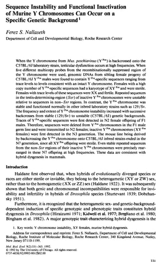
  • 8. 338 Nallaseth Y Pos-specific sequences. They were detected by hybridization of DNAs with probes A-D and (GATA)s. The types of progeny resulting from the background on which the Y ‘OSchro- mosome was either structurally and functionally unstable (C57BL/6J) or stable ( 129/ Sv) are schematically represented in figure 2. Four types of sibling C57BL/6J Y ‘OS females were classified on the basis of the estimated copy numbers of their Y ‘OS- specific sequence content relative to normal males. For simplification of analysis, the major Y ‘OS-specific sequence content of normal C57BL/6J Y ‘OSand 129/Sv Y ‘OS males hybridizing to probes A-D and (GATA)5 was rounded to 100 copies, or 100%. The four types of C57BL/6J Y ‘OSfemales identified were High Copy Y, Low Copy Y, Traces Y, and Normal females, which had, respectively, equal amounts, lo%, l%, and 0% of the Y “‘“-specific sequence content of males (fig. 2). Some of the phenotypic and karyotypic characteristics of these females are also listed (fig. 2 ) . Detection of unique-copy levels or traces of Y Pos-specific sequences in female DNA with any one probe established Traces Y identity for that female. Regardless of the presence or absence of Y ‘OS-specific sequences hybridizing to the five probes in these analyses, these progeny are likely to be distinct from inbred strains. One example of their difference was the elevated frequency of transmission of a 4.7-kb EcoRI frag- ment cross-hybridizing with probe B. It was transmitted at six-fold higher frequencies to daughters of C57BL/6J Y ‘OSmales than to daughters of inbred strain C57BL/6J males (author’s unpublished data). Molecular and genetic analysis of Traces Y and High Copy Y females are presented in the present paper. Levels of Y ‘OS-specific Sequences Found in Traces Y Females Transfer of unique-copy levels of Y ‘OS-specific sequences to autosomes or X chromosomes during meiosis is one result predicted by structural instability of the Y ‘OSchromosome. Getiomic DNAs from sibling female progeny of C57BL/6J Y ‘OS cnB1/8J M. poschiabinus Y-chromosome in female c57BlAJ baekgrolltld M. poschiavinus Y-chromosome in 129Sv background No No No YeS YeS FIG. 2.-Types of progeny from matings involving genetic backgrounds on which Y chromosome from Musposchiavinus was unstable (C57BL/6J) and stable ( 129/Sv). X and Y chromosomes are represented, respectively, by a hatched rectangle and a shaded rectangle with a blackened, box superimposed on it. The blackened box represents the Sxr region containing the Tdy locus that is translocated to XX’“’ males. (N) and X represent backcross generations and matings, respectively. (N) represents backcrossing the M. pos- chiuvinus Y chromosome for 3-7 generations onto the C57BL/6J genetic background and onto the 129/ Sv genetic background for > 18 generations. Unblackened boxes superimposed on either shaded rectangles or closed boxes represent extent of deletions and/or rearrangements of Y-specific sequences. Shaded boxes superimposed on hatched boxes represent unique-copy levels of Y-specific sequences that have putatively been transferred to either X chromosomes or autosomes. High Copy Y, Low Copy Y, Traces Y, and Normal females and males were classified on the basis of the estimated copy numbers of their Y-specific sequence content (see text ).
  • 9. Instability and Inactivation of Murine Y Chromosomes 339 males were tested for the presence of Y ‘OS-specific sequences by hybridization with probes A-C and (GATAh . Traces Y females and Normal females that were identified by this analysis are represented by T and F below their respective lanes in figure 3. XXSx’and Normal male DNAs are represented by S and M below their respective lanes in figure 3. Estimated levels of Y ‘OS-specific restriction fragments identified in each DNA are listed below the respective lanes in figure 3. Y Pos-specificEcoRI fragments hybridizing to probe A were of expected size (0.6 kb) and copy number (+++ ) in male DNAs (fig. 3A, lanes 5 and 6) and absent (- ) in Normal female DNAs (fig. 3A, lanes 3 and 4). X-linked internal standard EcoRI fragments were detected in all lanes (fig. 3A, lanes l-6). The lower levels of the internal standards in male DNAs relative to female DNAs reflected the fivefold lower amounts of male DNAs that were loaded on the gel. Probe A hybridized to - 1.O copy (+; see arrow in fig. 3A, lane 1) and -0.1 copies ( l/f; see arrow in fig. 3A, lane 2) of the Y ‘OS-specific 0.6-kb EcoRI fragment in two female DNAs and therefore identified them as Traces Y DNAs. Y ‘“-specific EcoRI fragments hybridizing to probe B were of expected size (4.3 kb, 3.7 kb, and 2.2 kb) and copy numbers (+++) in male DNAs (fig. 3B, lanes 5- 8) and were absent (- ) in Normal female DNAs (fig. 3B, lanes 1, 2, and 4). The internal standard 3.9-kb EcoRI fragment for probe B was detectable in all female DNAs (fig. 3B, lanes l-4). At the high exposure levels required to detect the 3.9-kb EcoRI fragment, Y ‘OS-specific EcoRI fragments in male DNAs hybridized to probe B were obscured. They were clearly visualized at lower exposure levels (fig. 3B, lanes 7 and 8). Probe B hybridized to unique-copy levels of the Y ‘“-specific 4.3-kb EcoRI fragment in one female DNA (+; see arrow in fig. 3B, lane 3) and therefore identified it as Traces Y DNA. Y ‘““-specific EcoRI fragments hybridizing to probe C were of expected size (6.6 kb) and copy number (+) in mixtures of male/female DNAs ( l/250) (fig. 3C, lane 9) and were absent ( - ) in normal female DNAs (fig. 3C, lanes 7 and 8). The internal standard 3.5-kb EcoRI fragment was detectable in all DNAs (fig. 3C, lanes 1 to 9). Probe C hybridized to unique-copy levels of the Y ““-specific 6.6-kb EcoRI fragment in six female DNAs (+; see thick arrow in fig. 3C, lanes l-6) and therefore identified them as Traces Y DNAs. A minor Y ‘OS-specific 9.7-kb EcoRI fragment of probe C was also detected in one Traces Y DNA (fig. 3C, lane 1). HueIII-digested DNA from females (fig. 3D, lanes l-4 and 7-10)) XXS”’males (fig. 3D, lanes 5 and 11)) and normal males (fig. 3D, lanes 6 and 12) were hybridized with end-labeled (GATA)5 oligonucleotide. Autoradiographs of the same filter were exposed to high (fig. 3D, lanes l-6) and low (fig. 3D, lanes 7-12) intensity levels. Hue111 fragments hybridizing to (GATA)S and specific for the Sxr region were of expected size ( -23 kb and r 100 kb) and total copy number (> 1,000) in XXS”’male DNAs (fig. 3D, lanes 5 and 11) and were absent in two Normal female DNAs (fig. 3D, lanes 1, 2, 7, and 8). Significantly lower copy-number levels of the 2 100-kb and -23-kb Hue111fragments hybridizing to (GATAk were present in Normal male DNA (fig. 3D, lanes 6 and 12). The internal standard Hue111 fragments for (GATA)s, ranging from -5.0 kb to -0.5 kb, were detected in all DNAs (fig. 3D, lanes l-6 and 7- 12). Relative to the total levels of 2 100-kb and -23-kb Hue111fragments in XXSxr male DNA, the (GATAh probe hybridized to unique-copy levels of the -23-kb Hue111 fragment in two female DNAs (see arrow in fig. 3D, lanes 3 and 4) and therefore identified them as Traces Y DNAs. At the high levels of exposure required to detect these -23-kb Hue111 fragments in Traces Y female DNAs, comigrating fragments in
  • 10.
  • 11.
  • 12.
  • 13. Instability and Inactivation of Murine Y Chromosomes 343 XX’“’ male and normal male (fig. 3D, lanes 5 and 6) DNAs were obscured. Both r lOO-kb and -23-kb Hue111 fragments were clearly visualized at lower levels of ex- posure of this autoradiograph (fig. 3D, lanes 11 and 12). Comparison of the internal standard Hue111 fragments of (GATA)s in Normal female DNAs (fig. 3D, lanes 1, 2, 7, and 8) and in Traces Y female DNAs (fig. 3D, lanes 3, 4, 9, and 10) showed that all restriction-endonuclease digestions were complete and that more Normal female DNAs than Traces Y female DNAs were loaded on the gel. These results excluded trivial technical explanations such as partial digests and underloading, for the -23- kb H&II fragment of (GATA)S being present in Traces Y female but absent in Normal female DNAs. It was conceivable that, despite precautions, contamination by either plasmid or male genomic DNAs may be trivial technical explanations for the presence of traces of Y-specific sequences in female DNAs. Both of these potential explanations were ruled out by the data presented here. Some, but not all, of the Y-linked probes identified Y ‘““-specific sequences in genomic DNA from the same Traces Y female. Genomic DNAs from the individual Traces Y females represented in lanes 1, 3, and 4 in figure 3A were, respectively, represented in lanes 1,2, and 3 in figure 3C. Both probe A (fig. 3A) and probe C (fig. 3C) identified Y ‘OS-specific sequences in one of these females (fig. 3A, lane 1, and fig. 3C, lane 1). Although the respective internal standards of both probes A and C were clearly visible, only probe C revealed Y ““-specific sequences in two of these females (compare fig. 3A, lanes 3 and 4, with fig. 3C, lanes 2 and 3). To further exclude contamination as a potential explanation, Traces Y genomic DNA on the samejilter was successively hybridized with probes A and C. Genomic DNA from the same individual Traces Y female hybridized with probes A, C, and (GATA)s are represented in lanes 1,4, 7, and 10 in figure 3E. Y Pos-specificsequences (see thick blackened arrow in fig. 3E) of,probes A (fig. 3E, lanes 1 and 3) and (GATA)S (fig. 3E, lanes 7 and 10) of expected size and copy number were detected in this Traces Y female DNA. The filter represented in lanes 1 and 2 of figure 3E was stripped of probe, and both it and a similarly treated filter of male genomic DNA (fig. 3E, lane 3) were rehybridized with probe C (fig. 3E, lanes 4-6). Although the internal standards of probe C (3.5 kb) and probe A (residual signals at 2.2 kb and 1.7 kb) were clearly detected, the expected Y ‘OS-specific 6.6-kb EcoRI fragment hybridizing to probe C was absent in this Traces Y female DNA (see unblackened arrow in fig. 3E, lanes 4 and 6). Furthermore, the -23-kb Hue111fragment hybridizing to (GATA)5 was present at unique-copy levels in this Traces Y female DNA (see thick blackened arrow in fig. 3E, lane 10) and was absent in Normal female DNA (fig. 3E, lane 11). The expected copy number of the -23-kb Hue111fragment was detected in coelectrophoresed male DNA (fig. 3E, lanes 9 and 12). Contamination by male DNA cannot explain detection of Y pOs-specific sequences in the same Traces Y DNA by only two of the three Y- linked probes. Since we have no plasmids of -23-kb size, detection of -23-kb Hue111 fragments in Y ‘OS-specific sequences hybridized with the (GATA)s oligonucleotide cannot be attributed to contamination by plasmids. In any case, most plasmid and other DNAs would be digested to small fragments, because of the high frequency of sites for HueIII. This result also suggested the random representation of loci from the Y ‘OSchromosome hybridizing to probes A-C and (GATA)s in Y Pos-specific sequences of Traces Y females. Sequences from the entire length of Y ‘OSchromosomes were transmitted to 29%-83% of Traces Y female progeny per generation (table 1; Nallaseth 1987 ). Karyotypic analysis of five independent Traces Y females that had 40 chromo-
  • 14. 344 Nallaseth somes failed to reveal a Y ‘OSchromosome, confirming that they were karyotypically XX. The presence of a Y ‘OSchromosome was confirmed in two of the male siblings of these females. In an identical mating, Either et al. ( 1982) reported 42 XX females. Y ‘OSchromosomes from High Copy Y Females of N4 and N5 Generations Sterile sex-reversed, karyotypically XY ‘OS(High Copy Y) females represent high proportions of N3 progeny of C57BL/6J Y ‘OSmales (Either et al. 1982; Nallaseth 1987 ). However, extensive karyotypic analysis failed to reveal gross structural changes of the Y ‘OSchromosome from these High Copy Y females (Either et al. 1982; author’s unpublished data). Mutations due to higher sequence instability in the testis-deter- mining Sxr region, which represents only 0.05% of the Y chromosome, may explain this discrepancy. Although the testis-determining Tdy locus has been convincingly localized to the Sxr region of the murine Y chromosome (Cattanach et al. 1982; McLaren and Monk 1982; Singh and Jones 1982; McLaren et al. 1988; Roberts et al. 1988), fragments hybridizing specifically to the murine Tdy locus have not been cloned (Page et al. 1987; Koopman et al. 1989; Palmer et al. 1989). An alternative approach tested for higher instability of repeated sequences in Sxr regions than in non-Sxr regions of inactive Y ‘OSchromosomes. Copy numbers and ratios of restriction frag- ments from Sxr regions and non-Sxr regions were compared by hybridizing High Copy Y female DNAs with each of probes (GATA)5 and A-D. EcoRI-digested genomic DNAs from High Copy Y (fig. 4A, lanes 1 and 2) and Normal females (fig. 4A, lane 3) and from inbred strain (fig. 4A, lane 4)) 129/ Sv Y ‘OS(fig. 4A, lane 5), and C57BL/6J Y ‘OS(fig. 4A, lane 6) males were hybridized with probes A-C and rDNA. As expected, the rDNA-specific and Y Pas-specificEcoRI fragments were, respectively, present and absent in Normal female DNA. The mul- ticopy 1.8-kb EcoRI fragment hybridizing to probe C was an RFLP that was char- acteristic of inbred strain (C57BL/6J) Y chromosomes. It was present at unique-copy levels in DNA from males with the Y ‘OSchromosome and in DNA from High Copy Y females. This confirmed the identity of the Y ‘OSchromosome in these females. In these High Copy Y female and male genomic DNAs, there were no differences in the mobilities of major EcuRI fragments hybridizing to probes A-C and rDNA (fig. 4A). Y Pas-specificEcoRI fragments from 3 1 High Copy Y female DNAs and from one of each of the three types of listed male DNAs were densitometrically quantitated, and their ratios were plotted as per the legend to figure 4C. Values of hybridization ratios of seven pairs of Y ‘OS-specific EcoRI fragments hybridizing to probes A-C (fig. 4A and fig. 4C, lower panel) did not differ significantly (>2.5 X) between either male or inbred-strain-male DNAs and the mean for High Copy Y female DNAs (fig. 4C, upper panel). Furthermore, in High Copy Y female DNAs, the range and the mean of the hybridization ratios of each of these seven pairs of Y Pos-specificEcoRI fragments also did not differ significantly (>2.5 X) (fig. 4C, lower panel). Probe D, however, which also hybridized to sequences from the non-Sxr region, identified maximal sequence instability of Y ‘OSchromosomes. All - 100 copies of Y Pos-specificEcoRI fragments (fig. SB, lanes 1 and 5). hybridizing to probe D shifted from 169 bp (see unblackened arrow in fig. 5B, lane 2) to 225 bp (see blackened arrow in fig. 5B, lane 2) in one High Copy Y female DNA. Only partial shifts were detected in High Copy Y female sibling DNAs (fig. 5B, lanes 2-4), in male DNAs (fig. 5B, lane 5) , and in 27 other High Copy Y female DNAs, also from N4 and N5 generations
  • 15. Instability and Inactivation of Murine Y Chromosomes 345 (data not shown). To accentuate instability of sequences hybridizing to probe D, only genomic DNAs displaying maximal 225-bp/ 169-bp hybridization ratios were densi- tometrically quantitated and plotted as per the legend to figure 4C. The mean of the hybridization ratios for probe D fragments in High Copy Y female DNAs (28.9) was 1l-22-fold higher than their hybridization ratios in male DNAs (0.4 and 0.8) (fig. 4C, upper panel ). In High Copy Y female DNAs the hybridization ratios for probe D fragments was 0.1-26, or 260-fold, which was 29-fold higher than their mean hy- bridization ratios ( -8.9) (fig. 4C, lower panel). However, only one of eight pairs of Y ‘OS-specific EcoRI fragments from the non- Sxr region displayed this high degree of instability. The remaining seven pairs of Y ‘OS- specific EcoRI fragments were stable in 31 High Copy Y female DNAs from three types of tissue (kidney, liver, and spleen) and from two backcross generations. Thus, non- Sxr regions of inactive Y ‘OSchromosomes from High Copy Y females of N4 and N5 generations displayed great stability during both meiotic and somatic cell division. In contrast, the Sxr regions of inactive Y ‘OSchromosomes from the aforemen- tioned High Copy Y females were significantly unstable. Genomic DNAs from these High Copy Y females (fig. 4B, lanes 1, 2, 4, and 5), a Normal female (fig. 4B, lane 3), a Normal N3 male (fig. 4B, lane 7 ),and XX’“’males (fig. 4B, lane 6 ) were digested with either HaeIII (fig. 4B) or AluI (data not shown) and hybridized with the end- labeled eicosanucleotide (GATA)5. Internal standards showed that digests were com- plete, and equal amounts of DNA were loaded. Significant variations in copy numbers and hybridization ratios of the 2 lOO-kb/ -23-kb Hue111(fig. 4B) and AZuI (data not shown) fragments hybridizing to (GATA)5 were detected within High Copy Y female DNAs and between High Copy Y female, male, and pooled XX’” male DNAs (see arrows in fig. 4B, lanes l-7). The Z-lOO-kb and -23-kb HaeIII and A/u1 fragments hybridizing to (GATA)s were ‘densitometrically quantitated and plotted as per the legend to figure 4C. XX’“’ males have only -0.05% of the Y chromosome including the Tdy locus (Cattanach et al. 1982; Evans and Burtenshaw 1982; Singh and Jones 1982). However, in pooled XXsx’ male genomic DNAs, the copy number of the 2 100-kb HaeIII fragments was at least 11-fold higher than that in genomic DNAs from High Copy Y females (fig. 4B, lanes 1, 2, 4, and 5) and males (fig. 4B, lane 7 ). The mean of the hybridization ratios of 2 lOO-kb/ -23-kb Hue111and AluI fragments hybridizing to (GATA)5 in High Copy Y female genomic DNAs was - 3.7 (fig. 4C, upper panel). It did not differ significantly from the hybridization ratios of the 2 1OO- kb/ -23-kb HaeIII fragments of (GATA)5, either in pooled XX’“’ male genomic DNAs ( - 3.8 ) or in genomic DNAs from an N3 male ( - 1.97 ) (fig. 4C, upper panel). In contrast, in High Copy Y female genomic DNAs, the hybridization ratios of 2 lOO- kb/-23-kb Hue111and A/u1 fragments of (GATAk were 166.5 and 100, respectively (fig. 4C, lower panel). These ranges were, respectively, 45-fold and 5l-fold higher than the mean hybridization ratios of these Hue111and AZuI fragments in High Copy Y female genomic DNAs. Phenotypic inactivity of Sxr regions of Y ‘OSchromosomes in High Copy Y females correlates well with the molecular instability of their repeated sequence content. It may be that the high variability and the copy-number reductions of sequences in the Sxr regions relative to sequences in the non-Sxr regions reflect the indirect selection for analysis of inactive Y ‘OSchromosomes, i.e., High Copy Y females.
  • 16.
  • 17. Instability and Inactivation of Murine Y Chromosomes 347 Hybrldi~i Ratlo 0 2 8 ?l ONPrnaD 6(4.3)/A 6(3.7)/A 8(4.3)/8(3.7) C(6.6)/A C(l.6)/A C(6.6)/B(4.3) C(6.6)/6(3.7) PIG. 4.-High Copy Y females (HC) of N4 and N5 backcross generations whose Y paachromosomes were more extensively altered in Sxr regions than in non-Sxr regions. Types of matings and identities of symbols or abbreviations are as in the legend to fig. 3. A, EcoRI-digested genomic DNAs (20 pg) from High Copy Y females (lanes 1 and 2), a Normal female (lane 3)) and males (lanes 4-6)) coelectrophoresed and hybridized with probes A-C and with ribosomal DNA (rDNA) as internal standard. High Copy Y females were from the N4 and N5 backcross generations. Males were ofthe inbred strain C57BL/6J (lane 4), 129/ Sv Y ‘Or(lane 5), and C57BL/6J Y ‘OS(lane 6). Major Y-specific EcoRI fragment sizes-and probes to which they hybridized-are listed to the right. Autoradiographs were exposed to intermediate-intensity levels. Lower exposures of autoradiographs with a linear range of signals were densitometrically quantitated to provide the data plotted in panel C. B, HaeIII-digested genomic DNAs ( 10 fig) from some of High Copy Y females (lanes 1, 2, 4, and 5), Normal females (lane 3), XX’“’ males (pooled DNAs; lane 6), and a Normal N3 male (lane 7)) hybridized with end-labeled (CATAh oligonucleotide. DNAs in lanes 1 and 2, lanes 3-5 and lanes 6 and 7 were coelectrophoresed. Autoradiographs were exposed to low-intensity levels (6-8 h at room temperature). These and other autoradiographs were densitometrically quantitated for the data plotted in panel C. C, rDNA-specific and Y ‘-- specific EcoRI fragments of rDNA, probes A-C (panel A and fig. 5A) and probe D (fig. 5B), densitometrically scanned. Areas below peaks were integrated, and the area for each Y ‘OJ-specificEcoRI fragment was normalized to the area for the 12-kb EcoRI fragment of rDNA (internal standard). Sxr region-specific rlOO-kb and -23-kb Hue111(panel B) and Ah1 (data not shown) fragments hybridizing to (GATA), were also densitometrically scanned. Areas below these two peaks were integrated and normalized to integrated areas below the fragments from -5.0 kb to -0.5 kb, which served as internal standards for the (GATA )5probe. Hybridization ratios of pairs of restriction fragments identified on the X-axis of the lower graph of panel C were calculated for each of the genomic DNAs identified in the upper graph of panel C. The mean of hybridization ratios of pairs of fragments was determined for High Copy Y female DNAs and for all male DNAs. The range from minimal to maximal hybridization ratios of pairs of fragments was determined for High Copy Y female DNAs. The polymorphic 1.8-kb EcoRI fragment hybridizing to probe C was only quantitated in DNA from inbred-strain males. Probes A-D were absent in XX% male DNA. All DNAs, unless stated otherwise, were extracted from livers. Probes A-D were hybridized to 31DNAs from N4 and N5 HC-Y female siblings and to 1DNA from an N3 male parent; three types of tissues (kidney, liver, and spleen) from each of eight High Copy Y females plus one type of tissue from seven High Copy Y females totaled 31 DNAs. (GATAh was hybridized to 15 DNAs from N4 and N5 High Copy Y female siblings and from 1DNA from an N3 male parent; two types of tissue (kidney and liver) from each of seven High Copy Y females plus one type of tissue from 1 High Copy Y female totaled 15 High Copy Y DNAs. 8S= C57BL/6J (XY B6)b; fB = C57BL6J Y ‘OJ(XY pos)b; = 1291s~ Y ‘OS(XY ‘“) b; 0 = Sxr (XX’“‘) 8; and n = High Copy Y (XY ‘OS)P. Restriction Analysis: Exclusion of Mechanisms of Extrachromosomal Transmission of Y ““-specific Sequences One explanation for the origin of Y ‘OS-specific sequences in Low Copy Y and Traces Y females is extrachromosomal transmission via circular elements, retrotrans- posons, or transposons. One common feature to all three of these mechanisms of
  • 18.
  • 19. Instability and Inactivation of Murine Y Chromosomes 349 izing to each probe in High Copy Y, Low Copy Y, and Traces Y females. Enzymes with four-base and six-base recognition sites were included (Nallaseth 1987). Ran- domization of restriction fragments characteristic of induction of retroviruses (Jenkins and Copeland 1985) or transposons (Bingham et al. 1982) and excision into extra- chromosomal sequences ( Smith and Vinograd 1972; Stanfield and Lengyell979; Cal- abreta et al. 1982; Krolewski et al. 1982; Potter 1984; Walbot and Cullis 1985) were not detected. Thus, it was concluded that these mechanisms do not account for gen- eration of Y ‘OS-specific sequences in these three types of females. Some of the remaining - 10% of these female DNAs that displayed Y ‘OS-specific EcoRI fragments of unex- pected sizes that hybridized to probes A-C were analyzed below. Two distinct mechanisms for losses of Y ‘OS-specific EcuRI fragments were defined by Traces Y and High Copy Y female DNAs. One of the effects on High Copy Y DNA further excluded extrachromosomal transmission of Y Pos-specificsequences as a possible mechanism. Major Y ‘OS-specific EcoRI fragments of expected size (0.6 kb) and copy number ( - 100) were present (+++) in M. poschiuvinus (Valende) male genomic DNA hy- bridized with probe A (fig. 6, lane 7) . Characteristic minor Y Pas-specificEcoRI fmg- ments (4.2 kb and 3.8 kb) were also present at - IO-fold lower copy number in M. poschiavinus (Valende) male genomic DNA hybridized with probe A. Both major and minor Y Pos-specific EcoRI fragments hybridizing to probe A remained absent (-) from Normal female genomic DNAs, even at high exposures (fig. 6, lanes 1 and 5). Internal standard EcoRI fragments (2.2 kb, 1.7 kb, and 0.8 kb) hybridizing to probe A were present in all DNAs and showed that restriction-endonuclease digestions of genomic DNAs were complete. Y ‘OS-specific EcoRI fragments of the expected size of 0.6 kb were detected in two Traces Y female genomic DNAs hybridized with probe A (see thick blackened arrows in fig. 6, lanes 4 and 6). However, in genomic DNAs from two Traces Y female siblings hybridized with probe A, an EcoRI fragment of the unexpected size of 5.4 kb was present at unique-copy levels (see blackened arrows in fig. 6, lanes 2 and 3). Both 0.6-kb and 5.4-kb EcoRI fragments were present in genomic DNA from a third Traces Y female hybridized with probe A (see blackened arrows in fig. 6, lane 4). Overexposure of autoradiographs of M. poschiavinus ( Valende) male genomic DNA hybridized with probe A failed to reveal a similar sized (5.4-kb) EcoRI fragment (data not shown). Therefore, the 5.4-kb EcoRI fragments were re- producibly generated during transfer of Y ‘OS-specific sequences to autosomes/ X chro- mosomes transmitted to two Traces Y female siblings (fig. 6, lanes 2 and 3) . Because double crossovers are rare events, at least one of the restriction sites flanking the 0.6- kb EcoRI fragment hybridizing to probe A must have been lost during transfer of Y Pas-specificsequences. In contrast to the above loss of restriction sites of a single copy fragment, precise mobility shifts of all - 100 copies of Y ‘OS-specific restriction fragments were detected in genomic DNAs from some High Copy Y females. These High Copy Y females were also from the N5 backcross generation. The multicopy Y Pas-specific sequences hybridizing to probes A and D were well defined by restriction endonuclease mapping and sequencing. The Y ‘OS-specific 600-bp EcoRI fragment of probe A and the Y ‘OS- specific 169-bp EcoRI fragment of probe D were contiguous and present at the fre- quency of once per - 14-kb HpaI repeat unit. Thus, these two probes hybridized to sequences spanning ( - 14 kb X - 100 copies Z) 1,400 kb of the Y ‘OSchromosome. Approximately 50% of the sequences within the 169-bp EcoRI fragment of probe D
  • 20.
  • 21. Instability and Inactivation of Murine Y Chromosomes 35I in High Copy Y females. Precise mobility shifts of the Y ‘““-specific 169-bp EcoRI fragment occurred at the high frequency of 8 of 48 XY ‘OSoffspring from the backcross generations N3-N7. Restriction analysis showed that these mobility shifts were limited to the 169-bp EcoRI fragment within the - 14-kb HpaI repeat unit (author’s unpub- lished data). Trivial technical explanations such as partial digests or aberrant electrophoresis cannot explain either the reproducibility and precision or the limitation of mobility shifts to one of five multicopy EcoRI fragments within a single lane. The molecular mechanisms causing mobility shifts must operate coordinately, precisely, and selectively on each of - 100 copies of the 169-bp EcoRI fragment interspersed over 2 1,400 kb of the Y ‘OSchromosome. Because all other Y ‘OS-specific repeats were intact and the mobility shifts were limited to 169 bp within the - 14-kb @aI repeat unit, intrachro- mosomal events rather than extrachromosomal transmission are the likely mechanisms. These two results showed that deletions and/or rearrangements of Y ‘OSchromosomes were likely to be associated with the transfer of Y ‘OS-specific sequences to the autosomes or X chromosomes that were transmitted to Traces Y females. Sequences First Deleted and Transmitted from Y ‘OSChromosomes during Meiosis in Fl Males The C57BL / 65 and 129/ Sv genetic backgrounds are, respectively, inductive and noninductive for dysfunction of Y Pos-encoded testis determination. A correlation has been detected among the presence in sibling females of traces of Y Pas-specificsequences and intact inactive Y ‘OSchromosomes, the selective instability of repeated sequences in the Sxr region, and nonfunctional testis determination. To test whether the aberrant recombinational mechanisms that randomly delete Y Pos-specificsequences from Y ‘OS chromosomes actually correlate with the disruption of testis determination, a genetic approach was taken. The Y ‘OSchromosome was backcrossed from the 129/Sv to the C57BL/6J genetic backgrounds, where it was structurally and functionally destabilized at high frequencies. If the transmission of Y ‘OS-specific sequences to XX females (i.e., as deletion products) and the functional inactivity of Y ‘OSchromosomes in their XY ‘OSfemale siblings result from related mechanisms, then four predictions can be made for successive backcross generations (N). First, traces of Y Pos-specificsequences should not be detected on the 129/Sv genetic background on which the testis-deter- mining Sxr region is fully functional. Second, traces of Y Pos-specificsequences should not be detected in somatic tissues of F 1hybrids of C57BL/ 6JQX 129/Sv Y ‘% Third, traces of Y ‘OS-specific sequences should first be detected in backcross generations preceding the backcross generation in which inactive Y ‘OSchromosomes of High Copy Y females are detected. Fourth, generating of traces (i.e., deletions) should have a cumulative effect on the functional inactivity of a population of Y ‘OSchromosomes in successive backcross generations. These four predictions of a relationship between the generation of Y ‘OS-specific trace sequences (i.e., deletion products) and the func- tional inactivation of the Sxr region on the Y ‘OSchromosome were examined and found to be correct. They are addressed below. To minimize the presence of non-129/Sv sequence content, a male of the 129/ Sv Y ‘OS(N18) strain was used to produce (C57BL/6JO X 129/Sv Y “‘8) Fl hybrid offspring. Genomic DNAs from F 1 hybrid females were analyzed for their Y Pas-specific sequence content, and their sibling Fl hybrid males were mated with pure-strain C57BL/6J females. Genomic DNAs of [C57BL/6J? X Fl (C57BL/6JQ X 129/Sv Y “V)] N2 female offspring were analyzed for their Y ‘““-specific sequence content.
  • 22. 352 Nallaseth Similarly, a backcross series was independently initiated with a mating of C57BL/6Jn X C57BL/6J Y ““S (N3), and genomic DNAs of N4-N7 female offspring were an- alyzed for their Y ‘OS-specific sequence content. The results of these matings are shown in figures 3-8, and they are summarized in table 1. Genomic DNAs from an inbred strain male (fig. 7A, lane 12, fig. 7B, lane 10) and female (fig. 7A, lane 11, and fig. 7B, lane 9)) hybridized with probe A, displayed Y-specific and cross-hybridizing EcoRI fragments of expected sizes and copy numbers. The cross-hybridizing EcoRI fragments of probe A were clearly detected in genomic DNAs from Fl females (fig. 7A, lanes 1- 10). However, the Y Pos-specific0.6-kb EcoRI fragment hybridizing to probe A was absent from these Fl female DNAs (fig. 7A, lanes 1- 10). Similarly, the respective presence and absence of cross-hybridizing and Y-specific EcoRI fragments of probe C in Fl female genomic DNAs was established (data not shown). Therefore, sequences from Y ‘OSchromosomes were not transferred to either autosomes or X chromosomes in germ lines of 129/Sv Y ‘OSmales. Fur- thermore, cross-hybridizing sequences of probes A and C were not amplified and rearranged so as to acquire electrophoretic mobilities of Y P”“-specificsequences during the somatic development of Fl progeny. High frequencies (75%; table 1) of genomic DNAs from N2 females hybridized with probe A (see thick arrow in fig. 7B, lanes l-8) revealed unique-copy levels of the Y ‘““-specific 0.6-kb EcoRI fragment. Genomic DNAs of these N2 females hy- bridized with probe C and (GATA)s also revealed unique-copy levels of Y ‘“-specific fragments (data not shown). Therefore, Y ‘OS-specific sequences werejirst transferred to autosomes or X chromosomes in germ lines of Fl males. Genetic heterozygosity was sufficient-and predominant proportions of C57BL/ 65 alleles were therefore not required-for generating traces of Y Pos-specificsequences (i.e., deletion products). Genetic heterozygosity of C57BL/6J genetic backgrounds was not sufficient for generating High Copy Y females which were first detected in N3 generations. Thus, the second and third predictions of a causal relationship between the generating of Y ‘OS-specific deletion products and functional inactivity of Y ‘OS chromosomes were observed to be true. The frequencies of Traces Y females containing Y Pos-specific sequences in the backcross generations N2, N4, N5, and N6 were, respectively, 75%, 52%, 29%, and 39% (table 1). A x2 analysis (x2 = 50.05 ) with 1 degree of freedom showed a significant probability (P < 0.00 1) that these traces of Y-specific sequences would be transmitted to XX females at frequencies compatible with normal pseudoautosomal exchange. In contrast, the frequencies of High Copy Y females in the backcross generations N2, N4, N5, and N6 respectively, were O%, 14%, 49%, and 3 1% (table 1). Deleting Y ‘OS- specific sequences from a population of Y ‘OSchromosomes apparently had a cumu- lative effect on the inactivation of their testis-determining (Sxr region) functions in subsequent backcross generations. Thus, the fourth prediction of a causal relationship also held true. The absolute requirement for the germ-line presence of a functional Y ‘OSchro- mosome for the transmission of traces of Y ‘OS-specific sequences to female offspring was also established. Female progeny of (C57BL/6J Y ‘OSNormal females (N4) X C57BL/6J pure-strain males) matings lacked any Y Pos-specificsequences (table 1; Nallaseth 1987). The absence of Y ‘OS-specific sequences in these Normal female (N5 ) progeny further excluded the possibility that amplifications and rearrangements of the cross-hybridizing sequences of Y-specific probes would allow them to acquire Y ‘OS- specific mobilities.
  • 23.
  • 24.
  • 25. Instability and Inactivation of Murine Y Chromosomes 355 Table 1 Types of C57BL/6J YposFemales and Their Frequencies/Generation PARENTAL MATING %OF FEMALE PROGENY No. OF (C57BL/6J Y”“) FEMALES ASSAYED GENERATION High Low (no. of OF Normal” Traces Copy Copy females OFFSPRING (F) Y Y Y excludedb) C57BL/6J (P) X 129/Sv Ypos (6) C57BL/6J (P) X Fl Y’“” (8) C57BL/6J (Q) X N3 Y“” (a) C57BL/6J (P) x N4 Ypos (8) C57BL/6J (0) X N5 Ypw (8) C57BL/6J YpoE(F) (N4) (9) x C57BL/6J Ye6 (B)e Fl 100 0 0 0 9 N2 25 75’ 0 0 9 (1) N4 14* 52 14 0 26 (5) N5 17* 29 49 0 31 (2) N6 0* 39 31 15 15 (2) N5 100 0 0 0 14 NOTE.-All female offspring of C57BL/6J (Q) X C57BL/6J Ypm(8) matings are classified as C57BL/6J Y& females. Ypw = Y chromosome of Mus poschiavinusondifferent genetic backgrounds. ’Progeny in which YPM-specificsequences were not detected with the three moderately repeated Y-specific probes denoted as F. bNot counted in the number of females assayed. cThree of four probes present at trace levels; fourth probe present at low copy levels. dData from Normal (or Traces Y) females in which the presence or absence of Y%pecilic sequences was not conclusively established are omitted. cY” = Y chromosome of C57BL/6J strains. invariant ( co.5 X ) in 3 1 genomic DNAs extracted from three types of tissue (kidney, liver, and spleen) from these High Copy Y females and from males with either Y ‘OS chromosomes or inbred strain Y chromosomes. However, EcoRI-digested’genomic DNAs from High Copy Y females of the N6 and N7 generation hybridized with probe B showed that the ratio of the 4.3-kb and 3.7-kb fragments was altered >30-fold. All copies of the major 3.7-kb and minor 2.2- kb Y ‘““-specific EcoRI fragments hybridizing to probe B were clearly absent in spleen DNA-but not in kidney or liver DNAs-from one High Copy Y female sibling of the N6 generation (see unblackened arrows in fig. 8A, lane 7). All other major Y ‘OS- specific EcoRI fragments hybridizing to probes A-C were identical in kidney, liver, and spleen DNAs from both N6 siblings (fig. 8A, lanes 2-6). Losses of these two fragments occurred in one of four and two of three High Copy Y females of the N6 and N7 generations, respectively. Although genomic DNA in lane 2 of figure 8A was smeared, major Y ‘OS-specific EcoRI fragments hybridizing to probes A-C were visible. In addition, ratios of the rDNA-specific ( 12-kb) EcoRI fragment to each of the major Y ‘“-specific EcoRI fragments of probes A-C differed significantly (i.e., more than fivefold) between genomic DNAs from High Copy Y females of the N6 generation (fig. 8A, lanes 2-6) and N4 and N5 generations (fig. 4A). The filter that was autoradiographed for figure 8A was previously probed with probe C and exposed to high levels (fig. 8B). Minor Y Pas-specific 180-bp EcoRI fmg- ments hybridizing to probe C were present in liver DNAs (see blackened arrow in fig. 8B, lanes 3 and 6) but were absent in kidney and spleen DNAs (fig. 8B, lanes 2,4, 5, and 7) from both of these High Copy Y female siblings of the N6 generation. The filter autoradiographed for figure 8A was stripped of probe and reannealed with probe D. All copies of the Y ‘OS-specific 169-bp EcoRI fragment hybridizing to probe D displayed mobility shifts from 169 bp to 110 bp in liver DNAs-but not in
  • 26. 356 Nallaseth kidney and spleen DNAs-from both of these High Copy Y female siblings of the N6 generation (data not shown). In EcoRIdigested genomic DNAs from livers of two High Copy Y female siblings of the N7 generation, the major 3.7-kb and the minor 2.2-kb fragments hybridizing to probe B were absent in one (see unblackened arrows in fig. 8C, lane 2) but were present in the other (fig. 8C, lane I). All other major Y Pas-specificEcoRI fragments hybridizing to probes A-C were indistinguishable in DNAs from these two N7 siblings (fig. 8B, lanes 1 and 2). Therefore, multicopy Y ‘OS-specific EcoRI fragments hybridizing to each of the three probes B-D underwent distinct types of precise structural changes in different tissues. The Y ‘OSchromosome was structurally destabilized during the somatic de- velopment of High Copy Y females. Trivial technical explanations for these mobility shifts included either partial and star activity of restriction endonucleases or aberrant electrophoretic conditions. Star activity was tested for and was empirically excluded; furthermore, these explanations were incompatible both with the precision and re- producibility of mobility shifts between different genomic DNAs and with their lim- itation to one or two of five multicopy Y ‘OS-specific EcoRI fragments of DNA within a single lane. Therefore, eventually, even stable repeated sequences on Y ‘OSchromosomes showed precise structural alterations at high frequencies. These Y ‘OSchromosomes were structurally destabilized in terminal backcross generations, suggesting that their maintenance on C57BL/6J backgrounds had some cumulative effect. Discussion Structural Destabilization: Functional Inactivation of Feral Y ‘OSChromosomes on C57BL/6J Inbred Backgrounds The data presented in the present paper demonstrated that the Y ‘OSchromosome from the feral species MUSposchiavinuswas structurally destabilized at high frequencies when it was backcrossed onto the C57BL/6J inbred genetic background. However, the Y ‘OSchromosome was stable on the 129/Sv inbred genetic background. Molecular and genetic analysis showed (a) that there was a strong correlation between the struc- tural instability of the Y ‘OSchromosome on C57BL/6J backgrounds and eventual functional inactivation of its Tdy locus and ( b) that the Y ‘OSchromosome functioned normally on 129/ Sv genetic backgrounds (fig. 2). Two results predicted by the structural destabilization of Y ‘OSchromosomes were confirmed, by using Y-linked multicopy sequences, by probing genomic DNAs from daughters of C57BL/6J Y ‘OSmales. In genomic DNAs from XX females, unique- copy levels or traces of Y ‘OS-specific sequences were present at high frequencies (29%- 75%/Y-chromosomal repeat sequence/generation). These were Traces Y females. In genomic DNAs from some of their XY ‘OSfemale siblings, multiple copies of Y ‘OS- specific restriction fragments either were absent or underwent precise coordinated mobility shifts. These were High Copy Y females with structurally destabilized Y ‘OS chromosomes. Trivial technical explanations for trace sequences in Traces Y females and for destabilized Y ‘OSchromosomes in High Copy Y females were excluded. Data for Low Copy Y females are not presented. In 80 N4 offspring from matings that were identical to those in the present study, Either et al. ( 1982) identified 42 XX females, 16 XY ‘OSfemales, and 22 XY ‘OSwith ovaries and/ or ovotestis. Five Traces Y and 2 High Copy Y females were karyotypically XX and XY ‘OS,respectively (author’s unpublished data). Given that 11 of 21 N4
  • 27. Instability and Inactivation of Murine Y Chromosomes 357 females analyzed in the present work were Traces Y females, at least some of the XX females in the study by Either et al. ( 1982) were also Traces Y females. Inability to detect either presence of Yposchromosomal fragments or gross structural changes of Y ‘OSchromosomes in karyotypes of XX and XY ‘OSfemales could be explained by two results from this work: ( 1) in High Copy Y females of the N4 or N5 generation, repeated sequence copy numbers and stabilities were more extensively reduced in Sxr regions containing the T&locus and representing (0.05% of Y ‘OSchromosomes than in non-Sxr regions; and (2) since fragments ~200 kb cannot be visualized in optical microscopes, both Traces Y and Low Copy Y females would escape detection. Although it would be ideal to show that deletions/rearrangements of the Tdy locus inactivate testis-determining functions of Y ‘OSchromosomes, current under- standing of this complex developmental pathway (Singh et al. 1984; Chandra 1985; Page et al. 1987; Koopman et al. 1989; Palmer et al. 1989; Schneider-Gadicke et al. 1989)) precludes such an unambiguous correlation. Instead, when a genetic approach was used, increased structural instability indirectly correlated with the functional in- activity of a population of Y ‘OSchromosomes backcrossed from 129/Sv onto C57BL/ 65 genetic backgrounds for seven successive generations. Since all XY ‘OSoffspring were sterile, this line ended in the N7 generation. Traces Y females were absent in Fl but were present at high frequencies in litters from N2 and subsequent generations. High Copy Y females were absent in N2 but appeared at increasing frequencies in the subsequent, N3-N7 generations. Y ‘OS-linked repeated sequences that were stable during both germ-line and somatic development in the N4 and N5 generations underwent precise structural destabilization at high frequencies during somatic development in the N6 and N7 generations. Thus, maintenance of Y ‘OSchromosomes on C57BL/6J backgrounds resulted in structural destabilization, which eventually inactivated func- tions of their Tdy and other loci. The inactivation of testis-determining functions of the Y ‘OSchromosome because of its sequence instability is predictably stochastic. Therefore, the mouse line being derived will end in sterile individuals after varying numbers of backcrosses of the Y ‘OSchromosome onto the C57BL/6J genome. By selecting for males lacking any overt external signs of sex reversal, it has been possible to slightly extend the number of backcross generations to NlO, before the line ends in sterile XY ‘OSindividuals (J. Barry Whitney III, personal communication). Others (Nagamine et al. 1987; Biddle and Nishioka 1988) have reported similar extensions of numbers of backcrosses,of Y ‘OSchromosomes onto C57BL/6J genetic backgrounds. However, the results presented in the present paper make it likely that C57BL/6J Y ‘OSmice in each of these laboratories is genotypically distinct. Stable X-linked transmission of Y ‘OS-specific sequences from the germ line of a Low Copy Y female mated with males of an inbred strain was tested for. Nontrans- mission of Y Pos-specific sequences from the Low Copy Y female may be explained by one of the following three observations: ( 1) There were 1O-fold differences of copy numbers of Y ‘OS-specific sequences in three tissues of this female. Their complete absence from ovaries may explain nontransmission of Y “‘“-specific sequences by this Low Copy Y female. (2) The results in the present paper showed that sequences on Y ‘OSchromosomes underwent structural destabilization during germ-line and somatic development. Their excision from chromosomes of developing ova or embryos may explain nontransmission of Y ‘OS-specific sequences from this Low Copy Y female. ( 3) Finally, other donor sequences or transgenes are also not transmitted from male (Wilkie and Palmiter 1987) and female (Rohan et al. 1990) germ lines of transgenic mice. In the case of the male mouse, the MyK 103 transgene had to be excised during
  • 28. 358 Nallaseth spermatogenesis ( Wilkie and Palmiter 1987) to allow formation of viable sperm. In- stability of other nucleotide sequences during somatic or embryonal development has also been reported (Kelly et al. 1989; Rohan et al. 1990). Loss of Genetic Regulation of Recombination: Possible Cause of Instability of Y ‘OSChromosomes Locus-specific and chromosome-specific genetic regulation of the five fundamental functions of eukaryotic chromosome biology-i.e., replication, repair, recombination (interchromosomal and intrachromosomal), condensation, and segregation-are well established in yeast, Neurospora, and Drosophila (Baker et al. 1976; Gatti et al. 1980; Strathern et al. 1981; Klapholtz et al. 1985; Surosky and Tye 1988). Recombination in all organisms is known to be under rigorous genetic regulation (Kucherlapati and Smith 1988). The inbred strain C57BL/6J is genetically distinct from both 129/Sv and most other inbred strains (Taylor 1972; Cattanach and Moseley 1973; Forejt and Ivinyi 1975; Fitch and Atchley 1985). It is likely that alleles controlling recombination in C57BL/6J and 129/Sv inbred strains are distinct, thus explaining the instability of Y pOschromosomes on C57BL/6J but not on 129/Sv genetic backgrounds. At least two distinct types of aberrant recombination events on Y ‘OSchromosomes were identified by Y ‘OS-specific sequences in Traces Y and High Copy Y females. Y Pos-specificsequences in Traces Y females were first transmitted from F 1 male germ lines. They were only partially representative of copies ( - 1/ 100) and types [A-C and (GATA)5] of interspersed repeated sequences from the entire length of Y ‘OS chromosomes. Losses of flanking restriction sites of these traces of Y Pos-specific se- quences were also detected. These characteristics of Y Pas-specific sequences are in- consistent with translocations, extrachromosomal elements, and recombinational hot spots. They are consistent with random interchromosomal recombination products which require limited sequence homology for heteroduplex formation, e.g., gene con- versions. The inferred mechanism for the transfer of Y Pas-specific sequences to XX females is the loss of suppression of XB6-Y‘OSinterchromosomal recombination. This inference is supported by other observations on X-Y recombination. X-Y interchro- mosomal pairing is known to be destabilized in offspring from matings between lab- oratory strains and wild mice (Cattanach and Moseley 1973; Forejt and Ivanyi 1975; Matsuda et al. 1983; Handel 1987). Many translocations from Y chromosomes are to X chromosomes (Singh and Jones 1982; Page et al. 1987; Petit et al. 1987), and unequal crossovers have been shown to occur in the murine pseudoautosomal region (Harbers et al. 1986). Y Pos-specificsequences in XY ‘OSfemales showed contrasting characteristics. Pre- cise reproducible losses or mobility shifts of all or most copies of multicopy Y ‘OS- specific restriction fragments occurred in a single generation during somatic and/or germ-line development. These losses and mobility shifts were restricted to specific interspersed repeated sequences and to specific restriction fragments within a repeated sequence. Because of the high precision and frequency of these events and because of the large chromosomal sizes represented by interspersed repeated sequences, these were highly coordinated mechanisms. As somatic X-Y pairing is absent in mammals, these events must involve either intrachromosomal mechanisms of recombination or some other sequence modification. The loss of control of recombination, which resulted in sequence instability, was not limited to combinations of the C57BL/6J inbred strain and feral Y ‘OSchromo- somes. Y chromosomes from the inbred strain C57BL/6J and another feral mouse,
  • 29. Instability and Inactivation of Murine Y Chromosomes 359 hf. domesticus (Centreville Light), were reciprocally backcrossed for two generations. Traces of Y-specific sequences and precise losses and mobility shifts of multicopy Y-linked sequences also resulted from both types of these matings (author’s unpub- lished data). The high frequencies and precision of highly coordinated changes of sequences on Y ‘OSchromosomes suggest that these changes were likely to result from mechanisms that were distinct from other mechanisms causing genetic instability. The loss of epi- genetic control of retroviral replication by a maternal resistance factor and FV restric- tion loci in SWR/J X RF/J murine hybrids results in the induction of retroviruses (Jenkins and Copeland 1985). Similarly, their lower frequencies and different types of aberrant recombination products suggest that the meiotic instability of the poly- morphic repeat sequence PR 1 and VrDNAs in the murine rDNA loci (Kuehn and Amheim 1983; Kominami et al. 1985), microrecombinations in the murine major histocompatibility locus (Geliebter and Nathenson 1988)) somatic instability of murine variable numbers of tandem repeats (VNTRs) (Kelly et al. 1989 ), and the rearrange- ment-induced premeiotically (RIP) in N. crassa (Selker et al. 1987) are mechanistically unrelated to the instability of the Y ‘OSchromosome. It is now recognized that unknown mechanisms result in two opposing effects on the repeated sequence fraction of the mammalian genome. Although there is a large extent of nucleotide sequence flux within it, structural and functional order are imposed on the mammalian genome (Goldman et al. 1984; Dover and Flavell 1984; Bemardi et al. 1985; Dover and Tautz 1986; Schmid and Shen 1986; Zuckerlandl 1986; Dover 1987, 1990; Korenberg and Rykowski 1988; Kucherlapati and Smith 1988 ). Fluxes of repeated sequences include such large genome-encompassing effects as their concerted evolution, retrotranspositioning (Dover and Flavell 1984; Baltimore 1985; Rogers 1985; Dover and Tautz 1986; Schmid and Shen 1986; Zuckerlandll986; Dover 1987, 1990)) and expansions and contractions of VNTRs (Dover 1987, 1990; Jeffreys et al. 1990). The rate, precision, and extent of these nucleotide sequence fluxes cannot be explained either by Mendelian inheritance or by stochastic, slow, and un- regulated processes such as drift, selection, and “selfishness” of DNA (Dover and Tautz 1986; Schmid and Shen 1986; Dover 1987, 1990). Loss of regulation of any of the unconventional mechanisms, either imposing structural and functional order on mammalian genomes or driving large nucleotide sequence fluxes through them, are potential mechanisms for the non-Mendelian transmission of Y p”s-specificsequences documented in the present paper. Loss of Y ‘OSChromosome Structure and Function as Consistent with Hybrid Dysgenesis Occurring in Mammals ’ When genomes of two evolutionarily diverged species are hybridized, a collection of genotypic and phenotypic dysfunctions are induced. Traits collectively characterizing hybrid dysgenesis are requirements of directionality of genetic crosses, induction of transposons, disruption of regulation of interchromosomal (meiotic) and intrachro- mosomal (mitotic) recombination, suppression of recombination at active loci, in- duction of recombination in inactive loci (and genomes), chromosome nondisjunction, segregation (or transmission-ratio ) distortion, gonadal dysfunction (sterility), high frequencies of mutations, and embryonal lethalities (Haldane 1922; Dobzhansky 1951; Tracey 1972; Forejt and Ivanyi 1975; Kidwell et al. 1977; Bregliano et al. 1980; Bingham et al. 1982; Handel 1987; Yannopoulos et al. 1987). Most of the genotypic and phenotypic traits defining hybrid dysgenesis in other species are also identified in
  • 30. 360 Nallaseth the C57BL/6J Y ‘OSmouse. Two of the most important traits characterizing hybrid dysgenesis are loss of control of recombination and testicular dysfunction, and they are specifically recognized in C57BL/6J Y ‘OSmice (for a list of these and other traits, see table 2 ). Many of these traits were also reproduced when the Y chromosome from another feral species, M. domesticus (Centreville Light), was backcrossed onto C57BL/ 65 inbred strains (author’s unpublished results). Like most inbred strains, C57BL/6J originated as a hybrid of M. musculus do- mesticus P X M. m. musculus d (Bishop et al. 1985). The C57BL/6J inbred-strain genome contains alleles that make it distinct from all other inbred strains (Taylor 1972; Cattanach and Moseley 1973; Forejt and Ivanyi 1975; Bonhomme et al. 1984; Fitch and Atchley 1985). The Y ‘OSchromosome itself originates from M. poschiavinus species, which is highly diverged from all laboratory strains, both in its karyotypic constitution (Gropp et al. 1972; Cattanach and Moseley 1973; Capanna 1982) and in its nuclear (Cattanach and Moseley 1973) and mitochondrial (Ferris et al. 1983) genomes. It is likely that (a) differences in C57BL/6J alleles controlling recombination and (b) the evolutionary divergence of Y ‘OSchromosomes act together to produce sequence instability of the Y ‘OSchromosome in this inbred strain. Table 2 Traits Characterizing Dysgenesis in Strains Derived from Mus musculus X M. po~chia~inus Hybrids Trait Reference(s) Genotypic: Directionality of genetic crosses: C57BL/6JQ X 129/Sv Ypo6$ causes dysgenesis .,.,_.,_...,_.__._.._........................ Induction of interchromosomal recombination: Y-linked sequences are transferred to other chromosomes (transferred sequences are derived from B1, R, and satellite repeats) and sequences from >6,000 kb are transferred from YPorchromosome Suppression of interchromosomal recombination in autosomal and X chromosomal loci Aneuploidy of autosomes and X chromosomes Increased frequency of transmission of 4.7-kb EcoRI autosomal/X chromosomal unique sequence ........................... Induction of mitotic instability of YpDschromosomes ........... Highfrequencyofmutations .............................. Phenotypic: Disruption of primary sex determination ..................... Disruption of oogenesis and spermatogenesis ................. Disruption of embryogenesis Disruption of secondary masculinization Present study Nallaseth 1987; present study Cattanach and Moseley 1973 Gropp et al. 1972; Cattanach and Moseley 1973; Capanna 1982; Magnuson et al. 1985 Nallaseth 1987 Nallaseth 1987 Cattanach and Moseley 1973 Either et al. 1982; present study Gropp et al. 1972; Cattanach and Moseley 1973; Capanna 1982; Either et al. 1982; Magnuson et al. 1985; Nallaseth 1987 Gropp et al. 1972; Cattanach and Moseley 1973; Magnuson et al. 1985 Either et al. 1982; Nallaseth 1987
  • 31. Instability and Inactivation of Murine Y Chromosomes 361 Two important differences are noted between the hybrid dysgenesis detected in C57BL/6J Y ‘OSmice and that in Drosophila (Kidwell et al. 1977; Bregliano et al. 1980; Bingham et al. 1982; Yannopoulos et al. 1987). First, transposon (or retroviral) excision does not explain the presence of Y ‘OS-specific sequences in females. Second, in addition to random recombination, precise, reproducible, and high-frequency se- quence instabilities are induced de novo in loci that are normally recombinationally suppressed on the Y chromosome during hybrid dysgenesis. Only imprecise recom- bination at chromosome breaks marking sites of transposon excision occurs during hybrid dysgenesis in Drosophila. Finally, since natural hybrid zones of M. musculus X M. poschiavinus have been identified (Gropp et al. 1972; Capanna 1982), these observations may have important implications for speciation both in these hybrids and in laboratory strains, which originated as hybrids (Bishop et al. 1985 ) . Acknowledgments I wish to thank J. Barry Whitney III, who initially provided 129/v Y ‘OSand C57BL/6J Y ‘OSmale mice that were later transferred to the colonies at the University of South Carolina, and Michael J. Potter for providing Mus poschiavinus (Valende ) and &f. domesticus (Centreville Light) mice. They were maintained by funds from NCI, contract NOl-CB-25594. I also gratefully acknowledge the many contributions of Peter J. Hornsby, Mel DePamphilis, Gerald Smith, Robert Erickson, Bruce Baker, Rollie Harp, Gloria Choice, Charlotte Joyner, and Sharon Perry that made this work possible. This work was partially supported by Public Health Service grant HD 17523 awarded to Michael J. Dewey. LITERATURE CITED BAKER,B. S., A. T. C. CARPENTER,R. E. ESPOSITO,and L. SANDLER.1976. The genetic control of meiosis. Annu. Rev. Genet. 10:53-134. BALTIMORE,D. 1985. Retroviruses and retrotransposons: the role of reverse transcription in shaping the eukaryotic genome. Cell 40:48 l-482. BERNARDI,G., B. OLFSSON,J. FILIPSKI,M. ZERIAL,J. SALINAS,G. CUNY, M. MEUNIER-RO- TIVAL,and F. RODIER. 1985. The mosaic genome of warm-blooded vertebrates. Science 229:1277-1281. BIDDLE,F. G., and Y. NISHIOKA. 1988. Assays of testis development in the mouse distinguish three classes of M. domesiicus-type Y chromosome. Genome 30:870-878. BINGHAM,P. M., M. G. KIDWELL,and G. M. RUBIN. 1982. The molecular basis of P-M hybrid dysgenesis: the role of the P element, a P-strain-specific transposon family. Cell 29:995- 1004. BISHOP,E., P. BOURSOT,B. BARON,F. BONHOMME,and D. HATAT. 1985. Most classical Mus musculus domesticus laboratory mouse strains carry a Mus musculus musculus Y chromo- some. Nature 315:70-72. BONHOMME,F., J. CATALAN,J. BRITTON-DAVIDIAN,V. M. CHAPMAN,K. MORIWAKI,E. NEVO, and L. THALER. 1984. Genetic diversity and evolution in the genus Mus. Biochem. Genet. 22~275-300. BREGLIANO,J. C., C. PICARD,A. BUCHETON,A. PELISSON, J. M. LAVIGE,and L. HERITIER. 1980. Hybrid dysgenesis in Drosophila melanogaster. Science 207:606-6 11. CALABRETTA,B., D. L. ROBBERSON,H. A. BARRERA-SALDANA,T. P. LAMBROU,and G. F. SAUNDERS. 1982. Genome instability in a region of human DNA enriched in Alu repeat sequences. Nature 296:219-225. CAPANNA,E. 1982. Robertsonian numerical variation in animal speciation: Mus musculus, an emblematic model. Pp. 155-I 77 in Mechanisms of speciation. Alan R. Liss, New York.
  • 32. 362 Nallaseth CATTANACH,B. M., E. P. EVANS,M. D. BURTENSHAW,and J. BARLOW. 1982. Male, female and intersex development in mice of identical chromosome constitution. Nature 300:445- 446. CATTANACH,B. M., and H. MOSELEY. 1973. Non-disjunction and reduced fertility caused by the tobacco mosaic mouse metacentric chromosomes. Cytogenet. Cell Genet. 12:264- 287. CHANDLEY,A. C., P. GOETZ, T. B. HARGREAVE,A. M. JOSEPH,and R. M. SPEED. 1984. On the nature and extent of XY pairing at meiotic prophase in man. Cytogenet. Cell Genet. 38: 241-247. CHANDRA,H. S. 1985. Sex determination: a hypothesis based on non-coding DNA. Proc. Natl. Acad. Sci. USA 82:1165-l 169. COOKE, H. J., and B. A. SMITH. 1986. Variability at the telomeres of the human X/Y pseu- doautosomal region. Cold Spring Harbor Symp. Quant. Biol. 51:2 13-2 19. DOBZHANSKY,T. 195 1. Genetics and the origins of species, 3d ed. Columbia University Press, New York. DOVER, G. A. 1987. Victims or perpetrators of DNA turnover? Nature 342347-348. -. 1990. Remodelling the involucrin gene and why we are not chimps. BioEssays 12:24 l- 243. DOVER, G. A., and R. B. FLAVELL. 1984. Molecular coevolution: DNA divergence and the maintenance of function. Cell 28:622-623. DOVER, G. A., and D. TAUTZ. 1986. Conservation and divergence in multigene families: alternatives to selection and drift. Philos. Trans. R. Sot. Lond. [Biol.] 312 :275-289. EICHER,E. M., and L. L. WASHBURN. 1986. Identification and chromosomal positions of genes involved in primary sex determination. Annu. Rev. Genet. 20:327-360. EICHER, E. M., L. L. WASHBURN,J. B. WHITNEY III, and K. E. MORROW. 1982. Mus pos- chiavinus Y-chromosome in the C57BL/6J murine genome causes sex reversal. Science 217: 535-537. EVANS,P., and M. D. BURTENSHAW.1982. Crossing-over between the X and Y chromosomes of male mice carrying the sex reversing (Sxr) factor. Nature 300:443-445. FERRIS, F., M. KASAHARA,H. TICHY, E. NEUFELD,U. RITTE, and A. C. WILSON. 1983. Mi- tochondrial DNA evolution in mice. Genetics 105:68 l-708. FITCH, W. M., and W. R. ATCHLEY. 1985. Evolution in inbred strains of mice appears rapid. Science 228:1169-l 175. FOREIT, J., and P. IVANYI. 1975. Genetic studies on male sterility of hybrids between laboratory and wild mice (Mus musculus L.) Genet. Res. 24: 189-206. GATT&M., S. PIMPINELLI,and B. S. BAKER. 1980. Relationships among chromatid interchanges, sister chromatid’exchanges and meiotic recombination in Drosophila melanogaster. Proc. Natl. Acad. Sci. USA 721575-1579. GELIEBTER,J., and S. G. NATHENSON.1988. Microrecombinations generate sequence diversity in the murine major histocompatibility complex: analysis of the Kbm3, Kbm4, Kbm” and Kbm” mutants. Mol. Cell. Biol. 8:4342-4352. GOLDMAN,M. A., G. P. HOLMQUIST,M. C. GRAY,L. A. CASTON,and A. NAG. 1984. Replication timing of genes and middle repetitive sequences. Science 224:686-692. GOODFELLOW,P. J., S. M. DARLING,N. S. THOMAS,and P. N. GOODFELLOW.1986. A pseu- doautosomal gene in man. Science 234:740-743. GROPP, A., H. H. WINKING, L. ZECH, and H. MULLER. 1972. Robertsonian chromosomal variation and identification of metacentric chromosomes in feral mice. Chromosoma 39: 265-288. HALDANE,J. B. S. 1922. Sex ratios and unisexual sterility in hybrid animals. J. Genet. 12: lOl- 109. HANDEL, M. A. 1987. Genetic control of spermatogenesis in mice. Results Prob. Cell Differ. 15:1-62. HARBERS,K., P. SORIANO,U. MULLER, and R. JAENISCH. 1986. High frequency of unequal
  • 33. Instability and Inactivation of Murine Y Chromosomes 363 recombination in pseudoautosomal region shown by proviral insertion in transgenic mice. Nature 324682-685. HINTON, C. W. 1979. Two mutators and their suppressors in Drosophila ananassue. Genetics 92:1153-l 171. HIRAIZUMI, Y. 1971. Spontaneous recombination in Drosophila melunoguster males. Proc. Natl. Acad. Sci. USA 68:268-270. JEFFREYS,A. J., R. NEUMANN,and V. WILSON. 1990. Repeat unit sequence variation in mini- satellites: a novel source of DNA polymorphism for studying variation and mutation by single molecule analysis. Cell 60:473-485. JENKINS,N. A., and N. G. COPELAND.1985. High frequency germline acquisition of ectropic MuLV proviruses in SWR/JXRF/J hybrid mice. Cell 43:8 1l-8 19. KELLY, R., G. BULFIELD,A. COLLICK,M. GIBBS,and A. J. JEFFREYS. 1989. Characterization of a highly unstable mouse minisatellite locus: evidence for somatic mutation during early development. Genomics 5:844-856. KIDWELL,M. G., J. F. KIDWELL,and J. A. SVED. 1977. Hybrid dysgenesis in Drosophila mel- unoguster: a syndrome of aberrant traits including mutation, sterility and male recombination. Genetics 86:813-833. KLAPHOLTZ,S., C. S. WADDELL,and R. E. ESPOSITO. 1985. The role of the SPOl 1 gene in meiotic recombination in yeast. Genetics 96:567-588. KOMINAMI,R., K. SUDO, H. YOSHIKURA,H. SUZUKI, K. MORIWAIU,J. HILGERS, and M. MURAMATSU. 1985. A polymorphic repetitive sequence PRl family: evidence for meiotic instability. J. Mol. Biol. 183:301-309. KOOPMAN,P., J. GUBBAY,J. COLLIGNON,and R. LOVELL-BADGE.1989. Zfy gene expression patterns are not compatible with a primary role in mouse gene determination. Nature 342: 940-942. KORENBERG,J. R., and M. C. RYKOWSKI. 1988. Human genome organization: Alu, lines and the molecular structure of metaphase chromosome bands. Cell 53:39 l-400. KROLEWSKI,J. J., A. H. BERTELSEN,M. Z. HUMAYUN,and M. G. RUSH. 1982. Members of the Alu family of interspersed repetitive DNA sequences are in the small circular DNA population of monkey cells grown in culture. J. Mol. Biol. 154:399-4 15. KUCHERLAPATI,R., and G. R. SMITH(eds.). 1988. Genetic recombination. American Society for Microbiology, Washington, D.C. KUEHN, M., and N. ARNHEIM. 1983. Nucleotide sequence of the genetically labile repeated elements 5’ to the origin of mouse rRNA transcription. Nucleic Acids Res. 11:21 l- 224. MCLAREN, A., and M. MONK. 1982. Fertile females produced by inactivation of an X-chro- mosome of ‘sex-reversed’ mice. Nature 3Ofh446-448. MCLAREN, A., E. SIMPSON, J. T. EPPLEN, R. STUDER, P. KOOPMAN, E. P. EVANS, and P. S. BURGOYNE. 1988. Location of the genes controlling H-Y antigen expression and testis determination on the mouse Y chromosome. Proc. Natl. Acad. Sci. USA 85:6442- 6445. MAGNUSON, T., S. DEBROT, J. DIMPEL, A. ZWEIG, T. ZAMORA, and C. J. EPSTEIN. 1985. The early lethality of autosomal monosomy in the mouse. J. Exp. Zool. 236:353-360. MATSUDA,Y., H. T. IMAI, K. MORIWAKI,and K. KOND~. 1983. Modes of inheritance of X- Y dissociation in inter-subspecies hybrids between BALB/c mice and Mus musculus mo- lossinus. Cytogenet. Cell Genet. 35209-2 15. NAGAMINE,C. M., T. TAKETO,and G. C. KOO. 1987. Studies on the genetics of t&z-l XY sex reversal in the mouse. Differentiation 33:223-23 1. NALLASETH, F. S. 1987. The role of the Y-chromosome in the establishment of murine hybrid dysgenesis and in the analysis of the nucleotide sequence organization, genetic transmission and evolution of repeated sequences. Ph.D. diss., University of South Car- olina, Columbia. NALLASETH,F. S., and M. J. DEWEY. 1986. Moderately repeated Y-chromosomal sequence
  • 34. 364 Nallaseth families represent distinct types of organization and change. Nucleic Acids Res. 14:5295- 5307. NALLASETH,F. S., R. P. LAWTHER,M. R. STALLCUP,and M. J. DEWEY. 1983. Isolation of recombinant bacteriophage containing male-specific mouse DNA. Mol. Gen. Genet. 190: 80-84. PAGE,D. C., R. MOSHER,E. SIMPSON,E. M. C. FISHER,G. MARDON,J. POLLACK,B. MCGILL- VRAY,A. DE LA CHAPPELLE,and L. G. BROWN. 1987. The sex-determining region of the human Y-chromosome encodes a finger protein. Cell 51: 109 1- 1104. PALMER,M. S., A. M. SINCLAIR,P. BERTA,N. A. ELLIS,P. N. GOODFELLOW,N. E. ABBAS, and M. FELLOWS. 1989. Genetic evidence that ZFY is not the testis-determining factor. Nature 342:937-939. PETIT, C., A. DE LA CHAPELLE,J. LEVILLIERS,S. CASTILLO,B. NOEL, and J. WEISSENBACH. 1987. An abnormal terminal X-Y interchange accounts for most but not all cases of human XX maleness. Cell 49:595-602. POTTER, S. S. 1984. Rearranged sequences of a human Kpn I element. Proc. Natl. Acad. Sci. USA 81:1012-1016. ROBERTS,C., A. WEITH, E. PASSAGE,J. L. MICHOT, M. G. MATTEI, and C. E. BISHOP. 1988. Molecular and cytogenetic evidence for the location of Tdy and Hya on the mouse Y- chromosome short arm. Proc. Natl. Acad. Sci. USA 85:6446-6449. ROHAN, R. M., D. KING, and W. I. FRELS. 1990. Direct sequencing of PCR-amplified junction fragments from tandemly repeated transgenes. Nucleic Acids Res. 18:6089-6095. ROGERS, J. H. 1985. Origin and evolution of retroposons. Intl. Rev. Cytol. 93:187-279. ROUYER,F., M.-C. SIMMLER,C. JOHNSSON,G. VERGNAUD,H. J. COOK,and J. WEISSENBACH. 1986. A gradient for sex linkage in the pseudoautosomal region of the human sex chromo- somes. Nature 319:219-295. SCHMID,C. W., and C.-K. J. SHEN. 1986. The evolution ofinterspersed repetitive DNA sequences in mammals and other vertebrates. Pp. 323-358 in R. J. MACINTYRE,ed. Molecular evo- lutionary genetics. Plenum, New York. SCHNEIDER-GADICKE,A:, P. BEER-R•MERO,L. G. BROWN,R. NUSSBAUM,and D. PAGE. 1989. ZFX has a gene structure similar to ZFY, the putative human sex determinant, and escapes X inactivation. Cell 57:1247-1258. SELKER,E. U., E. B. CAMBARERI,B. C. JENSEN,and K. R. HAACK. 1987. Rearrangement of duplicated DNA in specialized cells of Neurospora. Cell 51:741-752. SIMMLER,M.-C., C. JOHNSSON,C. PETIT, F. ROUYER,G. VERGNAUD,and J. WEISSENBACH. 1987. Two highly polymorphic minisatellites from the pseudoautosomal region of the human sex chromosomes. EMBO J. 6:963-969. SINGH, L., and K. W. JONES. 1982. Sex reversal in the mouse (Mus musculus) is caused by a recurrent non-reciprocal crossover involving the X and on aberrant Y chromosome. Cell 28:205-2 16. SINGH, L., C. PHILLIPS,and K. W. JONES. 1984. The conserved nucleotide sequences of Bkm, which define Sxr in the mouse, are transcribed. Cell 36: 11l-120. SMITH,C. A., and J. VINOGRAD. 1972. Small polydisperse circular DNA of HeLa cells. J. Mol. Biol. 69:163-178. STANFIELD, S. W., and J. A. LENGYEL. 1979. Small circular DNA of Drosophila melano- gaster: chromosomal homology and kinetic complexity. Proc. Natl. Acad. Sci. USA 76: 6142-6146. STRATHERN,J., E. JONES,and J. BROACH.198 1. Molecular biology of the yeast Saccharomyces: life cycle and inheritance. J. Mol. Biol. 69: 163- 178. STURTEVANT,A. H. 1939. High mutation frequency induced by hybridization. Proc. Natl. Acad. Sci. USA 25:308-3 10. SUROSKY,R. T., and B. K. TYE. 1988. Meiotic disjunction of homologs in S. cerevisiae is directed by pairing and recombination of the chromosome arms but not by pairing of the centromeres. Genetics 119~273-287.
  • 35. Instability and Inactivation of Murine Y Chromosomes 365 TAYLOR,B. A. 1972. Genetic relationships between inbred strains of mice. J. Hered. 63:83-86. TRACEY,M. L. 1972. Sex chromosome translocations in the evolution of reproductive isolation. Genetics 72:3 17-333. TRES, L. L. 1977. Extensive pairing of the XY bivalent in mouse spermatocytes as visualized by whole-mount electron microscopy. J. Cell Sci. 25: l- 15. WALBOT,V., and C. A. CULLIS. 1985. Rapid genomic change in higher plants. Annu. Rev. Plant Physiol. 36:367-396. WILKIE, T. M., and R. D. PALMITER. 1987. Analysis of the integrant in Myk-103 transgenic mice in which males fail to transmit the integrant. Mol. Cell. Biol. 7: 1646-1655. YANNOPOULOS,G., N. STAMATIS,M. MONASTIRIOTI,P. HATZOPOULOS,and C. LOUIS. 1987. Hobo is responsible for the induction of hybrid dysgenesis by strains of Drosophila mela- nogaster bearing the male recombination factor 23.5MRF. Cell 49:487-495. ZUCKERLANDL,E. 1986. Polite DNA: functional density and functional compatibility in ge- nomes. J. Mol. Evol. 2412-27. WALTER M. FITCH, reviewing editor Received February 6, 1991; revision received October 3, 1991. Accepted October 4, 199 1