SlideShare a Scribd company logo
1 of 70
Presented by :
Esraa Riad Albatrawy
CONTENTS:
Definition:
Classifications:
Etiology and clinical features:
Diagnosis:
Evaluation
Management:
Enamel white lesions:
• Subsurface enamel
demineralization that clinically
presents itself as “a milky white
opacity.
Carious
w l:
White
spot
lesions
Non carious WL
Traumatic Dental fluorosis
(developmental):
hypoplasia
hypocalcification
•White spot lesions usually look whitish opaque
•This whitish appearance is caused by localized light scattering
within lesion body.
•In contrast to sound enamel, which is relatively
translucent, in white spot lesions the light scattered
between the enamel crystals and porosities. (With different
refractive indices)
White Spot Lesions
CARIOUS WHITE
LESIONS
White spot lesion
prolonged
plaque
accumulation
(prolonged
ortho ttt)
colonization
of aciduric
bacteria
over time,
area of
demineralized
enamel.
active white
spot lesion
if not treated,
cavitated
carious lesion
Dental plaque buffering capacity and degree of calcium and phosphate
supersaturation will determine the demineralization process
Risk factors :
Inadequate oral hygiene .
Inappropriate diet (refined sugar, frequent snacks)
History of recent caries lesions .
Lack of adjunctive preventive measures ( fluoride )
Prolonged Orthodontic treatment time
(fixed appliances) > 36 months.
NON CARIOUS
WHITE LESIONS
1- TRAUMATIC HYPOMINERALIZATION:
Well demarcated opacities on the labial surface,
due to injury or infection of the deciduous teeth,
which has affected mineralization of the
permanent teeth .
More defined in shape.
Well differentiated from surrounding enamel.
Often located in the middle of the crown.
Randomly distributed .
Definition:
Clinical appearance:
Whit
By:
2-DENTAL FLUOROSIS:
Cosmetic
condition
Affected
teeth are less
susceptible
to caries
↑↑fluoride
intake during
tooth
formation
• Mottled enamel is a condition indicates the fluorosis characterized by minute white
flecks, or yellow or brown spots or areas, scattered irregularly or streaked over the
surface of a tooth
PATHOGENESIS:
(DELAY IN REMOVAL OF PROTEIN MATRIX)
CLINICAL FEATURES
the most commonly affected tooth is the premolar followed by the
second molar, maxillary incisor, canine, first molar and mandibular
incisors.
White /
yellowish
lesion
Not well
defined
Symmetrical
distribution
Enamel hypoplasia
is a defect of the teeth in which the enamel is deficient in
quantity,[caused by defective enamel matrix formation
during enamel development, as a result of inherited and
acquired systemic condition
Hypocalcified Enamel
defect due to malfunction of enamel calcification, therefore
enamel is of normal thickness but is extremely brittle, with an
opaque/chalky presentation. Teeth are prone to staining and rapid
wear, exposing dentine. Enamel appears less radio-opaque
compared to dentine on radiographs.
DEVELOPMENTAL:
DIAGNOSTIC METHODS
The ideal method for the detection ofWSLs should have a high level
of sensitivity (the ability to detect disease when present) and
specificity (the ability to confirm that disease is absent).
DIAGNOSTIC METHODS
 Visual examination
 Digital photographic examination
 Optical Non-Fluorescent Methods
 Light Scattering
 Fluorescence methods
 Fluorescent dye
 Ultraviolet
 Laser (DIAGNO Dent), CO2 laser
 QLF (quantitative light fluorescence)
 Transillumination
 Electrical Conductance – ECM
 Digital Radiography – DDR
 Thermography
 Multiphoton imaging
VISUAL EXAMINATION:
Prior to examination, scaling of the teeth will be done to
remove any plaque and debris and all tooth surfaces were
polished with pumice and prophylaxis cup.
Simple and inexpensive : no expensive or complex
equipment is required.
Validity : it is often difficult to clinically distinguish white spots
caused by demineralization and those that are due to other
causes, such as developmental hypoplasia or fluorosis.
OPTICAL NON-FLUORESCENT METHODS (LIGHT
SCATTERING)
Light scattering, which can be measured using theOptical Caries
Monitor(OCM)
They used a 100-watt white light as a light source and measured
backscatter with a densitometer.
Convenient and nondestructive method of enamel
demineralization.
Can be applied in the clinical environment
Technique sensitive and results can vary with the degree of wetness
or drying of the tooth.
OPTICAL FLUORESCENT METHODS:
1-FLUORESCENT DYE:
fluorescent dye used to highlight carious enamel .
Once the fluorescent dye has been applied the
specimen is examined under a suitable light source
convenient and nondestructive method of
enamel demineralization.
The disadvantage of these dyes is that slight
procedural variations can result in widely different
degrees of dye uptake.They are mainly used for
the detection and removal of carious dentine.
2-ULTRAVIOLET LIGHT:
Ultraviolet (UV) light used for the early detection of
carious lesions on the smooth surfaces.
Special precautions are required to protect the patient
and operator because UV radiation is harmful to the
eyes and skin.
Safer methods using light sources with a longer
wavelength have been developed.
3-LASER (DIAGNODENT):
The technology of the DIAGNOdent, uses a 655 nm diode laser for detection of
noncavitated, occlusal pit-and-fissure tooth decay, in addition to smooth surface
caries at an earlier stages .
The DIAGNOdent measures laser fluorescence within the mineral structure of the
tooth.The equipment was calibrated to calculate the difference in fluorescence
between the demineralised area and the surrounding sound enamel and thereby
quantify mineral loss and lesion size.
Healthy tooth structure exhibits little or no fluorescence, resulting in very low
scale readings on the display
Decayed tooth structure will exhibit fluorescence, proportionate to the degree of
lost tooth structure, resulting in elevated scale readings on the display of the
DIAGNOdent
RECOMMENDATIONS FOR TREATMENT ARE:
values of 30+
operative and
preventative
care.
4-QLF (QUANTITATIVE LIGHT
FLUORESCENCE):
QLF is a diagnostic method that relies on the autofluorescence of teeth when they are
exposed to high-intensity blue light.
QLF is a highly sensitive diagnostic test, with the most promising fluorescent method of
measuring demineralization in use today.
It is usefull in monitoring of mineral changes in incipient enamel lesions, and for the
evaluation of preventive measures in caries prone persons, such as orthodontic patients.
↑↑sensitivity and
specificity
Transillumination:
(fotI , difoti)
Digital Imaging Fiber-OpticTrans-Illumination (DIFOTI), did not
measure the depth, could only detect surface changes associated
with early demineralization as early as 2 weeks.
The fluorescence of the tooth is closely
related to the mineral content of the
enamel, with demineralization showing
less fluorescence.The use of fiber optic
light makes it possible to see smaller
superficial optical refraction white
lesions; it undergoes by passing an
intense light beam through the tooth
Fiber optic trans-illumination (FOTI)
and digital imaging fiber optic trans illumination (DIFOTI)
DIGITAL RADIOGRAPHY:
The X-ray micro-tomography is aimed to characterize the mineral density (MD) of
enamel white spot lesions (WSLs) calibrated with different density.
X-ray micro-tomography is a sensitive in vitro technique capable of characterizing and
quantifying of MD small non-cavitated WSLs. This method has a promising potential
for future carious and quantitative remineralization studies.
THERMOGRAPHY
The concept of thermography for the detection of early enamel caries
has been discovered by Kaneko in 1999.
It measures the lesion activity rather than its presence or absence.
This is based on the principle of change in thermal radiation energy that
occurs when fluid is lost from a lesion by evaporation just as in WSLs
e.g., Infrared thermography, Frequency-domain infrared photothermal
radiometry and modulated luminescence (PTR/LUM).
MULTIPHOTON IMAGING
Unlike conventional fluorescence imaging, it uses two infrared photons
simultaneously to excite a fluorescent compound in the tooth.
Caries will appear as a dark form within a bright fluorescing tooth.
. It also helps to collect information from carious lesions up to 500 μm of
depth
EVALUATION
Different methods
are used for
evaluation on
clinical
examination
Ekstrand assessment scale (1995)
The Nyvad system (1999)
The Dundee SelectableThreshold Method for
Caries Diagnosis (DSTM in 2000)
The InternationalCaries Detection and
Assessment System (ICDAS in 2004)
Ekstrand system Nyvad system DSTM system ICDAS system
0—no/slight changes in enamel
translucency after prolonged
air dry (5 s)
0—healthy tooth G—healthy tooth 0—sound
1—opacity/discoloration
distinctly visible after air
drying, hardly on wet surfaces
1—active (intact) W—white spot lesion
1—first visual change
in enamel
2—opacity/discoloration
distinctly visible without air
drying
2—active (surface
discontinuity)
B—brown spot lesion
2—distinct visual
change in enamel
3—localised enamel breakdown
in opaque or discoloured
enamel and/or greyish
discoloration from the
underlying dentine
3—active (cavitated) E—enamel cavitation
3—localised enamel
breakdown
4—cavitation in enamel
exposing the dentine
4—inactive (intact)
D—dentine lesion (non-
cavitated)
4—underlying dark
shadow from dentine
5—inactive (surface
discontinuity)
C—dentine cavity
5—distinct cavity with
visible dentine
6—inactive (cavity) P—pulp involvement
6—extensive distinct
cavity with visible
dentine
7, 8, 9—presence or
absence of caries which
might be active or
inactive in the filling or
A—arrested dentinal
decay
F—filled surfaces
contiguous with the upper
Management modalities
Preventive
modalities
ttt modalities
Obliteration Eradication Camouflaging Restoration
Preventive modalities
Dietary
control
Regular
plaque
removal
Fluoride
Other
measures
Chlorhexidine Xylitol
Carbamide
peroxide
These measures aim to influence etiological factors of the caries process , therefore not
only prevent lesion formation(primary prevention)
but also accomplish arrest and remineralization of caries lesion (secondary prevention)
Obliteration
•Sealing
Resin
infiltration
Remineralization
Eradication
Microabration
Camouflaging
Bleaching
Restoration
Direct
Indirect
treatment
SEALING:
Sealant applications to the enamel surfaces adjacent to orthodontic
brackets during orthodontic treatment to form a physical barrier for
acidic conditions
Durability of sealants may vary due to mechanical abrasion from
mastication and brushing.These factors cause sealant abrasion
so, sealant preservation should be evaluated with 3-, 5-month
periods and renewed if necessary
Repeated applications are recommended for preventingWSLs
formation effectively
RESIN INFILTRATION:
resin infiltration is a technique that has been available as a
commercial product since 2010.
The product (ICON, DMG) was initially developed as a
treatment for incipient interproximal caries and anterior
white spot lesions.
Clinical experience with this technique, however, revealed
that it is also effective maskinge in enamel discoloration of
non-carious origin.
MECHANISM OF ACTION:
Resin infiltration was developed as a technique to treat enamel caries.
The histopathology of enamel caries occurs as acid dissolves inter-crystalline spaces within
enamel.
Since the outermost 10-30 microns of enamel is more resistant to dissolution from the
presence of fluorapatite, a more porous subsurface forms.
The principle of resin infiltration for caries arrest is to occlude the porosity formed during
the caries process and prevent pathways for acid to further dissolve the tooth structure
The opaque appearance of the white spot lesion occurs because light is scattered within the
body of the white spot lesion.
Light scattering is caused when light interacts with two substances with
different refractive indices.The refractive index of enamel (1.62-1.65) is
different than that of air (1.00).
Infiltration of the lesions with an infiltrant that has a refractive index of
1.52 is able to mask the lesion.
Resin infiltration has also been shown to infiltrate hypomineralized
enamel of non-carious developmental origin.
The two basic steps to achieve this goal are to remove the less-
porous surface layer of enamel and allow resin to infiltrate the
internal enamel porosities through capillary movement.
A solution of 15% hydrochloric acid
applied for 90-120 seconds shown to
almost completely remove the 45-micron
thick surface layer of the lesion.
infiltrate resin into the porosities created
during dissolution of intercrystallite
enamel.
Resin infiltration may be an effective method of treatment for anterior tooth
discolorations of developmental origin assuming their depth is not too great not
exceed 0.2 to 0.3 mm in depth..
Multiple etching steps may be needed in order to achieve the desired
outcome.
Several clinical trials have reported the masking effects of resin
infiltration have remained unchanged at follow up times up to:
• 12 months for non-carious lesions
• 24-45 months for carious lesions
Sufficient time is needed for infiltrate to penetrate a porous network
because capillary penetration is a time dependent process.
A method to help determine the thickness of a stain is to transilluminate the tooth
with a dental transilluminator or light-curing unit (with proper eye protection). If
the lesion becomes significantly darker with transillumination, the lesion is likely
deeper within the enamel.
VIDEO:
• Ideally, re-mineralizing agents need to
rapidly precipitate on partially
demineralized tooth structure and
transform into a more stable, less acid-
soluble apatite than the hard tissue
replaced.
re-mineralizing systems
Non-fluorinated,
Fluorinated
Synthetic,
Natural agent
• Calcium phosphate based
• Crystalline
• Stabilized amorphous
• Un-stabilized amorphous
• Calcium sucrose phosphate
• Bioactive glass
• Tricalcium phosphate
• Nano hydroxyaptite
• Biomimetic materials
• Amelogenins
• Peptides
• Enamel matrix derivative (EMD)
synthetic
CRYSTALLINE CALCIUM PHOSPHATE RE-
MINERALIZING SYSTEMS
Have poor solubility of the calcium phosphate phases,
So it is difficult to achieve enamel remineralization.
It is a problem that excessive amounts of calcium phosphate phases present in
the mouth may cause calculus.
Example: calcium sodium phospho-silicates referred as bioactive glasses,
are a kind of crystalline calcium phosphates derivatives and show maximum re-
mineralizing potential .
it was reported that calcium sodium phospho-silicate paste has shown to
release ions and transform them into hydroxycarbonate apatite for up to
2 weeks.
UN-STABILIZED AMORPHOUS
CALCIUM PHOSPHATE RE-
MINERALIZING SYSTEMS
Unstabilized amorphous calcium phosphate (ACP) formulation is
developed by mixing calcium ions with phosphate ions to produce an ion
active phase so that it could be precipitated as quickly as ACP or, in the
presence of fluoride ions, amorphous calcium fluoride phosphate (ACFP).
The desired effect is dissolution into the saliva and to promote tooth
remineralization , but these unstabilized forms can cause dental calculus
STABILIZED AMORPHOUS CALCIUM
PHOSPHATE RE-MINERALIZING SYSTEMS
CPP-ACP is a bioactive agent with a base of milk products able to bind calcium and phosphate
ions to stabilize calcium phosphate in solution and to increase the level of calcium phosphate in
dental plaque.
CPP-ACP also adheres to hydroxyapatite, soft tissues, and supplies free calcium and phosphate
ion, thereby helping to maintain reducing demineralization and promote remineralization by
reforming into calcium phosphate crystals
It can interact with hydrogen ions from the surface of the tooth and so it can penetrate to
enamel’s subsurface layer in order to produce mineral gain
Antibacterial and buffering efficacy has also been emphasized as interfering the growth and
adherence of Streptococcus mutans and Streptococcus sobrinus to dental plaque
Examples: the casein phosphopeptide-stabilized amorphous calcium phosphate (CPP-ACP)
and casein phosphopeptide stabilized amorphous calcium fluoride phosphate complexes (CPP-
ACFP)
BIOACTIVE GLASS
TRICALCIUM PHOSPHATE
 is a calcium salt of phosphoric acid with the chemical
formula Ca3(PO4)2.
BIOMIMETIC MATERIALS
Amelogenins
Peptides
Enamel matrix derivative (EMD)
Dendrimers
RECENT APPROACH FOR REMINERALIZATION
The most recent approach for the remineralization is the
BIOMIMETIC REMINERALIZING AGENTS, such as the self-
assembling peptide P11-4(Curodont repair) .
designed to mimic enamel matrix proteins and forms a 3D matrix
that contributes to the de novo hydroxiapatite formation, which
has been used for remineralization of subsurface lesions.
has high affinity to calcium ions in saliva which also leads to the
formation of enamel crystals around the enamel matrix.
stabilize the calcium, phosphate and fluoride ions and to localize
these ions in non-cavitated carious lesions for controlled re-
mineralization
NATURAL REMINERALIZING AGENT:
I). Plant-derived
herbal extract.
Clove
Cinnamon
Ginger
Rosemary
Wheat grass
Siwak
II). Fruit extract
Grape seed extract
Cranberry extract
III). Food by
products
Egg shell
Fermented shrimp
Paste
IV). Sweetener
Honey
V) Others
Zamzam water
WHITENINGBLEACHING:
It is the process of lightening the color of enamel.
techniques
At home method, that requires an intraoral
device/tray to apply a gel of peroxide, which can be
done at home and is more cost-effective. It must be
kept in mind that major changes are not observed
before the 7th day.
In-office method, which requires a professional to
perform the procedure, uses photoactivation, where the
changes of colour in the enamel can be noticed from the
first session
MICRO ABRATION:
Used alone or with conjugation of resin infiltration or ACP paste.
Eliminate superficial staining or defects
Includes hydrochloric acid and pumice application to the enamel, which removes
approximately 100 μ from the surface layer.
Micro-abraded enamel surface appears polished because of no interprismatic space, and
it is more resistant to bacterial colonization and demineralization
Reduce the size of white spot lesions by 83%
it could be a treatment option for post-orthodontic demineralized enamel lesions ,While,
the first option should be conservative methods such as topical fluoride application.
However, there is a limitation to its use. It was reported that the technique is effective if
the lesion sizes do not exceed from 0.2 to 0.3 mm in depth.
ResinRestorations/ Indirect Restorations
Patients with cavitated lesions, or more severeWSL
who have already attempted more conservative
esthetic treatments without significant improvement
may benefit from the preparation of the affected tooth
surfaces and restoration with either
direct resin restorations or
indirect porcelain restorations.
THANK YOU
REFERENCES:
• https://watermark.silverchair.com/111611-
710_1.pdf?token=AQECAHi208BE49Ooan9kkhW_Ercy7Dm3ZL_9Cf3qfK
Ac485ysgAAAsgwg
• https://watermark.silverchair.com/041218-
274_1.pdf?token=AQECAHi208BE49Ooan9kkhW_Ercy7Dm3ZL_9Cf3qfK
Ac485ysgAAAsgwg
• file:///C:/Users/D%20AGHA%20SMART%20STORE/Downloads/Enamel_
microabrasion_associated_with_resin_infiltr.pdf
• https://www.intechopen.com/books/contemporary-approach-to-dental-
caries/incipient-caries-lesions-in-orthodontics
• https://www.intechopen.com/books/dental-caries-diagnosis-prevention-
and-management/management-of-white-spot-lesions

More Related Content

What's hot

Radiographic assessment of dental caries
Radiographic assessment of dental cariesRadiographic assessment of dental caries
Radiographic assessment of dental cariesDrMohamedEkram
 
Pedia space maintainers
Pedia space maintainersPedia space maintainers
Pedia space maintainersIAU Dent
 
Radiopaque Lesions
Radiopaque LesionsRadiopaque Lesions
Radiopaque LesionsMaryam Arbab
 
Anchorage in Orthodontics
Anchorage in OrthodonticsAnchorage in Orthodontics
Anchorage in OrthodonticsIAU Dent
 
Myofunctional appliances in orthodontic
Myofunctional appliances in orthodonticMyofunctional appliances in orthodontic
Myofunctional appliances in orthodonticbilal falahi
 
Management of traumatic dental injury of primary teeth
Management of traumatic dental  injury of primary teethManagement of traumatic dental  injury of primary teeth
Management of traumatic dental injury of primary teethDr. Akash Ardeshana
 
Molar incisor hypomineralization (MIH) ojus
Molar incisor hypomineralization (MIH) ojusMolar incisor hypomineralization (MIH) ojus
Molar incisor hypomineralization (MIH) ojusGbenga Ojuola
 
object Localization in intraoral radiographies
object Localization in intraoral radiographiesobject Localization in intraoral radiographies
object Localization in intraoral radiographieszohre rafi
 
Radiographic Differential Diagnosis 2009
Radiographic Differential Diagnosis 2009Radiographic Differential Diagnosis 2009
Radiographic Differential Diagnosis 2009IAU Dent
 
Surgery in orthodontics
Surgery in orthodonticsSurgery in orthodontics
Surgery in orthodonticsUjwal Gautam
 
Band and loop space maintainer
Band and loop space maintainerBand and loop space maintainer
Band and loop space maintainerDr.kritika singh
 
classification of malocclusion
classification of malocclusionclassification of malocclusion
classification of malocclusionWaqar Jeelani
 
Slow maxillary expansion /certified fixed orthodontic courses by Indian dent...
Slow maxillary expansion  /certified fixed orthodontic courses by Indian dent...Slow maxillary expansion  /certified fixed orthodontic courses by Indian dent...
Slow maxillary expansion /certified fixed orthodontic courses by Indian dent...Indian dental academy
 
predentate period pedo
 predentate period pedo predentate period pedo
predentate period pedoParth Thakkar
 

What's hot (20)

Fluorosis index
Fluorosis indexFluorosis index
Fluorosis index
 
Radiographic assessment of dental caries
Radiographic assessment of dental cariesRadiographic assessment of dental caries
Radiographic assessment of dental caries
 
Pedia space maintainers
Pedia space maintainersPedia space maintainers
Pedia space maintainers
 
Radiopaque Lesions
Radiopaque LesionsRadiopaque Lesions
Radiopaque Lesions
 
Anchorage in Orthodontics
Anchorage in OrthodonticsAnchorage in Orthodontics
Anchorage in Orthodontics
 
Pit and fissure sealents
Pit and fissure sealentsPit and fissure sealents
Pit and fissure sealents
 
White spot lesions
White spot lesions White spot lesions
White spot lesions
 
Myofunctional appliances in orthodontic
Myofunctional appliances in orthodonticMyofunctional appliances in orthodontic
Myofunctional appliances in orthodontic
 
extraction in orthodontics
extraction in orthodonticsextraction in orthodontics
extraction in orthodontics
 
Management of traumatic dental injury of primary teeth
Management of traumatic dental  injury of primary teethManagement of traumatic dental  injury of primary teeth
Management of traumatic dental injury of primary teeth
 
Molar incisor hypomineralization (MIH) ojus
Molar incisor hypomineralization (MIH) ojusMolar incisor hypomineralization (MIH) ojus
Molar incisor hypomineralization (MIH) ojus
 
object Localization in intraoral radiographies
object Localization in intraoral radiographiesobject Localization in intraoral radiographies
object Localization in intraoral radiographies
 
Radiographic Differential Diagnosis 2009
Radiographic Differential Diagnosis 2009Radiographic Differential Diagnosis 2009
Radiographic Differential Diagnosis 2009
 
Cvm method
Cvm methodCvm method
Cvm method
 
Surgery in orthodontics
Surgery in orthodonticsSurgery in orthodontics
Surgery in orthodontics
 
Band and loop space maintainer
Band and loop space maintainerBand and loop space maintainer
Band and loop space maintainer
 
classification of malocclusion
classification of malocclusionclassification of malocclusion
classification of malocclusion
 
Slow maxillary expansion /certified fixed orthodontic courses by Indian dent...
Slow maxillary expansion  /certified fixed orthodontic courses by Indian dent...Slow maxillary expansion  /certified fixed orthodontic courses by Indian dent...
Slow maxillary expansion /certified fixed orthodontic courses by Indian dent...
 
Self correcting anomalies
Self correcting anomalies Self correcting anomalies
Self correcting anomalies
 
predentate period pedo
 predentate period pedo predentate period pedo
predentate period pedo
 

Similar to White Lesions Classification Diagnosis Management

WHITE SPOT LESIONS IN ORTHODONTICS
WHITE SPOT LESIONS IN ORTHODONTICSWHITE SPOT LESIONS IN ORTHODONTICS
WHITE SPOT LESIONS IN ORTHODONTICSShehnaz Jahangir
 
[1][m] minimally invasive restorative dentistry
[1][m] minimally invasive restorative dentistry[1][m] minimally invasive restorative dentistry
[1][m] minimally invasive restorative dentistryebrahim alenzi
 
Fluorescence imaging in dental diagnostics: Current status and future prospects.
Fluorescence imaging in dental diagnostics: Current status and future prospects.Fluorescence imaging in dental diagnostics: Current status and future prospects.
Fluorescence imaging in dental diagnostics: Current status and future prospects.A-dec Australia
 
Detection and diagnosis of dental caries
Detection and diagnosis of dental cariesDetection and diagnosis of dental caries
Detection and diagnosis of dental cariesGhaith Abdulhadi
 
Jc on caries diagnostic tools
Jc on caries diagnostic toolsJc on caries diagnostic tools
Jc on caries diagnostic toolsdrvinodini
 
Cariesdiagnosis 111107222350-phpapp01
Cariesdiagnosis 111107222350-phpapp01Cariesdiagnosis 111107222350-phpapp01
Cariesdiagnosis 111107222350-phpapp01Joy Dutta
 
Minimally invasive caries therapy
Minimally invasive caries therapyMinimally invasive caries therapy
Minimally invasive caries therapyOsama Elkhalifa
 
1st.Chairside Investigations in dental care.pptx
1st.Chairside Investigations in dental care.pptx1st.Chairside Investigations in dental care.pptx
1st.Chairside Investigations in dental care.pptxswarnimakhichi
 
Preventive Dentistry and Early Caries Detection
Preventive Dentistry and Early Caries DetectionPreventive Dentistry and Early Caries Detection
Preventive Dentistry and Early Caries Detectionghidalawand
 
Pedia caries prevention
Pedia caries preventionPedia caries prevention
Pedia caries preventionIAU Dent
 
Advance method in detection of dental caries copy
Advance method in detection of dental caries   copyAdvance method in detection of dental caries   copy
Advance method in detection of dental caries copyNUHA ELKADIKI
 
Chairside investigations
Chairside investigationsChairside investigations
Chairside investigationsrumelaghoshbds
 
Recent Methodology in Management of Non-carious Lesions
Recent Methodology in Management of Non-carious LesionsRecent Methodology in Management of Non-carious Lesions
Recent Methodology in Management of Non-carious LesionsAlaaDokmak
 
Class 2 Caries & Diagnostic Aids in Endodontics
Class 2 Caries & Diagnostic Aids in EndodonticsClass 2 Caries & Diagnostic Aids in Endodontics
Class 2 Caries & Diagnostic Aids in EndodonticsDr. Ishaan Adhaulia
 
Lasers in Dentistry
Lasers in DentistryLasers in Dentistry
Lasers in DentistryAswini sekar
 

Similar to White Lesions Classification Diagnosis Management (20)

OPERATIVE .GROUP 7.pptx
OPERATIVE .GROUP 7.pptxOPERATIVE .GROUP 7.pptx
OPERATIVE .GROUP 7.pptx
 
WHITE SPOT LESIONS IN ORTHODONTICS
WHITE SPOT LESIONS IN ORTHODONTICSWHITE SPOT LESIONS IN ORTHODONTICS
WHITE SPOT LESIONS IN ORTHODONTICS
 
[1][m] minimally invasive restorative dentistry
[1][m] minimally invasive restorative dentistry[1][m] minimally invasive restorative dentistry
[1][m] minimally invasive restorative dentistry
 
Fluorescence imaging in dental diagnostics: Current status and future prospects.
Fluorescence imaging in dental diagnostics: Current status and future prospects.Fluorescence imaging in dental diagnostics: Current status and future prospects.
Fluorescence imaging in dental diagnostics: Current status and future prospects.
 
Detection and diagnosis of dental caries
Detection and diagnosis of dental cariesDetection and diagnosis of dental caries
Detection and diagnosis of dental caries
 
Jc on caries diagnostic tools
Jc on caries diagnostic toolsJc on caries diagnostic tools
Jc on caries diagnostic tools
 
Cariesdiagnosis 111107222350-phpapp01
Cariesdiagnosis 111107222350-phpapp01Cariesdiagnosis 111107222350-phpapp01
Cariesdiagnosis 111107222350-phpapp01
 
Minimally invasive caries therapy
Minimally invasive caries therapyMinimally invasive caries therapy
Minimally invasive caries therapy
 
1st.Chairside Investigations in dental care.pptx
1st.Chairside Investigations in dental care.pptx1st.Chairside Investigations in dental care.pptx
1st.Chairside Investigations in dental care.pptx
 
Preventive Dentistry and Early Caries Detection
Preventive Dentistry and Early Caries DetectionPreventive Dentistry and Early Caries Detection
Preventive Dentistry and Early Caries Detection
 
Pedia caries prevention
Pedia caries preventionPedia caries prevention
Pedia caries prevention
 
Advance method in detection of dental caries copy
Advance method in detection of dental caries   copyAdvance method in detection of dental caries   copy
Advance method in detection of dental caries copy
 
Caries diagnosis
Caries diagnosisCaries diagnosis
Caries diagnosis
 
Caries diagnosis
Caries diagnosisCaries diagnosis
Caries diagnosis
 
Chairside investigations
Chairside investigationsChairside investigations
Chairside investigations
 
Caries diagnosis
Caries diagnosisCaries diagnosis
Caries diagnosis
 
Recent Methodology in Management of Non-carious Lesions
Recent Methodology in Management of Non-carious LesionsRecent Methodology in Management of Non-carious Lesions
Recent Methodology in Management of Non-carious Lesions
 
Class 2 Caries & Diagnostic Aids in Endodontics
Class 2 Caries & Diagnostic Aids in EndodonticsClass 2 Caries & Diagnostic Aids in Endodontics
Class 2 Caries & Diagnostic Aids in Endodontics
 
Dental caries
Dental cariesDental caries
Dental caries
 
Lasers in Dentistry
Lasers in DentistryLasers in Dentistry
Lasers in Dentistry
 

Recently uploaded

Artifacts in Nuclear Medicine with Identifying and resolving artifacts.
Artifacts in Nuclear Medicine with Identifying and resolving artifacts.Artifacts in Nuclear Medicine with Identifying and resolving artifacts.
Artifacts in Nuclear Medicine with Identifying and resolving artifacts.MiadAlsulami
 
Call Girls Service in Bommanahalli - 7001305949 with real photos and phone nu...
Call Girls Service in Bommanahalli - 7001305949 with real photos and phone nu...Call Girls Service in Bommanahalli - 7001305949 with real photos and phone nu...
Call Girls Service in Bommanahalli - 7001305949 with real photos and phone nu...narwatsonia7
 
Hi,Fi Call Girl In Mysore Road - 7001305949 | 24x7 Service Available Near Me
Hi,Fi Call Girl In Mysore Road - 7001305949 | 24x7 Service Available Near MeHi,Fi Call Girl In Mysore Road - 7001305949 | 24x7 Service Available Near Me
Hi,Fi Call Girl In Mysore Road - 7001305949 | 24x7 Service Available Near Menarwatsonia7
 
Call Girls Doddaballapur Road Just Call 7001305949 Top Class Call Girl Servic...
Call Girls Doddaballapur Road Just Call 7001305949 Top Class Call Girl Servic...Call Girls Doddaballapur Road Just Call 7001305949 Top Class Call Girl Servic...
Call Girls Doddaballapur Road Just Call 7001305949 Top Class Call Girl Servic...narwatsonia7
 
Low Rate Call Girls Pune Esha 9907093804 Short 1500 Night 6000 Best call girl...
Low Rate Call Girls Pune Esha 9907093804 Short 1500 Night 6000 Best call girl...Low Rate Call Girls Pune Esha 9907093804 Short 1500 Night 6000 Best call girl...
Low Rate Call Girls Pune Esha 9907093804 Short 1500 Night 6000 Best call girl...Miss joya
 
Call Girl Coimbatore Prisha☎️ 8250192130 Independent Escort Service Coimbatore
Call Girl Coimbatore Prisha☎️  8250192130 Independent Escort Service CoimbatoreCall Girl Coimbatore Prisha☎️  8250192130 Independent Escort Service Coimbatore
Call Girl Coimbatore Prisha☎️ 8250192130 Independent Escort Service Coimbatorenarwatsonia7
 
Russian Call Girls in Chennai Pallavi 9907093804 Independent Call Girls Servi...
Russian Call Girls in Chennai Pallavi 9907093804 Independent Call Girls Servi...Russian Call Girls in Chennai Pallavi 9907093804 Independent Call Girls Servi...
Russian Call Girls in Chennai Pallavi 9907093804 Independent Call Girls Servi...Nehru place Escorts
 
Call Girl Chennai Indira 9907093804 Independent Call Girls Service Chennai
Call Girl Chennai Indira 9907093804 Independent Call Girls Service ChennaiCall Girl Chennai Indira 9907093804 Independent Call Girls Service Chennai
Call Girl Chennai Indira 9907093804 Independent Call Girls Service ChennaiNehru place Escorts
 
Call Girls Service Chennai Jiya 7001305949 Independent Escort Service Chennai
Call Girls Service Chennai Jiya 7001305949 Independent Escort Service ChennaiCall Girls Service Chennai Jiya 7001305949 Independent Escort Service Chennai
Call Girls Service Chennai Jiya 7001305949 Independent Escort Service ChennaiNehru place Escorts
 
CALL ON ➥9907093804 🔝 Call Girls Baramati ( Pune) Girls Service
CALL ON ➥9907093804 🔝 Call Girls Baramati ( Pune)  Girls ServiceCALL ON ➥9907093804 🔝 Call Girls Baramati ( Pune)  Girls Service
CALL ON ➥9907093804 🔝 Call Girls Baramati ( Pune) Girls ServiceMiss joya
 
Call Girls Service Pune Vaishnavi 9907093804 Short 1500 Night 6000 Best call ...
Call Girls Service Pune Vaishnavi 9907093804 Short 1500 Night 6000 Best call ...Call Girls Service Pune Vaishnavi 9907093804 Short 1500 Night 6000 Best call ...
Call Girls Service Pune Vaishnavi 9907093804 Short 1500 Night 6000 Best call ...Miss joya
 
College Call Girls Pune Mira 9907093804 Short 1500 Night 6000 Best call girls...
College Call Girls Pune Mira 9907093804 Short 1500 Night 6000 Best call girls...College Call Girls Pune Mira 9907093804 Short 1500 Night 6000 Best call girls...
College Call Girls Pune Mira 9907093804 Short 1500 Night 6000 Best call girls...Miss joya
 
Russian Call Girls in Pune Tanvi 9907093804 Short 1500 Night 6000 Best call g...
Russian Call Girls in Pune Tanvi 9907093804 Short 1500 Night 6000 Best call g...Russian Call Girls in Pune Tanvi 9907093804 Short 1500 Night 6000 Best call g...
Russian Call Girls in Pune Tanvi 9907093804 Short 1500 Night 6000 Best call g...Miss joya
 
Call Girl Service Bidadi - For 7001305949 Cheap & Best with original Photos
Call Girl Service Bidadi - For 7001305949 Cheap & Best with original PhotosCall Girl Service Bidadi - For 7001305949 Cheap & Best with original Photos
Call Girl Service Bidadi - For 7001305949 Cheap & Best with original Photosnarwatsonia7
 
Call Girl Bangalore Nandini 7001305949 Independent Escort Service Bangalore
Call Girl Bangalore Nandini 7001305949 Independent Escort Service BangaloreCall Girl Bangalore Nandini 7001305949 Independent Escort Service Bangalore
Call Girl Bangalore Nandini 7001305949 Independent Escort Service Bangalorenarwatsonia7
 
Call Girls Colaba Mumbai ❤️ 9920874524 👈 Cash on Delivery
Call Girls Colaba Mumbai ❤️ 9920874524 👈 Cash on DeliveryCall Girls Colaba Mumbai ❤️ 9920874524 👈 Cash on Delivery
Call Girls Colaba Mumbai ❤️ 9920874524 👈 Cash on Deliverynehamumbai
 
VIP Mumbai Call Girls Hiranandani Gardens Just Call 9920874524 with A/C Room ...
VIP Mumbai Call Girls Hiranandani Gardens Just Call 9920874524 with A/C Room ...VIP Mumbai Call Girls Hiranandani Gardens Just Call 9920874524 with A/C Room ...
VIP Mumbai Call Girls Hiranandani Gardens Just Call 9920874524 with A/C Room ...Garima Khatri
 
Call Girls Service In Shyam Nagar Whatsapp 8445551418 Independent Escort Service
Call Girls Service In Shyam Nagar Whatsapp 8445551418 Independent Escort ServiceCall Girls Service In Shyam Nagar Whatsapp 8445551418 Independent Escort Service
Call Girls Service In Shyam Nagar Whatsapp 8445551418 Independent Escort Serviceparulsinha
 
Aspirin presentation slides by Dr. Rewas Ali
Aspirin presentation slides by Dr. Rewas AliAspirin presentation slides by Dr. Rewas Ali
Aspirin presentation slides by Dr. Rewas AliRewAs ALI
 
Call Girls Yelahanka Just Call 7001305949 Top Class Call Girl Service Available
Call Girls Yelahanka Just Call 7001305949 Top Class Call Girl Service AvailableCall Girls Yelahanka Just Call 7001305949 Top Class Call Girl Service Available
Call Girls Yelahanka Just Call 7001305949 Top Class Call Girl Service Availablenarwatsonia7
 

Recently uploaded (20)

Artifacts in Nuclear Medicine with Identifying and resolving artifacts.
Artifacts in Nuclear Medicine with Identifying and resolving artifacts.Artifacts in Nuclear Medicine with Identifying and resolving artifacts.
Artifacts in Nuclear Medicine with Identifying and resolving artifacts.
 
Call Girls Service in Bommanahalli - 7001305949 with real photos and phone nu...
Call Girls Service in Bommanahalli - 7001305949 with real photos and phone nu...Call Girls Service in Bommanahalli - 7001305949 with real photos and phone nu...
Call Girls Service in Bommanahalli - 7001305949 with real photos and phone nu...
 
Hi,Fi Call Girl In Mysore Road - 7001305949 | 24x7 Service Available Near Me
Hi,Fi Call Girl In Mysore Road - 7001305949 | 24x7 Service Available Near MeHi,Fi Call Girl In Mysore Road - 7001305949 | 24x7 Service Available Near Me
Hi,Fi Call Girl In Mysore Road - 7001305949 | 24x7 Service Available Near Me
 
Call Girls Doddaballapur Road Just Call 7001305949 Top Class Call Girl Servic...
Call Girls Doddaballapur Road Just Call 7001305949 Top Class Call Girl Servic...Call Girls Doddaballapur Road Just Call 7001305949 Top Class Call Girl Servic...
Call Girls Doddaballapur Road Just Call 7001305949 Top Class Call Girl Servic...
 
Low Rate Call Girls Pune Esha 9907093804 Short 1500 Night 6000 Best call girl...
Low Rate Call Girls Pune Esha 9907093804 Short 1500 Night 6000 Best call girl...Low Rate Call Girls Pune Esha 9907093804 Short 1500 Night 6000 Best call girl...
Low Rate Call Girls Pune Esha 9907093804 Short 1500 Night 6000 Best call girl...
 
Call Girl Coimbatore Prisha☎️ 8250192130 Independent Escort Service Coimbatore
Call Girl Coimbatore Prisha☎️  8250192130 Independent Escort Service CoimbatoreCall Girl Coimbatore Prisha☎️  8250192130 Independent Escort Service Coimbatore
Call Girl Coimbatore Prisha☎️ 8250192130 Independent Escort Service Coimbatore
 
Russian Call Girls in Chennai Pallavi 9907093804 Independent Call Girls Servi...
Russian Call Girls in Chennai Pallavi 9907093804 Independent Call Girls Servi...Russian Call Girls in Chennai Pallavi 9907093804 Independent Call Girls Servi...
Russian Call Girls in Chennai Pallavi 9907093804 Independent Call Girls Servi...
 
Call Girl Chennai Indira 9907093804 Independent Call Girls Service Chennai
Call Girl Chennai Indira 9907093804 Independent Call Girls Service ChennaiCall Girl Chennai Indira 9907093804 Independent Call Girls Service Chennai
Call Girl Chennai Indira 9907093804 Independent Call Girls Service Chennai
 
Call Girls Service Chennai Jiya 7001305949 Independent Escort Service Chennai
Call Girls Service Chennai Jiya 7001305949 Independent Escort Service ChennaiCall Girls Service Chennai Jiya 7001305949 Independent Escort Service Chennai
Call Girls Service Chennai Jiya 7001305949 Independent Escort Service Chennai
 
CALL ON ➥9907093804 🔝 Call Girls Baramati ( Pune) Girls Service
CALL ON ➥9907093804 🔝 Call Girls Baramati ( Pune)  Girls ServiceCALL ON ➥9907093804 🔝 Call Girls Baramati ( Pune)  Girls Service
CALL ON ➥9907093804 🔝 Call Girls Baramati ( Pune) Girls Service
 
Call Girls Service Pune Vaishnavi 9907093804 Short 1500 Night 6000 Best call ...
Call Girls Service Pune Vaishnavi 9907093804 Short 1500 Night 6000 Best call ...Call Girls Service Pune Vaishnavi 9907093804 Short 1500 Night 6000 Best call ...
Call Girls Service Pune Vaishnavi 9907093804 Short 1500 Night 6000 Best call ...
 
College Call Girls Pune Mira 9907093804 Short 1500 Night 6000 Best call girls...
College Call Girls Pune Mira 9907093804 Short 1500 Night 6000 Best call girls...College Call Girls Pune Mira 9907093804 Short 1500 Night 6000 Best call girls...
College Call Girls Pune Mira 9907093804 Short 1500 Night 6000 Best call girls...
 
Russian Call Girls in Pune Tanvi 9907093804 Short 1500 Night 6000 Best call g...
Russian Call Girls in Pune Tanvi 9907093804 Short 1500 Night 6000 Best call g...Russian Call Girls in Pune Tanvi 9907093804 Short 1500 Night 6000 Best call g...
Russian Call Girls in Pune Tanvi 9907093804 Short 1500 Night 6000 Best call g...
 
Call Girl Service Bidadi - For 7001305949 Cheap & Best with original Photos
Call Girl Service Bidadi - For 7001305949 Cheap & Best with original PhotosCall Girl Service Bidadi - For 7001305949 Cheap & Best with original Photos
Call Girl Service Bidadi - For 7001305949 Cheap & Best with original Photos
 
Call Girl Bangalore Nandini 7001305949 Independent Escort Service Bangalore
Call Girl Bangalore Nandini 7001305949 Independent Escort Service BangaloreCall Girl Bangalore Nandini 7001305949 Independent Escort Service Bangalore
Call Girl Bangalore Nandini 7001305949 Independent Escort Service Bangalore
 
Call Girls Colaba Mumbai ❤️ 9920874524 👈 Cash on Delivery
Call Girls Colaba Mumbai ❤️ 9920874524 👈 Cash on DeliveryCall Girls Colaba Mumbai ❤️ 9920874524 👈 Cash on Delivery
Call Girls Colaba Mumbai ❤️ 9920874524 👈 Cash on Delivery
 
VIP Mumbai Call Girls Hiranandani Gardens Just Call 9920874524 with A/C Room ...
VIP Mumbai Call Girls Hiranandani Gardens Just Call 9920874524 with A/C Room ...VIP Mumbai Call Girls Hiranandani Gardens Just Call 9920874524 with A/C Room ...
VIP Mumbai Call Girls Hiranandani Gardens Just Call 9920874524 with A/C Room ...
 
Call Girls Service In Shyam Nagar Whatsapp 8445551418 Independent Escort Service
Call Girls Service In Shyam Nagar Whatsapp 8445551418 Independent Escort ServiceCall Girls Service In Shyam Nagar Whatsapp 8445551418 Independent Escort Service
Call Girls Service In Shyam Nagar Whatsapp 8445551418 Independent Escort Service
 
Aspirin presentation slides by Dr. Rewas Ali
Aspirin presentation slides by Dr. Rewas AliAspirin presentation slides by Dr. Rewas Ali
Aspirin presentation slides by Dr. Rewas Ali
 
Call Girls Yelahanka Just Call 7001305949 Top Class Call Girl Service Available
Call Girls Yelahanka Just Call 7001305949 Top Class Call Girl Service AvailableCall Girls Yelahanka Just Call 7001305949 Top Class Call Girl Service Available
Call Girls Yelahanka Just Call 7001305949 Top Class Call Girl Service Available
 

White Lesions Classification Diagnosis Management

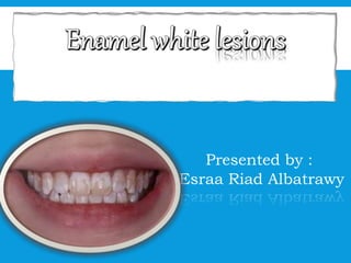
  • 1. Presented by : Esraa Riad Albatrawy
  • 2. CONTENTS: Definition: Classifications: Etiology and clinical features: Diagnosis: Evaluation Management:
  • 3. Enamel white lesions: • Subsurface enamel demineralization that clinically presents itself as “a milky white opacity.
  • 4. Carious w l: White spot lesions Non carious WL Traumatic Dental fluorosis (developmental): hypoplasia hypocalcification
  • 5. •White spot lesions usually look whitish opaque •This whitish appearance is caused by localized light scattering within lesion body. •In contrast to sound enamel, which is relatively translucent, in white spot lesions the light scattered between the enamel crystals and porosities. (With different refractive indices) White Spot Lesions
  • 7.
  • 8. prolonged plaque accumulation (prolonged ortho ttt) colonization of aciduric bacteria over time, area of demineralized enamel. active white spot lesion if not treated, cavitated carious lesion Dental plaque buffering capacity and degree of calcium and phosphate supersaturation will determine the demineralization process
  • 9.
  • 10. Risk factors : Inadequate oral hygiene . Inappropriate diet (refined sugar, frequent snacks) History of recent caries lesions . Lack of adjunctive preventive measures ( fluoride ) Prolonged Orthodontic treatment time (fixed appliances) > 36 months.
  • 12. 1- TRAUMATIC HYPOMINERALIZATION: Well demarcated opacities on the labial surface, due to injury or infection of the deciduous teeth, which has affected mineralization of the permanent teeth . More defined in shape. Well differentiated from surrounding enamel. Often located in the middle of the crown. Randomly distributed . Definition: Clinical appearance: Whit By:
  • 13. 2-DENTAL FLUOROSIS: Cosmetic condition Affected teeth are less susceptible to caries ↑↑fluoride intake during tooth formation • Mottled enamel is a condition indicates the fluorosis characterized by minute white flecks, or yellow or brown spots or areas, scattered irregularly or streaked over the surface of a tooth
  • 14. PATHOGENESIS: (DELAY IN REMOVAL OF PROTEIN MATRIX)
  • 15.
  • 16. CLINICAL FEATURES the most commonly affected tooth is the premolar followed by the second molar, maxillary incisor, canine, first molar and mandibular incisors. White / yellowish lesion Not well defined Symmetrical distribution
  • 17. Enamel hypoplasia is a defect of the teeth in which the enamel is deficient in quantity,[caused by defective enamel matrix formation during enamel development, as a result of inherited and acquired systemic condition Hypocalcified Enamel defect due to malfunction of enamel calcification, therefore enamel is of normal thickness but is extremely brittle, with an opaque/chalky presentation. Teeth are prone to staining and rapid wear, exposing dentine. Enamel appears less radio-opaque compared to dentine on radiographs. DEVELOPMENTAL:
  • 18. DIAGNOSTIC METHODS The ideal method for the detection ofWSLs should have a high level of sensitivity (the ability to detect disease when present) and specificity (the ability to confirm that disease is absent).
  • 19. DIAGNOSTIC METHODS  Visual examination  Digital photographic examination  Optical Non-Fluorescent Methods  Light Scattering  Fluorescence methods  Fluorescent dye  Ultraviolet  Laser (DIAGNO Dent), CO2 laser  QLF (quantitative light fluorescence)  Transillumination  Electrical Conductance – ECM  Digital Radiography – DDR  Thermography  Multiphoton imaging
  • 20. VISUAL EXAMINATION: Prior to examination, scaling of the teeth will be done to remove any plaque and debris and all tooth surfaces were polished with pumice and prophylaxis cup. Simple and inexpensive : no expensive or complex equipment is required. Validity : it is often difficult to clinically distinguish white spots caused by demineralization and those that are due to other causes, such as developmental hypoplasia or fluorosis.
  • 21. OPTICAL NON-FLUORESCENT METHODS (LIGHT SCATTERING) Light scattering, which can be measured using theOptical Caries Monitor(OCM) They used a 100-watt white light as a light source and measured backscatter with a densitometer. Convenient and nondestructive method of enamel demineralization. Can be applied in the clinical environment Technique sensitive and results can vary with the degree of wetness or drying of the tooth.
  • 22. OPTICAL FLUORESCENT METHODS: 1-FLUORESCENT DYE: fluorescent dye used to highlight carious enamel . Once the fluorescent dye has been applied the specimen is examined under a suitable light source convenient and nondestructive method of enamel demineralization. The disadvantage of these dyes is that slight procedural variations can result in widely different degrees of dye uptake.They are mainly used for the detection and removal of carious dentine.
  • 23. 2-ULTRAVIOLET LIGHT: Ultraviolet (UV) light used for the early detection of carious lesions on the smooth surfaces. Special precautions are required to protect the patient and operator because UV radiation is harmful to the eyes and skin. Safer methods using light sources with a longer wavelength have been developed.
  • 24. 3-LASER (DIAGNODENT): The technology of the DIAGNOdent, uses a 655 nm diode laser for detection of noncavitated, occlusal pit-and-fissure tooth decay, in addition to smooth surface caries at an earlier stages . The DIAGNOdent measures laser fluorescence within the mineral structure of the tooth.The equipment was calibrated to calculate the difference in fluorescence between the demineralised area and the surrounding sound enamel and thereby quantify mineral loss and lesion size. Healthy tooth structure exhibits little or no fluorescence, resulting in very low scale readings on the display Decayed tooth structure will exhibit fluorescence, proportionate to the degree of lost tooth structure, resulting in elevated scale readings on the display of the DIAGNOdent
  • 25. RECOMMENDATIONS FOR TREATMENT ARE: values of 30+ operative and preventative care.
  • 26. 4-QLF (QUANTITATIVE LIGHT FLUORESCENCE): QLF is a diagnostic method that relies on the autofluorescence of teeth when they are exposed to high-intensity blue light. QLF is a highly sensitive diagnostic test, with the most promising fluorescent method of measuring demineralization in use today. It is usefull in monitoring of mineral changes in incipient enamel lesions, and for the evaluation of preventive measures in caries prone persons, such as orthodontic patients.
  • 27. ↑↑sensitivity and specificity Transillumination: (fotI , difoti) Digital Imaging Fiber-OpticTrans-Illumination (DIFOTI), did not measure the depth, could only detect surface changes associated with early demineralization as early as 2 weeks. The fluorescence of the tooth is closely related to the mineral content of the enamel, with demineralization showing less fluorescence.The use of fiber optic light makes it possible to see smaller superficial optical refraction white lesions; it undergoes by passing an intense light beam through the tooth
  • 28. Fiber optic trans-illumination (FOTI) and digital imaging fiber optic trans illumination (DIFOTI)
  • 29. DIGITAL RADIOGRAPHY: The X-ray micro-tomography is aimed to characterize the mineral density (MD) of enamel white spot lesions (WSLs) calibrated with different density. X-ray micro-tomography is a sensitive in vitro technique capable of characterizing and quantifying of MD small non-cavitated WSLs. This method has a promising potential for future carious and quantitative remineralization studies.
  • 30. THERMOGRAPHY The concept of thermography for the detection of early enamel caries has been discovered by Kaneko in 1999. It measures the lesion activity rather than its presence or absence. This is based on the principle of change in thermal radiation energy that occurs when fluid is lost from a lesion by evaporation just as in WSLs e.g., Infrared thermography, Frequency-domain infrared photothermal radiometry and modulated luminescence (PTR/LUM).
  • 31. MULTIPHOTON IMAGING Unlike conventional fluorescence imaging, it uses two infrared photons simultaneously to excite a fluorescent compound in the tooth. Caries will appear as a dark form within a bright fluorescing tooth. . It also helps to collect information from carious lesions up to 500 μm of depth
  • 32. EVALUATION Different methods are used for evaluation on clinical examination Ekstrand assessment scale (1995) The Nyvad system (1999) The Dundee SelectableThreshold Method for Caries Diagnosis (DSTM in 2000) The InternationalCaries Detection and Assessment System (ICDAS in 2004)
  • 33. Ekstrand system Nyvad system DSTM system ICDAS system 0—no/slight changes in enamel translucency after prolonged air dry (5 s) 0—healthy tooth G—healthy tooth 0—sound 1—opacity/discoloration distinctly visible after air drying, hardly on wet surfaces 1—active (intact) W—white spot lesion 1—first visual change in enamel 2—opacity/discoloration distinctly visible without air drying 2—active (surface discontinuity) B—brown spot lesion 2—distinct visual change in enamel 3—localised enamel breakdown in opaque or discoloured enamel and/or greyish discoloration from the underlying dentine 3—active (cavitated) E—enamel cavitation 3—localised enamel breakdown 4—cavitation in enamel exposing the dentine 4—inactive (intact) D—dentine lesion (non- cavitated) 4—underlying dark shadow from dentine 5—inactive (surface discontinuity) C—dentine cavity 5—distinct cavity with visible dentine 6—inactive (cavity) P—pulp involvement 6—extensive distinct cavity with visible dentine 7, 8, 9—presence or absence of caries which might be active or inactive in the filling or A—arrested dentinal decay F—filled surfaces contiguous with the upper
  • 34.
  • 36. Preventive modalities Dietary control Regular plaque removal Fluoride Other measures Chlorhexidine Xylitol Carbamide peroxide These measures aim to influence etiological factors of the caries process , therefore not only prevent lesion formation(primary prevention) but also accomplish arrest and remineralization of caries lesion (secondary prevention)
  • 38. SEALING: Sealant applications to the enamel surfaces adjacent to orthodontic brackets during orthodontic treatment to form a physical barrier for acidic conditions Durability of sealants may vary due to mechanical abrasion from mastication and brushing.These factors cause sealant abrasion so, sealant preservation should be evaluated with 3-, 5-month periods and renewed if necessary Repeated applications are recommended for preventingWSLs formation effectively
  • 39.
  • 40. RESIN INFILTRATION: resin infiltration is a technique that has been available as a commercial product since 2010. The product (ICON, DMG) was initially developed as a treatment for incipient interproximal caries and anterior white spot lesions. Clinical experience with this technique, however, revealed that it is also effective maskinge in enamel discoloration of non-carious origin.
  • 41. MECHANISM OF ACTION: Resin infiltration was developed as a technique to treat enamel caries. The histopathology of enamel caries occurs as acid dissolves inter-crystalline spaces within enamel. Since the outermost 10-30 microns of enamel is more resistant to dissolution from the presence of fluorapatite, a more porous subsurface forms. The principle of resin infiltration for caries arrest is to occlude the porosity formed during the caries process and prevent pathways for acid to further dissolve the tooth structure The opaque appearance of the white spot lesion occurs because light is scattered within the body of the white spot lesion.
  • 42. Light scattering is caused when light interacts with two substances with different refractive indices.The refractive index of enamel (1.62-1.65) is different than that of air (1.00). Infiltration of the lesions with an infiltrant that has a refractive index of 1.52 is able to mask the lesion. Resin infiltration has also been shown to infiltrate hypomineralized enamel of non-carious developmental origin.
  • 43. The two basic steps to achieve this goal are to remove the less- porous surface layer of enamel and allow resin to infiltrate the internal enamel porosities through capillary movement. A solution of 15% hydrochloric acid applied for 90-120 seconds shown to almost completely remove the 45-micron thick surface layer of the lesion. infiltrate resin into the porosities created during dissolution of intercrystallite enamel.
  • 44. Resin infiltration may be an effective method of treatment for anterior tooth discolorations of developmental origin assuming their depth is not too great not exceed 0.2 to 0.3 mm in depth.. Multiple etching steps may be needed in order to achieve the desired outcome. Several clinical trials have reported the masking effects of resin infiltration have remained unchanged at follow up times up to: • 12 months for non-carious lesions • 24-45 months for carious lesions Sufficient time is needed for infiltrate to penetrate a porous network because capillary penetration is a time dependent process. A method to help determine the thickness of a stain is to transilluminate the tooth with a dental transilluminator or light-curing unit (with proper eye protection). If the lesion becomes significantly darker with transillumination, the lesion is likely deeper within the enamel.
  • 46. • Ideally, re-mineralizing agents need to rapidly precipitate on partially demineralized tooth structure and transform into a more stable, less acid- soluble apatite than the hard tissue replaced. re-mineralizing systems Non-fluorinated, Fluorinated Synthetic, Natural agent
  • 47. • Calcium phosphate based • Crystalline • Stabilized amorphous • Un-stabilized amorphous • Calcium sucrose phosphate • Bioactive glass • Tricalcium phosphate • Nano hydroxyaptite • Biomimetic materials • Amelogenins • Peptides • Enamel matrix derivative (EMD) synthetic
  • 48. CRYSTALLINE CALCIUM PHOSPHATE RE- MINERALIZING SYSTEMS Have poor solubility of the calcium phosphate phases, So it is difficult to achieve enamel remineralization. It is a problem that excessive amounts of calcium phosphate phases present in the mouth may cause calculus. Example: calcium sodium phospho-silicates referred as bioactive glasses, are a kind of crystalline calcium phosphates derivatives and show maximum re- mineralizing potential . it was reported that calcium sodium phospho-silicate paste has shown to release ions and transform them into hydroxycarbonate apatite for up to 2 weeks.
  • 49. UN-STABILIZED AMORPHOUS CALCIUM PHOSPHATE RE- MINERALIZING SYSTEMS Unstabilized amorphous calcium phosphate (ACP) formulation is developed by mixing calcium ions with phosphate ions to produce an ion active phase so that it could be precipitated as quickly as ACP or, in the presence of fluoride ions, amorphous calcium fluoride phosphate (ACFP). The desired effect is dissolution into the saliva and to promote tooth remineralization , but these unstabilized forms can cause dental calculus
  • 50. STABILIZED AMORPHOUS CALCIUM PHOSPHATE RE-MINERALIZING SYSTEMS CPP-ACP is a bioactive agent with a base of milk products able to bind calcium and phosphate ions to stabilize calcium phosphate in solution and to increase the level of calcium phosphate in dental plaque. CPP-ACP also adheres to hydroxyapatite, soft tissues, and supplies free calcium and phosphate ion, thereby helping to maintain reducing demineralization and promote remineralization by reforming into calcium phosphate crystals It can interact with hydrogen ions from the surface of the tooth and so it can penetrate to enamel’s subsurface layer in order to produce mineral gain Antibacterial and buffering efficacy has also been emphasized as interfering the growth and adherence of Streptococcus mutans and Streptococcus sobrinus to dental plaque Examples: the casein phosphopeptide-stabilized amorphous calcium phosphate (CPP-ACP) and casein phosphopeptide stabilized amorphous calcium fluoride phosphate complexes (CPP- ACFP)
  • 51.
  • 53. TRICALCIUM PHOSPHATE  is a calcium salt of phosphoric acid with the chemical formula Ca3(PO4)2.
  • 54.
  • 56. RECENT APPROACH FOR REMINERALIZATION The most recent approach for the remineralization is the BIOMIMETIC REMINERALIZING AGENTS, such as the self- assembling peptide P11-4(Curodont repair) . designed to mimic enamel matrix proteins and forms a 3D matrix that contributes to the de novo hydroxiapatite formation, which has been used for remineralization of subsurface lesions. has high affinity to calcium ions in saliva which also leads to the formation of enamel crystals around the enamel matrix. stabilize the calcium, phosphate and fluoride ions and to localize these ions in non-cavitated carious lesions for controlled re- mineralization
  • 57.
  • 58.
  • 59. NATURAL REMINERALIZING AGENT: I). Plant-derived herbal extract. Clove Cinnamon Ginger Rosemary Wheat grass Siwak II). Fruit extract Grape seed extract Cranberry extract III). Food by products Egg shell Fermented shrimp Paste IV). Sweetener Honey V) Others Zamzam water
  • 60.
  • 61. WHITENINGBLEACHING: It is the process of lightening the color of enamel. techniques At home method, that requires an intraoral device/tray to apply a gel of peroxide, which can be done at home and is more cost-effective. It must be kept in mind that major changes are not observed before the 7th day. In-office method, which requires a professional to perform the procedure, uses photoactivation, where the changes of colour in the enamel can be noticed from the first session
  • 62.
  • 63. MICRO ABRATION: Used alone or with conjugation of resin infiltration or ACP paste. Eliminate superficial staining or defects Includes hydrochloric acid and pumice application to the enamel, which removes approximately 100 μ from the surface layer. Micro-abraded enamel surface appears polished because of no interprismatic space, and it is more resistant to bacterial colonization and demineralization Reduce the size of white spot lesions by 83% it could be a treatment option for post-orthodontic demineralized enamel lesions ,While, the first option should be conservative methods such as topical fluoride application. However, there is a limitation to its use. It was reported that the technique is effective if the lesion sizes do not exceed from 0.2 to 0.3 mm in depth.
  • 64.
  • 65.
  • 66.
  • 67.
  • 68. ResinRestorations/ Indirect Restorations Patients with cavitated lesions, or more severeWSL who have already attempted more conservative esthetic treatments without significant improvement may benefit from the preparation of the affected tooth surfaces and restoration with either direct resin restorations or indirect porcelain restorations.
  • 70. REFERENCES: • https://watermark.silverchair.com/111611- 710_1.pdf?token=AQECAHi208BE49Ooan9kkhW_Ercy7Dm3ZL_9Cf3qfK Ac485ysgAAAsgwg • https://watermark.silverchair.com/041218- 274_1.pdf?token=AQECAHi208BE49Ooan9kkhW_Ercy7Dm3ZL_9Cf3qfK Ac485ysgAAAsgwg • file:///C:/Users/D%20AGHA%20SMART%20STORE/Downloads/Enamel_ microabrasion_associated_with_resin_infiltr.pdf • https://www.intechopen.com/books/contemporary-approach-to-dental- caries/incipient-caries-lesions-in-orthodontics • https://www.intechopen.com/books/dental-caries-diagnosis-prevention- and-management/management-of-white-spot-lesions

Editor's Notes

  1. Esraa riad
  2. First q answers
  3. Aside from the white spot lesion being the key for the early diagnosis of caries lesions, the intact surface layer has been a confusing aspect in the confirmation of demineralisation. The reason behind the presence of intact surface layer is because of: no doubt bacterial acids dissolves the surface as well as the subsurface enamel But, the calcium and phosphate ions produced from subsurface dissolution diffuse outward towards the surface. Hence, the surface enamel is in type of equilibrium with mineral being lost into plaque due to low pH, but being remineralised from the saliva. If the cariogenic environment continues, eventually the rate of transfer from the surface enamel into plaque becomes greater than the rate of precipitation, and the surface enamel collapse lead to frank cavitations.
  4. The ideal method for the detection of WSLs should have a high level of sensitivity (the ability to detect disease when present) and specificity (the ability to confirm that disease is absent).
  5. systems that use the natural fluorescence that occurs in tooth enamel. The light emission coefficient of a caries lesion is higher than healthy enamel resulting in lower fluorescence in caries lesions
  6. OPTICAL FLUORESCENT METHODS: 1-FLUORESCENT DYE:
  7. Cavo, diagnodent
  8. The fluorescence of the tooth is closely related to the mineral content of the enamel, with demineralization showing less fluorescence. This relationship allows for the quantification of demineralization or remineralisation at one time point or overtime.
  9. Fiber optic trans-illumination (FOTI) and digital imaging fiber optic trans-illumination (DIFOTI)
  10. MD= mineral density Picture of micro-ct showing md
  11. As the surface of a carious lesion may act as a barrier to resin infiltration, several preliminary studies evaluated different acid solutions for removal of the surface layer.