1. Molecular genetic diversity and characterization of conjugation genes in
the fish parasite Ichthyophthirius multifiliis
Elisabeth MacColl a
, Matthew D. Therkelsen a
, Tshering Sherpa a
, Hannah Ellerbrock a
, Lily A. Johnston a
,
Ravi H. Jariwala a
, WeiShu Chang a
, James Gurtowski b
, Michael C. Schatz b
, M. Mozammal Hossain c
,
Donna M. Cassidy-Hanley c
, Theodore G. Clark c,⇑
, Wei-Jen Chang a,⇑
a
Department of Biology, Hamilton College, Clinton, NY 13323, USA
b
Simons Center for Quantitative Biology, Cold Spring Harbor Laboratory, Cold Spring Harbor, NY 11724, USA
c
Department of Microbiology and Immunology, College of Veterinary Medicine, Cornell University, Ithaca, NY 14853, USA
a r t i c l e i n f o
Article history:
Received 15 September 2014
Revised 14 February 2015
Accepted 22 February 2015
Available online 2 March 2015
Keywords:
Sexual reproduction
Ciliophora
Hypotrich
IES
Phylogeny
Barcoding
a b s t r a c t
Ichthyophthirius multifiliis is the etiologic agent of ‘‘white spot’’, a commercially important disease of
freshwater fish. As a parasitic ciliate, I. multifiliis infects numerous host species across a broad geographic
range. Although Ichthyophthirius outbreaks are difficult to control, recent sequencing of the I. multifiliis
genome has revealed a number of potential metabolic pathways for therapeutic intervention, along with
likely vaccine targets for disease prevention. Nonetheless, major gaps exist in our understanding of both
the life cycle and population structure of I. multifiliis in the wild. For example, conjugation has never been
described in this species, and it is unclear whether I. multifiliis undergoes sexual reproduction, despite the
presence of a germline micronucleus. In addition, no good methods exist to distinguish strains, leaving
phylogenetic relationships between geographic isolates completely unresolved. Here, we compared
nucleotide sequences of SSUrDNA, mitochondrial NADH dehydrogenase subunit I and cox-1 genes, and
14 somatic SNP sites from nine I. multifiliis isolates obtained from four different states in the US since
1995. The mitochondrial sequences effectively distinguished the isolates from one another and divided
them into at least two genetically distinct groups. Furthermore, none of the nine isolates shared the same
composition of the 14 somatic SNP sites, suggesting that I. multifiliis undergoes sexual reproduction at
some point in its life cycle. Finally, compared to the well-studied free-living ciliates Tetrahymena ther-
mophila and Paramecium tetraurelia, I. multifiliis has lost 38% and 29%, respectively, of 16 experimentally
confirmed conjugation-related genes, indicating that mechanistic differences in sexual reproduction are
likely to exist between I. multifiliis and other ciliate species.
Ó 2015 Elsevier Inc. All rights reserved.
1. Introduction
With high virulence and an extremely broad host range,
Ichthyophthirius multifiliis (also known as Ich), is one of the most
important disease agents of farm-raised fish. Despite this, methods
for prevention and treatment of I. multifiliis infection (ichthyoph-
thiriasis) are currently limited, necessitating better understanding
of fundamental aspects of the parasite’s biology including its life
cycle and population structure (Dickerson, 2006; Matthews, 1994).
By all accounts, I. multifiliis has a relatively simple life cycle with
no intermediate hosts. The cycle’s three morphologically and func-
tionally distinct stages have been well described and consist of
infectious, free-swimming theronts, host-associated trophonts,
and encysted tomonts that divide mitotically off the fish.
Theronts burrow into the epithelial layers of the host and rapidly
transform into trophonts that feed on host tissue. Trophonts grow
to several hundred microns in diameter, becoming visible to the
eye as the characteristic white spots associated with disease.
After 7–10 days, trophonts mature, escape from the fish, and attach
to a solid surface becoming tomonts. Tomonts then secrete a
gelatinous cyst wall and produce in the range of 100–1000 daugh-
ter cells by multiple rounds of asexual division. Within 20–24 h,
theronts burst from the cyst and restart the infectious cycle
(Matthews, 2005).
Although the first full description of I. multifiliis was published
more than a century ago (Fouquet, 1876), and the first major out-
break of ichthyophthiriasis in the US was reported soon thereafter
(Stiles, 1893), we still know little about how disease outbreaks are
http://dx.doi.org/10.1016/j.ympev.2015.02.017
1055-7903/Ó 2015 Elsevier Inc. All rights reserved.
⇑ Corresponding authors.
E-mail addresses: tgc3@cornell.edu (T.G. Clark), wchang@hamilton.edu (W.-J.
Chang).
Molecular Phylogenetics and Evolution 86 (2015) 1–7
Contents lists available at ScienceDirect
Molecular Phylogenetics and Evolution
journal homepage: www.elsevier.com/locate/ympev
2. connected. This is largely due to a lack of genetic tools to distin-
guish strain differences. Currently, the most common method of
strain identification involves serotyping with antisera against
parasite immobilization antigens (i-antigens), a class of abundant
surface membrane proteins that vary between isolates (Dickerson
et al., 1993). However, the underlying basis of serotype variation
is not known, and it is entirely possible that isolates that are sero-
logically different are more closely related than those that are sero-
logically identical. An alternative approach using an 822 nt region
of the mitochondrial cytochrome c oxidase subunit I (cox-1) gene
that was successful in barcoding different species of Tetrahymena
failed to provide much resolution on the phylogenetic relation-
ships among seven isolates of I. multifiliis collected in the state of
Georgia (Kher et al., 2011).
Recent sequencing of the I. multifiliis macronuclear genome
(Coyne et al., 2011) could play an important role in developing
new and more effective genetic markers to define strain differences
among parasite isolates. Such markers could also be very helpful in
unraveling questions surrounding sexual reproduction, a poorly
understood process in this species. Although sexual reproduction
is the primary mechanism for achieving genetic diversity in most
eukaryotes, it has not yet been directly observed in many parasites
(Schurko et al., 2008), such as important human pathogens
Entamoeba histolytica (Weedall et al., 2012), Giardia intestinalis
(Xu et al., 2012a), Giardia duodenalis (Takumi et al., 2012), and
Trichomonas vaginalis (Conrad et al., 2012), which primarily utilize
asexual reproduction once successful adaptations to host and envi-
ronmental niches have been established (Heitman, 2006; Weedall
et al., 2015). To date, sexual reproduction has not been conclusive-
ly observed in I. multifiliis, neither in environmental samples nor
captive laboratory strains.
Despite this, a number of observations suggest that I. multifiliis
may undergo sexual reproduction as part of its life cycle. First,
long-term cultures of I. multifiliis are often difficult to establish
via serial passage on fish, a problem which could be attributed to
senescence due to failed sexual reproduction (Matthews et al.,
1996). Second, I. multifiliis carries a germline micronucleus, which
in other ciliate species undergoes meiosis to form a germinal
pronucleus during sexual conjugation. Occasionally, more than a
single micronucleus can be identified (particularly in theronts),
although it is unclear whether these have undergone meiosis or
serve in germline exchange; no further nuclear events, such as
those found in other ciliates, have been observed in I. multifiliis
(Matthews et al., 1996). Finally, recent studies by Chi et al. have
provided the first molecular evidence suggesting that I. multifiliis
might be capable of sexual reproduction by identifying meiosis-
specific genes in its genome (Chi et al., 2014).
In the present study, we compare nucleotide sequences of the
SSUrDNA, mitochondrial NADH dehydrogenase subunit I (nad1_b)
and cox-1 genes, and 14 somatic SNP sites from nine geographically
distinct I. multifiliis isolates. We show that the two mitochondrial
sequences, nad1_b and cox-1, contain sufficient information to
discern these isolates at the molecular level, and resolve their phy-
logenetic relationships. Furthermore, combining results of phylo-
genetic analyses derived from mitochondrial sequences and
patterns of somatic SNP data, it is clear that the population of
I. multifiliis has been undergoing extensive sexual reproduction.
2. Materials and methods
2.1. I. multifiliis, DNA extraction and sequencing
Nine isolates of I. multifiliis were harvested from infected fish
and maintained clonally (except for G15) in the lab following
established protocols (Cassidy-Hanley et al., 2011). The fish, and
therefore the isolates, were obtained from different locations,
beginning in 1995. Each isolate was named with a letter(s) repre-
senting the state of origin and a sequential number (Table 1). For
example, G5 was the fifth isolate obtained from the state of
Georgia (Cassidy-Hanley et al., 2011).
Except for the G15, Ark7, and Ark9 isolates (which used
tomonts), DNA was extracted from theronts following protocols
published elsewhere (Cassidy-Hanley et al., 2011). Sequences of
PCR products and plasmids were determined using the Sanger
sequencing method (Genewiz, South Plainfield, NJ). For isolate
G15, DNA was extracted using the High Pure PCR Template
Preparation kit (Roche, Indianapolis, IN) following manufacturer’s
protocols and was sequenced using both PacBio and Illumina tech-
nologies (UCSD IGM Genomics Center, La Jolla, CA). A detailed pro-
tocol on assembling the G15 genome is provided in Supplementary
Materials.
2.2. SNP site identification
To identify potential SNP sites, we aligned both 454 and Sanger
sequencing reads to the G5 reference genome (Coyne et al., 2011)
using SMALT (Postingl, 2013). The alignments were then filtered
and sorted by a set of criteria (minimum base quality 50, minimum
mapping quality 25, and minimum coverage of 6 for Sanger and 8
for 454) using SAMtools (Li et al., 2009). A list of potential SNP sites
was then identified using VarScan (Koboldt et al., 2012), and a frac-
tion of these were further examined visually in IGV Viewer
(Thorvaldsdottir et al., 2013). Since we wanted to investigate
SNPs in intragenic regions but were not sure about the quality of
gene annotations in the I. multifiliis genome, we ultimately chose
14 SNPs that could be distinguished by restriction enzyme diges-
tions and resided in genes with homologs in the closely related
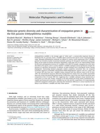
Tetrahymena thermophila (Eisen et al., 2006) (Fig. 1). Of the 14
SNPs, 9 were synonymous substitutions, two were located in
intronic regions, one was in the 50
UTR, another was a non-synony-
mous substitution, and the last one displayed no variants in all
nine strains and was on the third position of a codon. Each of the
14 SNP sites was found on a different scaffold (Supplemental
Table S1).
2.3. PCR, cloning, and restriction fragment length polymorphisms
PCR was performed in 1X GoTaq Green Master Mix (Promega,
Madison, WI), 0.2 lM of each primer, and 2–20 ng of I. multifiliis
DNA in a 50 ll reaction volume. Cycling conditions were: 95 °C
2 min followed by 35 cycles of 95 °C 30 s, 50 °C (or another
Table 1
Histories and characteristics of nine strains of I. multifiliis.
Strain
Name
Location of Isolation Serotype Date Host
G5 Fish farm, Georgia D 1995 Channel
catfish
G15 Supermarket, Athens,
Georgia
D 2011 Red parrot
fish
NY3 Petstore, Ithaca, NY D 2004 Oscar
NY4 Petstore, Ithaca, NY G 2004 Freshwater
shark
NY7 Supermarket, New
Hartford, NY
Unknowna
2010 Oscar
Ark5 Central Arkansas D 2005 Channel
catfish
Ark7 Stoneville, MS Unknowna
2008 Channel
catfish
Ark9 Lonoke, AR Unknowna
2008 Golden shiner
Ark10 Stuttgart, AR D 2011 Blue catfish
a
Serotype could not be determined by existing antibodies.
2 E. MacColl et al. / Molecular Phylogenetics and Evolution 86 (2015) 1–7
3. specified annealing temperature) 1 min, and 72 °C 1.5 min. A final
extension at 72 °C for 7 min was added to the end of PCR. Primer
sequences are provided in Table S2. PCR products were purified
and eluted using the DNA Clean & Concentrator Kit (Zymo
Research, Irvine, CA) and were either cloned into the pGEM-T
Easy Vector (Promega) and sequenced, directly sequenced, or sub-
jected to restriction enzyme digestion (New England Biolab,
Ipswich, MA). Digested PCR products were electrophoresed on
agarose gels, and images were taken using a ChemiDoc
XRS + system (Bio-Rad, Hercules, CA).
2.4. Sequence and phylogenetic analyses
All new sequences were deposited in GenBank: cox-1 (GenBank:
KJ690547–KJ690555), nad1_b (GenBank: KJ690556–KJ690564),
SSUrDNA (GenBank: KJ690565–KJ690572). Sequences were viewed
and manipulated in Jalview (Waterhouse et al., 2009) and/or
BioEdit (Hall, 1999). Since maximum parsimony trees inferred
from nad1_b (509 nt) and cox-1 (1493 nt) sequences yielded the
same topology (Supplemental Fig. S1), we concatenated sequences
of both genes and reconstructed phylogenetic trees of the nine
I. multifiliis isolates using maximum parsimony (MP) criterion with
10,000 bootstrapping replicates (Wilgenbusch et al., 2003), maxi-
mum likelihood (ML) method with TIM1 + I model predetermined
by JModelTest (Darriba et al., 2012) and 1000 bootstrapping repli-
cates (Guindon et al., 2003). For the MrBayes analysis (MB,
(Ronquist et al., 2012)), GTR + I + G model was used in two simul-
taneous, independent runs, each with seven heated chains and one
cold chain. A total of 2,500,000 MCMC steps were run with a sam-
pling frequency of 1000, and by the end of MCMC the standard
deviation of split frequencies reached 0.0057. A burn-in of 25%,
or 625, was used to generate both parameters and the consensus
tree.
3. Results
3.1. Molecular characterization of I. multifiliis
To help identify suitable molecular markers to distinguish
I. multifiliis isolates and determine their phylogeny, we sequenced
two mitochondrial regions and the 18S SSUrDNA sequence from
nine different isolates sampled from four states (Arkansas,
Georgia, Missouri, and New York) starting in 1995 (Table 1). The
first mitochondrial region was 1493 nt in length comprising
approximately 72% of the cox-1 coding sequences, of which 27
were parsimony-informative sites (hereafter cox-1). The other
mitochondrial region we surveyed was 509 nt in length containing
the entire NADH dehydrogenase subunit 1 gene and its flanking
sequences, as well as the first 70 nt of the juxtaposed apocyco-
chrome b gene. Collectively, we refer to this second region as the
nad1_b locus, which contained 11 parsimony-informative sites.
The 18S SSUrDNA locus we sequenced was 1702 bp in length.
When mitochondrial sequences from the different parasite iso-
lates were compared, isolates NY3 and Ark9 were found to share
the same mitochondrial haplotype. Here, a mitochondrial haplo-
type refers to a unique combination of SNPs on the surveyed mito-
chondrial regions. In contrast to NY3 and Ark9, isolates NY4 and
G15 shared a second mitochondrial haplotype in both the cox-1
and nad1_b loci (Fig. 2). Topologies of maximum-parsimony trees
inferred from cox-1 and nad1_b sequences were identical, suggest-
ing that the two mitochondrial regions have evolved in a similar
way (Supplemental Fig. S1). We, therefore, concatenated nucleo-
tide sequences from these two mitochondrial regions for further
phylogenetic analyses.
3.2. At least two distinct groups of I. multifiliis were present in the US
Tree topologies inferred from the concatenated mitochondrial
sequences using maximum-parsimony, Bayesian, and maximum-
likelihood methods were similar, and clearly suggested that differ-
ent isolates fell into at least two genetically distinct groups (Fig. 2).
The first group (NY3 and Ark9) showed substantial genetic varia-
tion from the second group (Ark7, G5, NY4, G15, Ark5, and
Ark10) in the two mitochondrial loci, indicating long-term inde-
pendent evolution since the two groups diverged. NY7 was more
closely related to the second group than to the first, but it indepen-
dently accumulated considerable variations, which distinguished it
from either of the two groups. Within the second group, NY4, G15,
Ark5, and Ark10 were more closely related to each other than to
Ark7 and G5, which formed a sister clade.
We compared our cox-1 sequences to a shorter region of the
same gene previously sequenced from six independent isolates
from the state of Georgia by Kher et al. (containing 797 nt, and
16 parsimony-informative sites) (Kher et al., 2011). Based on that
comparison, the six Georgia isolates (designated G2, G3, G4, G6,
and G7) were found to more closely resemble the second group
from our study than the first (data not shown). Specifically, G3
and Ark7 shared the same haplotype, and G6 and Ark5 shared
another haplotype in this shorter cox-1 region. Sequences from
G2, G5, G7, G15, and NY4 were also identical in this region.
In contrast to the multiple variable sites in the two mitochon-
drial loci, we observed only a single nucleotide substitution in
the SSUrDNA locus among the nine isolates. Although we did not
expect that intraspecies relationships could be resolved using
SSUrDNA sequences (Brunk et al., 1990; Jerome et al., 1996), our
SSUrDNA sequences differed significantly from one reported in
Fig. 1. SNP compositions of nine I. multifiliis isolates. SNP site sequences were determined by restriction fragment length polymorphisms and/or sequencing. Sequences are
presented in IUPAC nucleotide code where K = G or T, R = A or G, W = A or T, Y = C or T.
E. MacColl et al. / Molecular Phylogenetics and Evolution 86 (2015) 1–7 3
4. 1995 (Wright et al., 1995). While some of these differences might
be explained by a higher sequencing error rate using radio-isotope
labeled ddNTPs in the 1990s (10 out of 11 sites in discrepancy were
indels/insertions), the differences could indicate the presence of an
I. multifiliis lineage that diverged much earlier. Further experi-
ments will be needed to test this hypothesis.
3.3. Sexual reproduction in I. multifiliis
To address the issue of sexual reproduction in I. multifiliis, where
the process may be rare or obscure, we adopted a schema proposed
by Ramesh, Malik and Logsdon that utilizes a molecular toolbox
with which to scan genomes for the presence of meiosis-specific
genes (Ramesh et al., 2005). Chi et al. applied this concept and
reported the presence of numerous meiosis-specific and meiosis-
related genes in I. multifiliis (Chi et al., 2014). Nevertheless, gene
models of several of these meiosis and conjugation related genes
(see Section 3.4) were either questionable or missing functional
domain(s), raising the possibility that these sequences represented
pseudogenes. For example, we could not generate a SPO11 gene
model containing the full length catalytic topoisomerase domain
at its 30
end, using the Wise2 package (data not shown) (Birney
et al., 2004). Chi et al. used a different approach, and the SPO11
model they proposed also lacked a full length topoisomerase
domain (Chi et al., 2014). Furthermore, we have not been able to
detect SPO11 transcripts using RT–PCR (Chang, unpublished
results) in different life stages of I. multifiliis and therefore suspect
that sexual conjugation can proceed without the presence of this
and other conjugation-related genes (see below).
3.4. Results from bioinformatic analyses suggest that some
conjugation-related genes are missing in the I. multifiliis genome
Due to the findings presented in Section 3.3, we decided to
search the I. multifiliis genome for homologs to genes that have
been experimentally shown to participate in conjugation in the
Fig. 2. Maximum likelihood tree constructed by concatenating nad1_b (509 nt) and cox-1 (1,493 nt) sequences. Bootstrap values (maximum-parsimony/Bayesian/maximum-
likelihood) are shown next to supported branches with branch lengths drawn to reflect genetic distances. NY3 and Ark9 shared identical sequences in these two loci, as did
NY4 and G15.
Table 2
The presence of 16 conjugation-related genes in I. multifiliis inferred from blast results.
Tetrahymena homolog Ichthyophthirius Paramecium References
Twi1 (TTHERM_01161040) IMG5_148140 + Mochizuki et al. (2002)
Dcl1 (TTHERM_00284230) +a,c
+ Mochizuki et al. (2005)
Hen1 (TTHERM_00433810) – + (Kurth and Mochizuki, 2009)
Giw1 (TTHERM_01276320) – À Noto et al. (2010)
Ema1 (TTHERM_00088150) IMG5_004170b
+ Aronica et al. (2008)
CnjB (TTHERM_01091290) IMG5_053760b,c
+ Bednenko et al. (2009)
Wag1 (TTHERM_00299879) – À Bednenko et al. (2009)
Ezl1 (TTHERM_00335780) IMG5_205160b
+ Liu et al. (2007)
Pdd1 (TTHERM_00125280) IMG5_193570 + Coyne et al. (1999)
Die5 (TTHERM_00686240) +a,c
+ Matsuda et al. (2010)
Tpb2 (TTHERM_01107220) – + Cheng et al. (2010) and Baudry et al. (2009)
Tku80 (TTHERM_00492460) IMG5_099960b
+ Lin et al. (2012)
Asi2 (TTHERM_00191480) +a,c
+ Yin et al. (2010)
Cda12 (TTHERM_00013410) Àd
+ Zweifel et al. (2009)
Zfr1 (TTHERM_01285910) – + Xu et al. (2012b)
Fen1 (TTHERM_00780850) IMG5_140760 + Cole et al. (2008)
a
Fragments matched by tblastn search (e-value < 10À8
).
b
Tblastn result suggests a more extensive match than the current gene model to the query.
c
Matched query region < 50% of query length.
d
A reciprocal best hit was detected with an e-value of 10À3
.
4 E. MacColl et al. / Molecular Phylogenetics and Evolution 86 (2015) 1–7
5. model ciliates T. thermophila and Paramecium tetraurelia. Ideally, if
sexual conjugation is present and the pathway is largely conserved
in I. multifiliis, we should expect to find most conjugation-related
genes. Our inventory included 13 genes involved in DNA rear-
rangements that occur when a new somatic nucleus, the macronu-
cleus, is formed during conjugation (Twi1 – Asi2, Table 2), and
three genes involved in the formation of conjugation junctions.
We used T. thermophila protein sequences and did both blastp
searches against predicted I. multifiliis proteome, and a tblastn
search against the I. multifiliis genome. Reciprocal blastp was also
carried out to help ensure orthology. Information on homologs in
P. tetraurelia was obtained from Tetrahymena Genome Database
Wiki (Stover et al., 2012) or from blastp search results against
ParameciumDB (Arnaiz et al., 2007). Using an e-value cutoff of
10À8
in both forward and reciprocal blast, we found less than half
(7 out of 16) of the homologs’ gene models (and thus predicted
protein sequences) to be present in the I. multifiliis genome.
Three genes, Dcl1, Die5, and Asi2, showed promising tblastn hits
in the I. multifiliis genome, but the length coverages all fell below
50% and again raised the question as to whether these potential
hits in I. multifiliis reflected remnants of pseudogenes.
3.5. SNP data and mitochondrial sequences strongly support that
I. multifiliis reproduces sexually
Lastly, we assessed sexual reproduction in I. multifiliis through
an entirely different approach, namely, population genetics.
Fourteen potential somatic SNP sites located on 14 different genes
(Fig. 1, Supplemental Table S1) were first identified by aligning
sequencing reads from the I. multifiliis genome project to the refer-
ence assembly (see Section 2.2). We then determined genetic var-
iations in these 14 sites in the nine I. multifiliis isolates and found
heterozygosities—in one or more isolates—at all but one site, locat-
ed in the MCM2/3/5 gene (Fig. 1). No two isolates shared an iden-
tical composition across the 14 SNP sites. Between NY3 and Ark9,
which shared the same cox-1 and nad1_b sequences, 6 out of 14
SNP sites were of different composition. NY4 and G15 were anoth-
er pair sharing identical cox-1 and nad1_b sequences, but they dif-
fered at 8 SNP sites. The high heterogeneity found on these 14
somatic SNPs in the nine I. multifiliis isolates strongly suggests that
I. multifiliis has been reproducing sexually, not exclusively clonally.
4. Discussion
In this study, we present a molecular toolkit that can be used to
effectively distinguish isolates of the fish parasite I. multifiliis and
help determine their phylogenetic relationships. By surveying a
longer cox-1 sequence (compared to (Kher et al., 2011)), and by
including another mitochondrial locus, nad1_b, we show that nine
I. multifiliis isolates sampled from four states since 1995 could be
sorted into at least two genetically distinct groups (Fig. 2).
Although not completely unexpected, neither serotypes nor geo-
graphical locations of these isolates reflected their phylogenies
(Figs. 1 and 2). For example, NY3 and G15 were both serotype D
but were distantly related. In addition, while NY3 and NY4 were
both isolated from Ithaca, NY in the same year, this location was
uninformative as the isolates were from different pet stores and
derived from separate lineages. Our toolkit now allows for both sci-
entists and the aquaculture industry to better understand connec-
tions between ichthyophthiriasis outbreaks, and the distributions
and migrations of this parasite.
In addition to genotyping mitochondrial haplotypes in I. multi-
filiis, we also surveyed SNP information on 14 somatic genes. Our
SNP data and mitochondrial sequences strongly indicate that
I. multifiliis has been undergoing sexual reproduction. This
contrasts with a number of other parasite species that preferential-
ly reproduce clonally (Heitman, 2006). Our data also indicate that
sexual reproduction in I. multifiliis proceeds without exchanging
mitochondria, as we never found more than one mitochondrial
haplotype in any of the nine strains. This suggests that sexual
reproduction in I. multifiliis may resemble that in other ciliates,
such as Tetrahymena and Paramecium, where haploid nuclei are
exchanged through a temporal junction without exchanging mito-
chondria, and argues against a mechanism in which mating cells
fuse to form a single zygote (Matthews et al., 1996). However,
when we surveyed the I. multifiliis genome for homologs to 16
genes known to play a role in conjugation in the model ciliates
T. thermophila and P. tetraurelia, many appeared absent suggesting
that the underlying mechanisms involved in sexual reproduction
in these species may differ. The 16 genes in this case play roles
in generating and transporting scan RNAs (Twi1, Dcl1, Hen1, and
Giw1), in removing non-coding germline sequences (Ema1, CnjB,
Wag1, Ezl1, Pdd1, Die5, Tpb2, and Tku80), in DNA endoreplication
(Asi2), in membrane trafficking (Cda12), and in formation of the
conjugation junction (Zfr1 and Fen1) during conjugation (Table 2).
By a loose criterion of counting positive matches in blast
searches based only on e-values, we found that I. multifiliis has lost
38% of its conjugation-related genes (6 out of 16) compared to
T. thermophila, and 29% (4 out of 14, after taking out Giw1 and
Wag1) compared to P. tetraurelia. In particular, a subset of genes
involved in the process of removing non-coding internal eliminat-
ed sequences (IESs) from the new, developing somatic DNA was
not detected in the I. multifiliis genome. The subset included genes
encoding for the Hen1 protein responsible for methylating scan
RNA (Kurth and Mochizuki, 2009), the Giw1 protein for transport-
ing Twi1-small RNA complex into the nucleus (Noto et al., 2010),
the Wag1 protein for interacting with Twi1 (Bednenko et al.,
2009), and the transposase Tpb2 for cutting IESs (Cheng et al.,
2010). Hen1 methylates the 30
ends of short scan RNA, but not
other classes of small RNAs, to assist IES removal. Loss of the
Hen1 gene in T. thermophila did not lead to a lethal phenotype,
but the removal of IESs was impaired and resulted in inefficient
production of progeny cells (Kurth and Mochizuki, 2009).
Domesticated transposases (piggyBac class of Tpb2 in T. thermophi-
la and piggyMac in P. tetraurelia; TBE in O. trifallax) have been
shown to be directly involved in the cleavage of IESs in the three
well-studied ciliate species (Vogt et al., 2013). Silencing these
transposases using RNAi affected the DNA rearrangement processes.
In T. thermophila, the conjugation process arrested (Cheng et al.,
2010), and in P. tetraurelia less than 5% of progeny cells were viable
(Baudry et al., 2009). In O. trifallax, aberrantly rearranged DNA was
detected when the expression of TBE transposases was knocked
down during conjugation (Nowacki et al., 2009). Interestingly,
the transposon scan we conducted using Transposon-PSI (Haas,
2010) against I. multifiliis genome failed to identify any piggyBac
transposases (data not shown), agreeing with the observations that
Coyne and colleagues made when they published the genome
(Coyne et al., 2011). These findings suggest that the I. multifiliis
germline genome may contain fewer or no IESs, as compared to
T. thermophila, P. tetraurelia, and O. trifallax. However, other genes
in I. multifiliis may complement the loss of these genes. For
instance, I. multifiliis may use a different class of transposase to
substitute piggyBac transposases. CnjB has shown overlapping
activities with Wag1 in T. thermophila (Bednenko et al., 2009),
and CnjB alone may be sufficient to overcome the loss of Wag1 in
I. multifiliis, even though its gene model is subject to questioning
(Table 2). Similarly, the loss of SPO11 (if it were not functional in
I. multifiliis) may be complemented by other proteins that can
induce double strand breaks in meiosis (Ramesh et al., 2005;
Malik et al., 2007, 2008), as observed in the putatively sexual
organism Dictyostelium discoideum (Flowers et al., 2010).
E. MacColl et al. / Molecular Phylogenetics and Evolution 86 (2015) 1–7 5
6. I. multifiliis has not only lost genes involved in IES removal, but
also homologs that are essential for cytokinesis (Cda12) (Zweifel
et al., 2009) and the formation of the conjugation junction (Zfr1)
(Xu et al., 2012b) in T. thermophila. Knocking down Cda12 expres-
sion led to arrested conjugation, and Zfr1 knockout cells aborted
conjugation 9–10 h post conjugation. Combined with the observa-
tions stated above, the mechanism of I. multifiliis sexual conjuga-
tion may differ, at least somewhat, from the well-documented
processes in other ciliate species. This may account for the current
inability to observe I. multifiliis sexual conjugation via microscopic
examinations. An alternative explanation may be that sexual con-
jugation occurs rarely and only in a few cells, which makes observ-
ing such an event difficult under microscope. Functions of genes
that are lost are complemented by other proteins as discussed
above.
Our study provides the first effective way to characterize
I. multifiliis populations at the molecular level. Furthermore, these
methods provide unique population genetics data that strongly
suggest this parasite reproduces sexually. The existence of sexual
reproduction in this species adds to our knowledge of how para-
sites generate genetic diversity to cope with host defenses and
may help us locate potential targets to control ichthyophthiriasis
outbreaks. Also, in I. multifiliis, where the genome is estimated to
contain $66% fewer genes than its closest free-living species
T. thermophila (Coyne et al., 2011), the evolution of both parasitism
and the genome needs to be examined in more detail.
Acknowledgments
The authors would like to thank Hamilton students Eli Bunzel,
Sydney Feinstein, Rachel Green, Yingbin Mei, Sheila Mwangi,
Teng Teng, Dominic Veconi, and Kassandra Zaila for testing
experimental procedures and sharing data; Steven Young for soft-
ware support; Drs. Robert Coyne, Thomas G. Doak, Linda Sperling,
and Meng-Chao Yao for their insightful discussions; Dr. Brian Haas
for the assistance on using Transposon-PSI; Dr. David Straus from
USDA and Dr. Harry Dickerson from University of Georgia for pro-
viding I. multifiliis samples. This work was supported by Hamilton
College Summer Research Funds; the Casstevens Family Fund to
MDT; National Science Foundation (MRI-0959297); and Research
Corporation Cottrell College Award (20976).
Appendix A. Supplementary material
Supplementary data associated with this article can be found, in
the online version, at http://dx.doi.org/10.1016/j.ympev.2015.02.
017.
References
Arnaiz, O., Cain, S., Cohen, J., Sperling, L., 2007. ParameciumDB: a community
resource that integrates the Paramecium tetraurelia genome sequence with
genetic data. Nucleic Acids Res. 35, D439–D444.
Aronica, L., Bednenko, J., Noto, T., DeSouza, L.V., Siu, K.W., Loidl, J., Pearlman, R.E.,
Gorovsky, M.A., Mochizuki, K., 2008. Study of an RNA helicase implicates small
RNA-noncoding RNA interactions in programmed DNA elimination in
Tetrahymena. Genes Dev. 22, 2228–2241.
Baudry, C., Malinsky, S., Restituito, M., Kapusta, A., Rosa, S., Meyer, E., Betermier, M.,
2009. PiggyMac, a domesticated piggyBac transposase involved in programmed
genome rearrangements in the ciliate Paramecium tetraurelia. Genes Dev. 23,
2478–2483.
Bednenko, J., Noto, T., DeSouza, L.V., Siu, K.W., Pearlman, R.E., Mochizuki, K.,
Gorovsky, M.A., 2009. Two GW repeat proteins interact with Tetrahymena
thermophila argonaute and promote genome rearrangement. Mol. Cell. Biol. 29,
5020–5030.
Birney, E., Copley, R., 2004. Wise2 package.
Brunk, C.F., Kahn, R.W., Sadler, L.A., 1990. Phylogenetic relationships among
Tetrahymena species determined using the polymerase chain reaction. J. Mol.
Evol. 30, 290–297.
Cassidy-Hanley, D.M., Cordonnier-Pratt, M.M., Pratt, L.H., Devine, C., Mozammal
Hossain, M., Dickerson, H.W., Clark, T.G., 2011. Transcriptional profiling of stage
specific gene expression in the parasitic ciliate Ichthyophthirius multifiliis. Mol.
Biochem. Parasitol. 178, 29–39.
Cheng, C.Y., Vogt, A., Mochizuki, K., Yao, M.C., 2010. A domesticated piggyBac
transposase plays key roles in heterochromatin dynamics and DNA cleavage
during programmed DNA deletion in Tetrahymena thermophila. Mol. Biol. Cell
21, 1753–1762.
Chi, J., Mahe, F., Loidl, J., Logsdon, J., Dunthorn, M., 2014. Meiosis gene inventory of
four ciliates reveals the prevalence of a synaptonemal complex-independent
crossover pathway. Mol. Biol. Evol. 31, 660–672.
Cole, E.S., Anderson, P.C., Fulton, R.B., Majerus, M.E., Rooney, M.G., Savage, J.M.,
Chalker, D., Honts, J., Welch, M.E., Wentland, A.L., et al., 2008. A proteomics
approach to cloning fenestrin from the nuclear exchange junction of
tetrahymena. J. Eukaryot. Microbiol. 55, 245–256.
Conrad, M.D., Gorman, A.W., Schillinger, J.A., Fiori, P.L., Arroyo, R., Malla, N., Dubey,
M.L., Gonzalez, J., Blank, S., Secor, W.E., et al., 2012. Extensive genetic diversity,
unique population structure and evidence of genetic exchange in the sexually
transmitted parasite Trichomonas vaginalis. PLoS Negl Trop. Dis. 6, e1573.
Coyne, R.S., Nikiforov, M.A., Smothers, J.F., Allis, C.D., Yao, M.C., 1999. Parental
expression of the chromodomain protein Pdd1p is required for completion of
programmed DNA elimination and nuclear differentiation. Mol. Cell 4, 865–872.
Coyne, R.S., Hannick, L., Shanmugam, D., Hostetler, J.B., Brami, D., Joardar, V.S.,
Johnson, J., Radune, D., Singh, I., Badger, J.H., et al., 2011. Comparative genomics
of the pathogenic ciliate Ichthyophthirius multifiliis, its free-living relatives and
a host species provide insights into adoption of a parasitic lifestyle and
prospects for disease control. Genome Biol. 12, R100.
Darriba, D., Taboada, G.L., Doallo, R., Posada, D., 2012. JModelTest 2: more models,
new heuristics and parallel computing. Nat. Methods 9, 772.
Dickerson, H.W., 2006. Ichthyophthirius multifiliis and cryptocaryon irritans (phylum
ciliophora). In: Woo, P.T.K. (Ed.), Fish Diseases and Disorders, Protozoan and
Metazoan Infections, vol. 1. CABI, Wallingford, UK, pp. 116–153.
Dickerson, H.W., Clark, T.G., Leff, A.A., 1993. Serotypic variation among isolates of
Ichthyophthirius multifiliis based on immobilization. J. Eukaryot. Microbiol. 40,
816–820.
Eisen, J.A., Coyne, R.S., Wu, M., Wu, D., Thiagarajan, M., Wortman, J.R., Badger, J.H.,
Ren, Q., Amedeo, P., Jones, K.M., et al., 2006. Macronuclear genome sequence of
the ciliate Tetrahymena thermophila, a model eukaryote. PLoS Biol. 4, e286.
Flowers, J.M., Li, S.I., Stathos, A., Saxer, G., Ostrowski, E.A., Queller, D.C., Strassmann,
J.E., Purugganan, M.D., 2010. Variation, sex, and social cooperation: molecular
population genetics of the social amoeba Dictyostelium discoideum. PLoS
Genet. 6, e1001013.
Fouquet, D., 1876. Note sur une espece d’Infusoires parasites des poissons d’eau
douce. Arch de Zool Exper et Gen 5, 157–165.
Guindon, S., Gascuel, O., 2003. A simple, fast, and accurate algorithm to estimate
large phylogenies by maximum likelihood. Syst. Biol. 52, 696–704.
Haas, B.J., 2010. TransposonPSI: An Application of PSI-Blast to Mine (Retro-)
Transposon ORF Homologies. (<http://transposonpsi.sourceforge.net/>, 2014).
Hall, T.A., 1999. BioEdit: a user-friendly biological sequence alignment editor and
analysis program for Windows 95/98/NT. Nucleic Acids Symp. Ser. 41, 95–98.
Heitman, J., 2006. Sexual reproduction and the evolution of microbial pathogens.
Curr. Biol. 16, R711–R725.
Jerome, C.A., Lynn, D.H., 1996. Identifying and distinguishing sibling species in the
Tetrahymena pyriformis complex (Ciliophora, Oligohymenophorea) using PCR/
RFLP analysis of nuclear ribosomal DNA. J. Eukaryot. Microbiol. 43, 492–497.
Kher, C.P., Doerder, F.P., Cooper, J., Ikonomi, P., Achilles-Day, U., Kupper, F.C., Lynn,
D.H., 2011. Barcoding Tetrahymena: discriminating species and identifying
unknowns using the cytochrome c oxidase subunit I (cox-1) barcode. Protist
162, 2–13.
Koboldt, D.C., Zhang, Q., Larson, D.E., Shen, D., McLellan, M.D., Lin, L., Miller, C.A.,
Mardis, E.R., Ding, L., Wilson, R.K., 2012. VarScan 2: somatic mutation and copy
number alteration discovery in cancer by exome sequencing. Genome Res. 22,
568–576.
Kurth, H.M., Mochizuki, K., 2009. 20
-O-methylation stabilizes Piwi-associated small
RNAs and ensures DNA elimination in Tetrahymena. RNA 15, 675–685.
Li, H., Handsaker, B., Wysoker, A., Fennell, T., Ruan, J., Homer, N., Marth, G., Abecasis,
G., Durbin, R., 1000 Genome Project Data Processing Subgroup, 2009. The
sequence alignment/map format and SAMtools. Bioinformatics 25, 2078–2079.
Lin, I.T., Chao, J.L., Yao, M.C., 2012. An essential role for the DNA breakage-repair
protein Ku80 in programmed DNA rearrangements in Tetrahymena
thermophila. Mol. Biol. Cell 23, 2213–2225.
Liu, Y., Taverna, S.D., Muratore, T.L., Shabanowitz, J., Hunt, D.F., Allis, C.D., 2007.
RNAi-dependent H3K27 methylation is required for heterochromatin formation
and DNA elimination in Tetrahymena. Genes Dev. 21, 1530–1545.
Malik, S.B., Ramesh, M.A., Hulstrand, A.M., Logsdon Jr., J.M., 2007. Protist homologs
of the meiotic Spo11 gene and topoisomerase VI reveal an evolutionary history
of gene duplication and lineage-specific loss. Mol. Biol. Evol. 24, 2827–2841.
Malik, S.B., Pightling, A.W., Stefaniak, L.M., Schurko, A.M., Logsdon Jr, J.M., 2008. An
expanded inventory of conserved meiotic genes provides evidence for sex in
Trichomonas vaginalis. PLoS ONE 3, e2879.
Matsuda, A., Shieh, A.W., Chalker, D.L., Forney, J.D., 2010. The conjugation-specific
Die5 protein is required for development of the somatic nucleus in both
Paramecium and Tetrahymena. Eukaryot. Cell 9, 1087–1099.
Matthews, R.A., 1994. Ichthyophthirius multifiliis fouquet 1876: infection and
protective responses within the fish host. In: Pike, A.W., Lewis, J.W. (Eds.),
Parasitic Diseases of Fish. Samara Publishing, Dyfed, UK, p. 17.
6 E. MacColl et al. / Molecular Phylogenetics and Evolution 86 (2015) 1–7
7. Matthews, R.A., 2005. Ichthyophthirius multifiliis Fouquet and Ichthyophthiriosis in
Freshwater Teleosts. Adv. Parasitol. 59, 159–241.
Matthews, R.A., Matthews, B.F., Ekless, L.M., 1996. Ichthyophthirius multifiliis:
observations on the life-cycle and indications of a possible sexual phase. Folia
Parasitol. 43, 203–208.
Mochizuki, K., Gorovsky, M.A., 2005. A Dicer-like protein in Tetrahymena has
distinct functions in genome rearrangement, chromosome segregation, and
meiotic prophase. Genes Dev. 19, 77–89.
Mochizuki, K., Fine, N.A., Fujisawa, T., Gorovsky, M.A., 2002. Analysis of a Piwi-
related gene implicates small RNAs in genome rearrangement in tetrahymena.
Cell 110, 689–699.
Noto, T., Kurth, H.M., Kataoka, K., Aronica, L., DeSouza, L.V., Siu, K.W., Pearlman, R.E.,
Gorovsky, M.A., Mochizuki, K., 2010. The Tetrahymena argonaute-binding
protein Giw1p directs a mature argonaute–siRNA complex to the nucleus. Cell
140, 692–703.
Nowacki, M., Higgins, B.P., Maquilan, G.M., Swart, E.C., Doak, T.G., Landweber, L.F.,
2009. A functional role for transposases in a large eukaryotic genome. Science
324, 935–938.
Postingl, H., 2013. SMALT, Wellcome Trust Sanger Institute. (<http://www.sanger.
ac.uk/resources/software/smalt/>, 2014).
Ramesh, M.A., Malik, S.B., Logsdon Jr, J.M., 2005. A phylogenomic inventory of
meiotic genes; evidence for sex in Giardia and an early eukaryotic origin of
meiosis. Curr. Biol. 15, 185–191.
Ronquist, F., Teslenko, M., van der Mark, P., Ayres, D.L., Darling, A., Hohna, S., Larget,
B., Liu, L., Suchard, M.A., Huelsenbeck, J.P., 2012. MrBayes 3.2: efficient Bayesian
phylogenetic inference and model choice across a large model space. Syst. Biol.
61, 539–542.
Schurko, A.M., Logsdon Jr, J.M., 2008. Using a meiosis detection toolkit to
investigate ancient asexual ‘‘scandals’’ and the evolution of sex. BioEssays
30, 579–589.
Stiles, C.W., 1893. Report on a parasitic protozoan observed on fish in the aquarium.
Bull. U.S. Fish Comm. 13, 173–190.
Stover, N.A., Punia, R.S., Bowen, M.S., Dolins, S.B., Clark, T.G., 2012. Tetrahymena
Genome database Wiki: a community-maintained model organism database.
Database (Oxford), bas007.
Takumi, K., Swart, A., Mank, T., Lasek-Nesselquist, E., Lebbad, M., Caccio, S.M.,
Sprong, H., 2012. Population-based analyses of Giardia duodenalis is consistent
with the clonal assemblage structure. Parasit. Vect. 5, 168–3305-5-168.
Thorvaldsdottir, H., Robinson, J.T., Mesirov, J.P., 2013. Integrative Genomics Viewer
(IGV): high-performance genomics data visualization and exploration. Brief
Bioinf. 14, 178–192.
Vogt, A., Goldman, A.D., Mochizuki, K., Landweber, L.F., 2013. Transposon
domestication versus mutualism in ciliate genome rearrangements. PLoS
Genet. 9, e1003659.
Waterhouse, A.M., Procter, J.B., Martin, D.M., Clamp, M., Barton, G.J., 2009. Jalview
version 2 – a multiple sequence alignment editor and analysis workbench.
Bioinformatics 25, 1189–1191.
Weedall, G.D., Hall, N., 2015. Sexual reproduction and genetic exchange in parasitic
protists. Parasitology 142 (Suppl 1), S120–S127.
Weedall, G.D., Clark, C.G., Koldkjaer, P., Kay, S., Bruchhaus, I., Tannich, E., Paterson,
S., Hall, N., 2012. Genomic diversity of the human intestinal parasite Entamoeba
histolytica. Genome Biol. 13, R38–2012-13-5-r38.
Wilgenbusch, J.C., Swofford, D., 2003. Inferring evolutionary trees with PAUP⁄. Curr.
Protoc. Bioinf. Chapter 6 (Unit 6.4).
Wright, A.D., Lynn, D.H., 1995. Phylogeny of the fish parasite Ichthyophthirius and
its relatives Ophryoglena and Tetrahymena (Ciliophora, Hymenostomatia)
inferred from 18S ribosomal RNA sequences. Mol. Biol. Evol. 12, 285–290.
Xu, F., Jerlstrom-Hultqvist, J., Andersson, J.O., 2012a. Genome-wide analyses of
recombination suggest that Giardia intestinalis assemblages represent different
species. Mol. Biol. Evol. 29, 2895–2898.
Xu, J., Tian, H., Wang, W., Liang, A., 2012b. The zinc finger protein Zfr1p is localized
specifically to conjugation junction and required for sexual development in
Tetrahymena thermophila. PLoS ONE 7, e52799.
Yin, L., Gater, S.T., Karrer, K.M., 2010. A developmentally regulated gene, ASI2, is
required for endocycling in the macronuclear anlagen of Tetrahymena.
Eukaryot. Cell 9, 1343–1353.
Zweifel, E., Smith, J., Romero, D., Giddings Jr, T.H., Winey, M., Honts, J., Dahlseid, J.,
Schneider, B., Cole, E.S., 2009. Nested genes CDA12 and CDA13 encode proteins
associated with membrane trafficking in the ciliate Tetrahymena thermophila.
Eukaryot. Cell 8, 899–912.
E. MacColl et al. / Molecular Phylogenetics and Evolution 86 (2015) 1–7 7