2. Leishmaniasis
• Worldwide Zoonotic Disease caused by protozoan: Leishmania & Viannia
• Endemic: Central & S. America, Africa, Indo-Pak,
• Among animals, Dog, rodents are major reservoirs
• Human is also principal reservoir
• People of all age group are at risk.
• Some types of Leishmania parasites: blood transfusions or contaminated
needles (needle sharing).
•Congenital transmission (PW to her baby).
3. 3
20 different species
6 important,
Leishmania donovani,
L. tropica,
L. major,
L. infantum
L. braziliensis and
L. mexicana.
L. donovani, L. tropica and L. major are
transmitted by sand flies of the genus
Phlebotomus, while L. braziliensis and L.
mexicana are transmitted by the sand
fly genus Lutzomyia.
Leishmaniasis
4. 4
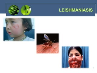
There are 4 main types of the disease:
In cutaneous forms, skin ulcers usually form
on exposed areas: face, arms and legs. These
usually heal within a few months, leaving
scars.
Diffuse cutaneous leishmaniasis produces
disseminated and chronic skin lesions
resembling those of lepromatous leprosy. It is
difficult to treat.
In mucocutaneous forms, lesions can
partially or totally destroy mucous membranes
of the nose, mouth and throat cavities and
surrounding tissues.
Visceral leish, also known as kala azar, is
characterized by high fever, substantial weight
loss, swelling of the spleen and liver, and
anaemia. If left untreated, the disease can have
a fatality rate as high as 100% within two years.
Leishmaniasis
5. Types of
Cutaneous :
• Most common
•Numerous skin sores (1-4
w)
• Self healing (disfigure)
• BASED ON CLINICAL :
ZCL
ACL
Leishman
Visceral (Kala Azar):
• Most serious
• Deaths if untreated
• Northern Areas
Types of Leishmaniasis in Pakistan
6. 6
Leishmania spp. cycle b/w
vertebrate hosts and sand fly vectors
in either promastigote or amastigote
form.
Promastigotes are slightly
elongated and contain a single
nucleus with an anterior flagellum
originating from a kinetoplast while
amastigotes are slightly round to
oval, still contain a single nucleus
and kinetoplast, but retain only a
rudimentary flagellum.
Both stages are capable of
replication via binary fission but not
within the same host.
Life Cycle of Leishmaniasis
7. Macrophages: 1st Line
Defense and promptly
engulf invading organisms.
Amastigotes : Binary
Fission and released
in circulation system
8.
9.
10.
11.
12. Pathogenesis
• Leishmaniasis is a slowly progressive disease
that can take up to 7 years to become clinically
apparent. Even then, signs are frequently
nonspecific and a diagnosis of Leishmania is
seldom considered.
• Dogs are most commonly infected with L.
infantum (L. donovani complex) which is
responsible for visceral leish.
• However, up to 90% of infected dogs present
with both visceral and cutaneous lesions.
• However, many dogs appear naturally resistant
to this parasite and may remain asymptomatic
despite known infection.