Preliminary Study on Monitoring Drug Resistance of Colon Cancer with Intravox...
Optoporation Poster final Draft
1. Acknowledgements
-NSF PRISM grant DMS-0928053 (Dix Pettey,
P.I.), which funds the Mathematics in Life
Sciences Program.
-We acknowledge the Christopher S. Bond Life
Sciences Center for supporting this work
- We thank Dr. Viator and the Viator Lab:
(Adam Eshein and Kiran Bhattacharyya) for
advice and guidance.
Optoporation: Selective Permeabilizaton of Cell Membranes with Gold Nanopartcles
Dustin Johnson, Matt Molengraft, Adam Eshein, Kiran Bhattacharyya, and Dr. John Viator
Introduction
The process of porating cells, or optoporation, has
the potential to become a valuable tool for many
different fields of work. Optoporation is the process
of forming different sized pores in cells by attaching
nanoparticles to the cell, that will absorb laser
pulses and subsequently cause poration in the cell
wall. This method can potentially be applied in
several different areas of work, including genetics
and cancer research. The purpose of this study was
to determine the parameters related to cell viability
after optoporation. More specifically, the main
targets were to discover how different parameters
(such as laser energy, pulses, amount of
nanoparticles added, etc.) affected the poration rate
of the cells and their re-sealing speed.
Method
In this study, all tests followed the same
fundamental procedure of tagging each respective
type of cell (white blood or breast cancer) with a
fluorescent dye and adding nanoparticles to the
dish full of tagged cells. After waiting, special dye
(Fluorescent Brighter) was added. This dye was
used because it is not permeable through the cell
membrane. Therefore, the cell must be porated for
the dye to enter into the cell. Finally, the cells were
shot with a 532 nm laser with varied energies
ranging from 15-20 mJ.
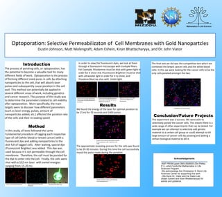
White Blood Cells Died with
Proflavine
Cancer Cells Dyed Rhodamine Shooting cells With laser
Results
We found the energy of the laser for optimal poration to
be 15 mJ for 70 seconds and 1400 pulses.
Breast cancer cells By themselves Porated
by Nanoparticles with rhodamine and
FLBR
Picture of breast cancer cells Showing
their healing process
Competitive test with overlay
In order to view the fluorescent dyes, we look at them
through a fluorescent microscope with multiple filters.
For Example, Rhodamine must be shot with green light in
order for it shine red, Fluorescent Brightner must be shot
with ultraviolet light in order for it to shine, and
Provaline Must be shot with Violet light.
Cancer cells through fluorescent
microscope showing Rhodamine
excitation.
Cancer cells through fluorescent
microscope showing Fluorescent
Brightener excitation.
Overlay of Cancer cells with
Fluorescent Brighter and
Rhodamine
Conclusion/Future Projects
The experiment was a success. We were able to
selectively porate the cancer cells. This means there is a
wide range of other experiments that can be tested. For
example we can attempt to selectivly add genetic
material to a certain cell group or could attempt to kill
large amount of cancer cells by porating and adding a
certain biological material to kill it.
The approximate resealing process for the cells was found
to be 20-30 minutes. During this time the cell successfully
reseal the pores made during the poration.
The final test we did was the competitive test which we
combined the breast cancer cells and the white blood
cells. In this we were looking for the cancer cells to be the
only cells porated amongst the two.