Planning of Nuclear Medicine Facility
•Download as PPTX, PDF•
2 likes•2,212 views

A presentation on Planning of Nuclear Medicine facility and regulatory requirements as a prerequisite.

Recommended
Recommended
More Related Content
What's hot
What's hot (20)
Similar to Planning of Nuclear Medicine Facility
Similar to Planning of Nuclear Medicine Facility (20)
Recently uploaded
Recently uploaded (20)
Planning of Nuclear Medicine Facility
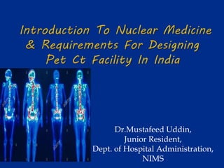
- 1. Dr.Mustafeed Uddin, Junior Resident, Dept. of Hospital Administration, NIMS
- 2. Introduction to Nuclear medicine How is it different from radiology & imageology ? Applications of Nuclear medicine Categories of Nuclear medicine labs Planning of Nuclear Medicine Unit Radioactive waste disposal Personnel Radiation protection Radiation Safety
- 3. Process where in an unstable nuclide emits charged particles or EMR to attain a stable state. This could last from picoseconds to longer times resulting in more emissions (m) Units of radioactivity Curie , Becquerel (SI) measure the r.a decay as dps or cps
- 4. Nuclear medicine is concerned with diagnostic and therapeutic uses of artificially produced radioisotopes. Nuclear medicine procedures are of two types: 1. In-vivo procedures in which radioisotopes are administered to patients, and 2. In-vitro procedures where radioactivity is added to the samples collected from the patient. Again, in-vivo tests are classified into: (a) imaging procedures, and (b) non-imaging procedures.
- 5. An imaging procedure, which is more popularly known as scintigraphy, provides an image of the distribution of administered radioactivity in the organ or tissue of interest at any given time. Non-imaging procedures are aimed at measurements of gross radioactivity in the organ of interest at any particular time. Serial measurements provide a time- activity curve
- 6. -Transmission imaging - Xrays /sound waves - Anatomical imaging - ExcellentResolution FORM -Emission imaging - Radioactivity - Functional imaging - Less resolution FUNCTION Radiology vs NM
- 7. (i) imaging of various organs such as thyroid, liver, brain, bone, kidneys etc., (ii) thyroid function studies, and therapy of thyroid disorders, (iii) investigations of central nervous system, (iv) absorption studies in gastroenterology, (v) nuclear hematology, e.g., blood volume studies, iron kinetics, etc., (vi) renal function studies, (vii) nuclear cardiology, (viii) in-vitro studies like radioimmunoassay (RIA) of various hormones
- 11. Molecular imaging using short lived positron emitters tagged to organic molecules to detect the patho physiologic abnormalities at cellular level. 18FDG --- 110min 13NH3(Ammonia) --- 10min 15O --- 2min 82Rb --- 72 sec
- 12. On the basis of the nature of work carried out and the facilities required Category 1: Laboratories performing in-vitro radio assays with ready to use kits. Category 2: Laboratories carrying out radio-labeling of ligands with preparation of kits and using them for in- vitro radioassays Category 3: Laboratories performing in-vivo non-imaging procedures and in-vitro assays Category 4: As category 3, but also performing in-vivo static/dynamic imaging procedures . Radionuclide Therapy: Categories 3 and 4 nuclear medicine laboratories can also undertake treatment of thyrotoxicosis with l31I and polycythemia vera with 32P, with a few added facilities.
- 13. MODEL OF CARE The model of care will depend on level of services provided as defined in the service plan and the presence or otherwise of PET as a sub- component of the Nuclear Medicine Unit. In large centers, it will be a discrete unit. If there are only one or two gamma cameras, it may be a discrete sub-unit of Medical Imaging.
- 14. LOCATION A ground floor site is preferred but if this cannot be achieved, consideration should be given to units above, below and adjoining the proposed location with regard to radiation shielding requirements, the weight of equipment and associated shielding and access for equipment and radioactive isotopes. The Unit should not act as a thoroughfare to other units of the healthcare facility
- 15. Functional Areas Reception / Administration Waiting areas for outpatients and inpatients, including toilets patient holding, observation and recovery area treatment areas including gamma camera rooms, specialized scanning imaging rooms (SPECT, PET, PET/CT, bone densitometry), stress testing facilities support areas including utilities, staff station Hot Lab / Radioactive Waste Store staff areas including offices and amenities
- 17. A TYPICAL LAYOUT OF A NUCLEAR MEDICINE LABORATORY FOR DIAGNOSTIC STUDIES (IN‐VIVO) IMAGING) TOTAL AREA =150‐160 SQ. M. Note: All the walls /partitions of the Nuclear Medicine laboratory should be made of 9” brick or 6” concrete. Fume Hood to be installed; if required Decontamination Room is optional. The active toilet may be used for personnel decontamination by providing a shower and wash basin.
- 18. Fig: A TYPICAL LAYOUT PLAN FOR A 2‐BEDDED ISOLATION WARD FOR HOSPITALISATION OF PATIENTS TREATED WITH LARGE DOSE OF I‐131. Note: All the walls /partitions of the facility should be made of concrete only, the thickness of which will depend on the area of the room, position of the bed and the occupancies all around. The ducting line of the fume hood should be shown in the plan ,Decontamination Room is optional. The active toilet may be used for personnel decontamination by providing a shower
- 19. Fig: DUAL DELAY TANK SYSTEM FOR COLLECTION AND SAFE DISPOSAL OF RADIOACTIVE WASTE FROM ISOLATION WARD Note: The capacity of the delay tank for a single
- 20. PATIENT WAITING Waiting areas should allow separation of dosed and un-dosed patients, particularly as some patients may need to wait for 45 minutes after dosing for uptake. It is also preferable to separate dosed patients from relatives and visitors to the unit which may include young adults, pregnant women and children. Dosed patients should have access to drinking water and toilet facilities without having to access general waiting areas. Outpatients should be separated from inpatients for privacy reasons with separate entrances
- 21. GAMMA CAMERA The gamma camera is a device used in Nuclear Medicine to image gamma radiation emitting radioisotopes to view and analyze images of the human body or the distribution of medically injected, inhaled, or ingested radionuclide emitting gamma rays, producing a two dimensional image.
- 22. VIEWING AND REPORTING AREA A dedicated room with dimmable lighting will be required for viewing and reporting on scans. Each workstation should accommodate imaging screens, computers for access to imaging and patient information systems, writing and shelving space for reference materials. The number of reporting stations will depend on service level, number of scanning rooms and the staff establishment
- 23. HOT LAB / DISPENSARY AND RADIOACTIVE WASTE STORE Radioactive radiopharmaceuticals are stored and prepared ready for administration to the patient in the Hot Lab. A lead screen barrier is required for the dispensary area. A radioactive waste storage area may also be incorporated into or adjacent to this space. Provide radiation shielding as advised by AERB Consultants. The Waste Store will require a sink and basin with hands-free taps for hand washing and equipment decontamination.
- 24. Generator produced 99mTc, 68 Ga ,82Rb Cyclotron produced 18F,11C,13N,15O Reactor produced 131 I , 99Mo,123I,201Tl
- 25. - Pure Gamma emitter - Easily available - 6hrs half life-Low radiation burden - Flexible chemistry - No anaphylaxis Almost 99% of diagnostic work in the NM departments is with 99mTc labeled tracers
- 28. is a radioactive compound used in NM , which does not have any pharmacological effect, bcos in most cases they are used in tracer amounts and does not show any dose response relation ship like conventional drugs
- 29. Hot component - Isotope 99m TECHNITIUM 131Iodine 201 Thallium 67 Gallium 111Indium 123 Iodine Cold component Chemical substances tagged with isotopes to study the organ of interest. - DTPA-kidneys - MDP -bones - S-Colloid-liver -MAA - lungs -sestaMIBI-heart/tumor
- 31. Delay and decay – short lived radio isotopes Dilute and disperse – small quantity radioactivity Concentrate and contain – large quantity radioactivity
- 32. The radionuclides used in nuclear medicine are mostly short lived, the half life being in the range from a few hours to a few days. Normally, "Tc99 and 131 I are the two major radionuclides that are used for nuclear medicine procedures in large quantities. Other radionuclides used at relatively low levels of activities are 5lCr, 57Co, S8Co, S9Fe, 32P and 125I and a few others. Because of the short half lives, the disposal of radioactive waste is done by the simple method of decay and disposal.
- 33. • Licensed Physician and Technologist • Radiation Safety officer RSO I,II,III Mandatory
- 34. ALARA – As Low As Reasonably Achievable
- 35. Please inform the doctor if you are pregnant or breast feeding यदि आप गर्भवती है या स्तनपान कर रहे हैं तो डॉक्टर को ूचितत करं مہربانی براہ کریں مطلع کو ڈاکٹر تو ہو والی پالنے دودھ یا ہیں حاملہ آپ اگر మీరు గరభవతి లేదా బిడ్డకు పాలు ఇస్త ు నట్ల యితే డాకటర్ కు తెలియ చేయండి CAUTION
- 36. Avoid Contamination Wear gloves, use tissue paper De -contaminate when necessary Reduce exposure Time, Distance, Shielding
- 37. sheilding
- 38. BIOGRAPH-16 SIEMENS BIOGRAPH-16 Functional & Anatomical Imaging
- 39. 1.5 cm infiltrating ductal CA Very dense breasts Negative mammogram PET/CT for initial staging COMPREHENSIVE METASTATIC WORKUP Primary Liver lesions Vertebral body
- 40. Introduction Site & Layout Plan approval Submission of regulatory consent form Pre-commissioning Inspection Approval of commissioning/Routin e Operation Work practice Waste Disposal Radiation Protection aspects Shielding calculations Personnel monitoring devices Radiation safety issues Operating suggestion Personnel monitoring services for staff members Radiation monitoring devices Staff requirement
- 41. Presently, every well-established hospital would like to procure Medical Cyclotron or positron emission tomography-computed tomography (PETCT) facility in their NM department. The various stages of approval of PET-CT facility by the Atomic Energy Regulatory Board (AERB) and important steps that one has to know/follow before planning for this new facility are summarized in the following sections
- 42. The user has to submit to AERB two copies each of the proposed layout plan, site plan, and elevation drawing of the facility indicating the floor, nature of occupancy around, above and below, if any, has to be submitted in “B3” size paper (353×500 mm2) along with the application form AERB/RSD/NMF/SLA (downloadable from www.aerb.gov.in). The user has to clearly indicate the dimension of each of the rooms associated with the facility in the proposed layout plan of the NM department.
- 44. The user has to submit the details of the completion of the construction work as per the approved plan, installation of equipments, procurement of radiologic protection accessories, enrollment of radiation workers in Personal Monitoring Services and availability of qualified staff as per AERB Safety Code AERB/SC/MED-IV (Rev-1 2001), shall be intimated to Head, RSD, AERB, by submitting the Regulatory Consent form no. AERB/444-NM/RC-FORM (downloadable from www.aerb.gov. in)
- 45. In this stage, the precommissioning inspection of the facility will be carried out by AERB official(s) to ensure that the construction of NM facility is as per the approved plan and also to verify the information provided in stage “2” mentioned above
- 46. On ensuring the compliance of the requirement as specified in AERB safety Code SC/MED-IV (Rev.1. 2001) for the safe handling of radioactive material in the approved NM facility, the authorization for the procurement of radioactive material indigenously or no objection certificate (NOC) for procurement of radioactive material from abroad will be issued for the stipulated time period.
- 47. In NM facility, the radiopharmaceutic formulation should be prepared, handled, administered to the patients, and disposed of in a safe manner taking into account adequate radiation protection measures Radioisotopes should be stored, used, and transported safely and securely all the time. Any unusual event that has resulted or has the potential to result in overexposure to the workers or public should be reported to the AERB.
- 48. In India, radioactive waste management is governed by the Atomic Energy (Safe Disposal of Radioactive Wastes) Rules, 1987, GSR-125 issued under the Atomic Energy Act, 1962. In NM, having PET-CT facility, mostly short- lived radioisotopes C-11, N-13, O-15, F-18, and others are used in unsealed form. The half-lives of these radioisotopes range from few minutes to few hours There are 2 main approaches to the disposal of radioactive waste. One is characterized as “dilute and disperse” and the other as “confine and contain.”
- 49. NM facility with PET-CT employs relatively large activities of high-energy photon emitting radioisotopes. This coupled with the current dose limits for members of the public, can result in a shielding requirement. Even modest reductions in the radiation levels at 511 keV require significant amounts of shielding. A thorough and site-specific evaluation has to be made for each facility
- 50. Determine the expected workload (number of patients per week) of the facility, maximum radioactivity to be administered per procedure, and CT workload (total mAs and kVp) per patient. Determine the occupancies of areas within the facility and in adjacent, uncontrolled areas. Include consideration of occupancies at floors above and below the facility. Determine the location of the place where all the initial activities of radioisotopes are to be dispensed. This also includes the injected patient working area. Obtain the isodose curves of the transmission sources in a PET-CT scanner
- 51. Automated dose dispenser Dose calibrator with thick lead shielding to reduce technologist exposure Tungsten syringe shields to reduce finger dose Extra thick L-block table-top shields (5 cm of lead compared with 1.2 cm of lead in standard NM applications) Syringe carriers for transporting the dose from one room to another Remotely actuated syringes that keep the syringe totally enclosed in a shield while the operator delivers the dose by pushing on an extension rod
- 52. Maximize separation from the patient after injection and minimize the time spent with them If the patient is ambulatory, allow as much separation as feasible when they are escorted to the scanner room In the hot laboratory, particular attention should be paid to minimize the time required to handle the dose during the assay and verification steps
- 53. Every staff member, involved in radioactive work, such as radiopharmacy, radiochemistry, dispensing of radioactivity, radiopharmaceutic dose administration, patient imaging, and others, should be covered with personnel monitoring services. For availing this facility, one may approach the Head, CD and R Section, RP and AD, BARC, Anushakti Nagar, Mumbai. Presently, 3 such laboratories are accredited as given below. 1. M/s Avanttech Laboratories (P) Ltd. # 76, 7th Street, Ground Floor, Porur Garden, Phase – I, Chennai – 600 095. 2. M/s Renentech Laboratories Pvt. Ltd. C-106, Synthofine Industrial Estate,Off Aarey Road, Goregaon (E) Mumbai – 40 063. 3. M/s Ultratech Laboratories Pvt. Ltd. 12/15, Priyadarshini, Parisar (W) Bhilai, C.G. - 490020.
- 54. In the NM department having the Medical Cyclotron and PET facilities, following are the monitoring equipments required: GM-based survey meter/ionization chamber– based survey meter Contamination monitor Pocket dosimeters Isotope dose calibrator
- 55. Since the quantity of radionuclide that is being handled is significant and the energy of associated radiation is high, It is absolutely necessary to have an automatic smaller dosage dispensing unit, fume hood, and L-Bench to handle smaller dose for quality assurance tests, lead bricks, shielding devices made of tungsten (syringe shield, source container, transport container, and others), remote handling devices (de-capper, cap sealer, long vial holder, pair of tongs, and others.
- 56. Nuclear medicine physician : MBBS plus A post graduate degree/diploma in NM recognized by the Medical Council of India or National Board of Examination. Nuclear medicine technologist Qualifications : A Bachelor’s degree in NM technology from a university; or A Bachelor’s degree in Science from a university; and Bachelor degree/Post graduate degree/diploma in NM technology from a university
- 57. Radiological safety officer :A candidate with the following qualification is eligible to appear in the RSO Certification Examination conducted by the AERB. Qualifications for RSO Level-II A post graduate degree/diploma in Nuclear Medicine recognized by the Medical Council of India or National Board of Examination, Ministry of Health and Family Welfare; Or A degree/post graduate diploma/post graduate degree in Nuclear Medicine Technology from an institution or university
- 61. Lets use it for the benefit of mankind
- 62. Guidelines For Starting A Nuclear Medicine Laboratory : Excerpts from a booklet published by Bhabha Atomic Research Centre, India. Tandon P. Regulatory requirements for designing PET-CT facility in India. Indian Journal of Nuclear Medicine : IJNM : The Official Journal of the Society of Nuclear Medicine, India. 2010;25(2):39-43. doi:10.4103/0972-3919.72684. Chapter 27, : Indian health Facility Guidelines,2014 www.aerb.gov.in www.barc.gov.in