
Recommended
More Related Content
What's hot
What's hot (20)
Similar to BIOLOGICAL OXIDATION L4 (Oxidative phosphorylation)
Similar to BIOLOGICAL OXIDATION L4 (Oxidative phosphorylation) (20)
Recently uploaded
Recently uploaded (20)
BIOLOGICAL OXIDATION L4 (Oxidative phosphorylation)
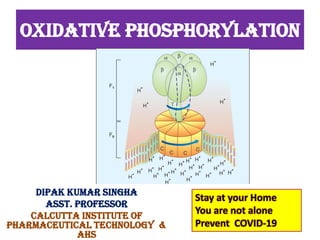
- 1. OXIDATIVE PHOSPHORYLATION DIPAK KUMAR SINGHA ASST. PROFESSOR CALCUTTA INSTITUTE OF PHARMACEUTICAL TECHNOLOGY & AHS Stay at your Home You are not alone Prevent COVID-19
- 2. BIOLOGICAL OXIDATION and OXIDATIVE PHOSPHORYLATION The students must be able to answer questions on the following topics: ➢ Stages of Oxidation of Foodstuffs ➢ Redox potentials ➢ Biological oxidation ➢ Enzymes and co-enzymes involved biological oxidation ➢ High energy compounds ➢ Organization of electron transport chain ➢ Oxidative Phosphorylation ➢ Chemiosmotic theory ➢ Proton pump ➢ ATP synthesis ➢ Inhibitors of ATP synthesis
- 3. Biological Oxidation • The transfer of electrons from the reduced co-enzymes through the respiratory chain to oxygen is known as biological oxidation.
- 4. OXIDATIVE PHOSPHORYLATION Energy released during this process is trapped as ATP. This coupling of oxidation with Phosphorylation is called oxidative Phosphorylation. In the body, this oxidation is carried out by successive steps of dehydrogenations. The transport of electrons through the ETC is linked with the release of free energy. The process of synthesizing ATP from ADP and Pi coupled with the electron transport chain is known as oxidative phosphorylation. The complex V of the inner mitochondrial membrane is the site of oxidative phosphorylation.
- 5. Energetics of Oxidative Phosphorylation The E0 ׳and G0 ׳of biological oxidation may be calculated as follows: ½ O2 + 2H+ → H2O (E׳ 0 = + 0.82) NAD+ + H+ + 2e → NADH (E׳ 0 = – 0.32) When these two equations are computed; ½ O2 + NADH + H+ → H2O + NAD+ (E׳ 0 = 1.14 V) ΔG0 ׳ = – nF E׳ 0 = –2 × 23.06 × 1.14 = –52.6 kcal/mol The free energy change between NAD+ and water is equal to 53 kcal/mol. This is so great that, if this much energy is released at one stretch, body cannot utilize it. Hence, with the help of ETC assembly, the total energy change is released in small increments so that energy can be trapped as chemical bond energy, ATP (Fig.20.2).
- 7. P : O Ratio The P : O ratio refers to the number of inorganic phosphate molecules utilized for ATP generation for every atom of oxygen consumed. More appropriately, P : O ratio represents the number of molecules of ATP synthesized per pair of electrons carried through ETC. The mitochondrial oxidation of NADH with a P : O ratio of 3 can be represented by the following equation : NADH + H+ +1/2O2 + 3ADP+ 3Pi------ NAD+ +3ATP+4H2O • P : O ratio of 2 is assigned to the oxidation of FADH2. (Note : Although yet to be proved beyond doubt, some workers suggest a P : O ratio of 2.5 for NADH + H+, and 1.5 for FADH2, • based on the proton translocation)
- 8. P : O Ratio The P:O ratio is defined as the number of inorganic phosphate molecules incorporated into ATP for every atom of oxygen consumed. When a pair of electrons from NADH reduces an atom of oxygen (½ O2), 2.5 mol of ATP are formed per 0.5 mol of O2 consumed. In other words, the P:O ratio of NADH oxidation is 2.5; The P:O value of FADH2 is 1.5.
- 9. Sites of ATP Synthesis phosphorylation in ETC There are three reactions in the ETC that are exergonic to result in the synthesis of 3 ATP molecules three sites of ATP formation in ETC are 1. Oxidation of FMNH2 by coenzyme Q. 2. Oxidation of cytochrome b by cytochrome c1 . 3. Cytochrome oxidase reaction. Each one of the above reactions representsa coupling site tor ATP production. There are only two coupling sites for the oxidation of FADH2 (P : O ratio 2), since the first site is bypassed
- 10. Sites of ATP Synthesis raditionally, the sites of ATP synthesis are marked, as site 1, 2 and 3, as shown in Figure 20.13. But now it is known that ATP synthesis actually occurs when the proton gradient is dissipated, and not when the protons are pumped out (Fig. 20.15). Fig. 20.15: Summary of ATP synthesis. One mitochondrion is depicted, with inner and outer memberanes. ETC complexes will push hydrogen ions from matrix into the intermembrane space. So, intermediate space has more H+ (highly acidic) than matrix. So, hydrogen ions tend to leak into matrix through Fo. Then ATPs are synthesized. I, II, III, IV = components of ETC
- 11. Energetics of oxidative phosphorylation The transport of electrons from redox pair NAD+/NADH (Eo = - 0.32) to finally the redox pair |OrlflzO (Eo = + 0.82) may be simplified and representedin the following equation to, * NADu + H* ----+ H2o + NAD+ The redox potential difference between these two redoxp ai rsi s 1.14 V, which is equivalentto an energy 52 Cal/mol. Three ATP are svnthesizedi n the ETC when NADH is oxidized which equals to 21.9 Cal (each ATP = 7.3 Cal). The efficiency of energy conservation is calculated as 21.9x100 = 42%to52% Therefore, when NADH is oxidized, about 42'/. of energy is trapped in the form of 3 ATP and the remaining is lost as heat. The heat liberation is not a wasteful process/ since it allows ETC to go pn continuously to Benerate t ATP. Further, this heat is necessary to maintain hody temperature.
- 12. MEGHANISM OF OXIDATIVE PHOSPHORYLATION • Several hypotheses have been put forth to explain the process of oxidative phosphorylation. • The most important among them-namely, chemical coupling, and chemiosmotic. • Chemical coupling hypothesis • A series of phosphorylated high-energy intermediates are first produced which are utilized for the synthesis of ATP. • Believed to be analogous to the substrate level phosphorylation that occurs in glycolysis or citric acid cycle. • This hypothesis lacks experimental evidence,
- 13. 1.Chermical coupling hypothesis This hypothesis was put forth by Edward Slater (1953). According to chemical coupling hypothesis during the course of electron transfer in respiratory chain, a series of phosphorylated high- energy intermediates are first produced which are utilized for the synthesis of ATP. These reactions are believed to be analogous to the substrate level phosphorylation that occurs in glycolysis or citric acid cycle. However, this hypothesis lacks experimental evidence, since all attempts, so far, to isolate any one of the high-energy intermediates have not been successful
- 14. 2.CHEMIOSMOTIC THEORY The coupling of oxidation with phosphorylation is termed oxidative phosphorylation. Peter Mitchell in 1961 (Nobel prize, 1978) proposed this theory to explain the oxidative phosphorylation. The transport of protons from inside to outside of inner mitochondrial membrane is accompanied by the generation of a proton gradient across the membrane. Protons (H+ ions) accumulate outside the membrane, creating an electrochemical potential difference (Fig. 20.15). This proton motive force drives the synthesis of ATP by ATP synthase complex (Fig. 20.14).
- 15. Fig. 20.14: ATP synthase. Protons from outside pass through the pore of Fo into the matrix, when ATP is synthesized
- 16. Proton Pump and ATP Synthesis The proton pumps (complexes I, III and IV) expel H+ from inside to outside of the inner membrane. So, there is high H+ concentration outside the inner membrane. This causes H+ to enter into mitochondria through the channels (Fo); this proton influx causes ATP synthesis by ATP synthase. A summary is shown in Figure 20.15. The pH outside the mitochondrial inner membrane is 1.4 units lower than inside. Further, outside is positive relative to the inside (+0.14 V) (Fig. 20.15). The proton motive force (PMF) is 0.224 V corresponding to a free energy change of 5.2 kcal/mol of protons.
- 17. ATP Synthase (Complex V) It is a protein assembly in the inner mitochondrial membrane. It is sometimes referred to as the 5th Complex (Figs 20.14 and 20.15). Proton pumping ATP synthase (otherwise called F1-Fo ATPase) is a multisubunit transmembrane protein. It has two functional units, named as F1 and Fo. It looks like a lollipop since the membrane embedded Fo component and F1 are connected by a protein stalk. Fo Unit: The “o” is pronounced as “oh”; and not as “zero”. The “o” stands for oligomycin, as Fo is inhibited by oligomycin. Fo unit spans inner mitochondrial membrane. It serves as a proton channel, through which protons enter into Mitochondria (Fig. 20.14). Fo unit has 4 polypeptide chains and is connected to F1. Fo is water insoluble where as F1 is a water soluble peripheral membrane protein.
- 18. F1 Unit: It projects into the matrix. It catalyzes the ATP synthesis (Fig. 20.14). F1 unit has 9 polypeptide chains, (3 alpha, 3 beta, 1 gamma, 1 sigma, 1 epsilon). The alpha chains have binding sites for ATP and ADP and beta chains have catalytic sites. ATP synthesis requires Mg++ ions. Mechanism of ATP synthesis: Translocation of protons carried out by the Fo catalyzes the formation of phospho-anhydride bond of ATP by F1. Coupling of the dissipation of proton gradient with ATP synthesis (oxidative phosphorylation) is through the interaction of F1 and Fo. Binding Change Mechanism The binding change mechanism proposed by Paul Boyer (Nobel prize, 1997) explains the synthesis of ATP by the proton gradient. The ATP synthase is a “molecular machine”, comparable to a “water-driven hammer, minting coins”. Fo is the wheel; flow of protons is the water fall and the structural changes in F1 lead to ATP coin being minted for each turn of the wheel. The F1 has 3 conformation states for the alpha-beta functional unit: O state—Does not bind substrate or products L state—Loose binding of substrate and products T state—Tight binding of substrate and products
- 19. According to this theory, the three beta subunits (catalytic sites), are in three functional states: O form is open and has no affinity for substrates. L form binds substrate with sluggish affinity. T form binds substrate tightly and catalyzes ATP synthesis. As protons translocate to the matrix, the free energy is released, and this is harnessed to interconvert these 3 states. The bond is synthesized in the T state and ATP is released in the O state. The sequence of events is as follows: 1. ADP and Pi bind to L binding site 2. L to T conversion is by energy driven conformational change that catalyzes the formation of ATP 3. T state reverts to O state when ATP is released 4. L state is regenerated for further ADP binding. For the complete rotation of F1 head through the 3 states, 10 protons are translocated
- 20. Inhibitors of ATP Synthesis Site Specific Inhibitors (Table 20.5; Fig. 20.17) Inhibitors of Oxidative Phosphorylation i. Atractyloside inhibits the translocase whereas oligomycin inhibits the Fo (Table 20.5). ii. Ionophores are lipid-soluble compounds that increase the permeability of lipid bilayers to certain ions. There are two types of ionophores; mobile ion carries (e.g. valinomycin) and channel formers (e.g. gramicidin). Valinomycin allows potassium to permeate mito-chondria and dissipate the proton gradient. iii. The toxicity of cyanide is due to its inhibitory effect on the terminal cytochrome, which brings cellular respiration to a standstill.
- 21. Uncouplers of Oxidative Phosphorylation • Uncouplers will allow oxidation to proceed, but the energy instead of being trapped by phosphorylation is dissipated as heat. This is achieved by removal of the proton gradient. (Table 20.5; Fig. 20.17).