
Recommended
More Related Content
What's hot
What's hot (20)
Similar to Immunoglobulin Functions and Classes
Similar to Immunoglobulin Functions and Classes (20)
More from Dhileeban Maharajan
Recently uploaded
Recently uploaded (20)
Immunoglobulin Functions and Classes
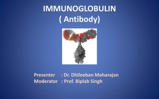
- 1. IMMUNOGLOBULIN ( Antibody) Presenter : Dr. Dhileeban Maharajan Moderator : Prof. Biplab Singh
- 2. ANTIBODIES React specifically with the antigen that stimulated their production Globulins : alpha, beta & gamma (immunoglobulin) “first line of defence” Synthesized only by B cell lineage Mediators of humoral immunity Two types: Membrane bound, Secreted Extremely diverse & Specific
- 3. FUNCTIONS 1. Neutralization of microbial toxins 2. Activation of complement system 3. Opsonization of pathogens Enhanced phagocytosis 4. Antibody dependent cell mediated cytotoxicity 5. Antibody mediated mast cell activation
- 4. IMMUNOGLOBULINS Five classes of immunoglobulins: i. Immunoglobulin G (IgG) ii. Immunoglobulin M (IgM) iii. Immunoglobulin A (IgA) iv. Immunoglobulin E (IgE) v. Immunoglobulin D (IgD)
- 5. Classes of immunoglobulins and their heavy chains and subclasses CLASS HEAVY CHAIN SUBCLASSES IgG Gamma (γ) γ1,γ2,γ3,γ4 IgM Mu (µ) None IgA Alpha (α) α1,α2 IgD Delta (δ) None IgE Epsilon (ε) None
- 6. STRUCTURE OF IMMUNOGLOBULINs Glycoproteins & complex structure of four polypeptide chains Two identical heavy chains (typically 50,000–70,000 Da each & longer) Two identical light chains (25,000Da each & shorter) This gives immunoglobulin an overall ‘Y’ or ‘T’ shape Crystallographic analyses
- 7. PARATOPE Antigen-binding site Part of antibody which recognizes and binds to an antigen (Epitope). Small region (5 to 10 amino acids) of the Variable region (Fab region) Contains parts of the heavy and light chains
- 8. Heavy chain Made up of 420–440 amino acids Held together by one to five disulfide (S—S) bonds Bound to a light chain by disulfide bond & noncovalent bonds These interactions form the basic four-chain (H–L)2 antibody structure
- 9. Light chains Made up of 220–240 amino acids Structurally and chemically similar in all classes 2 types: kappa (κ) and lambda (λ) They differ in their amino acids present in constant region Present in human serum in a ratio of 2:1
- 10. Variable region Amino-terminal half of the light or heavy chain Consisting of 100–110 amino acids Different for each class of Igs The variable regions of both light and heavy chains consist of three highly variable regions known as hypervariable regions Function Antigen binding site
- 11. Constant region The carboxyl-terminal half of the molecule Contains two basic amino acid sequences The Fc fragment, found to crystallize under low ionic conditions, is present in the constant region of heavy chain Functions Activation of the complement, binding to cell surface receptors, placental transfer, etc. The constant region of the light chain has no biological function.
- 12. Hinge region Flexible amino acid stretch Present between heavy chains linked by disulfide bonds High content of proline and hydrophobic residues Flexibility assists inititiation of the complement cascade Gamma, delta and alpha chains have hinge region Mu and epsilon chains do not have hinge region
- 13. Papain Cleavage Papain cleaves just above the interchain disulfide bonds linking the heavy chains, resulting in production of two identical Fab fragments (monovalently binds to the antigen) and one Fc fragment
- 14. Pepsin Cleavage Pepsin cleaves just below these bonds, thereby generating different digestion products producing an Fc fragment and two Fab fragments [F(ab)2], which upon exposure to reducing conditions are separated into Fab monomeric units
- 15. Antigen determinants • 3 major determinants: – Isotypes – Allotypes – Idiotypes
- 16. Isotypes A particular constant region of the light-or heavy-chain of the Ig which is used to classify them present in all members of a species Heavy chain isotype markers: µ,γ,α,δ and ε for IgM, IgG, IgA, IgD, and IgE, respectively Light chain isotype markers: κ and λ
- 17. Allotypes Allelic differences in both the variable and constant regions of immunoglobulin Am on α heavy chains, Gm on γ heavy chains, and Km on κ light chains >25 Gm types ,3 Km and 2 Am on IgA have been described Allotype markers are absent on µ, δ, ε heavy chains and on λ light chains
- 18. Idiotypes A specificity that is associated with the variable region Idiotype markers are found on the hypervariable region of the Ig Idiotypes are specific for each antibody molecule Anti-idiotypic antibodies produced against Fab fragments prevent antigen–antibody interaction
- 19. Biosynthesis of Immunoglobulins • B lymphocytes and plasma cells take part in the synthesis of immunoglobulins • Resting B cells synthesize only small amounts of Igs that mainly get incorporated into cell membranes but plasma cells (abundant cytoplasm & rich in ER) produce and secrete large amounts of immunoglobulins • In separate polyribosomes, the heavy chain & light chains are produced and later joined either as H-L hemimolecule or as H2 & L2, Later they associate to form complete molecule
- 20. NUCLEUS ER POLYRIBOSOMES HEAVY CHAINS PLASMA CELL GLYCOSYLATION
- 21. Immunoglobulin G Molecular weight of 150,000 Da half-life of 23 days (longest among all the Igs) most abundant - about 80% of the total serum immunoglobulin Four IgG subclasses based on γ chain isotypes : IgG1, IgG2, IgG3, and IgG4 o IgG1, IgG3 & IgG4: cross placental barrier o IgG3, IgG1 & IgG2: complement activation o IgG1 & IgG3: mediate opsonisation (high affinity for Fc receptors of phagocytes)
- 22. Biological activity In response to infection, IgG antibodies appear late after appearance of IgM antibodies, but persists for a longer period Protection against the microorganisms Distributed equally in the intra and extra- vascular compartments Only Ig that crosses the placenta & confers natural passive immunity to the newborns Takes part in precipitation, complement fixation & neutralization of toxins and viruses and binds to microorganisms and facilitates the process of phagocytosis
- 24. Immunoglobulin M Constitute about 5–8% of total serum Ig Intra-vascular distribution Heavy molecule with molecular weight varying from 900k to 1,000k Da Half-life of 5 days basically a pentamer, composed of five Ig subunits & J-chain 2 µ heavy chains & 2 κ/λ light chain Heavy chains are larger than those of IgG by about 20,000 Da, corresponding to an extra domain on the constant region
- 26. Biological activities Pentameric IgM has high valency (10 antigen binding sites) thus efficient in binding antigens with many repeating epitopes, such as viral particles and RBCs Efficient than IgG in activating complement Good complement activation (two Fc regions in close proximity) First Ig produced in primary response; deficiency leads to septicaemia Also act as a B cell receptor
- 27. Presence of IgM antibody in serum indicates recent infection (short lived and disappear early) First Ig to be synthesized by a neonate in about 20 weeks of age; since it is not able to cross placenta, its presence indicates intra-uterine infection The detection of IgM antibodies in serum is useful for the diagnosis of congenital infections, such as syphilis, rubella, toxoplasmosis, etc.
- 28. Immunoglobulin A 2nd most abundant Ig (10–15% of serum IG) Half-life of 6–8 days Consists of α heavy chain (58-kDa & 470 amino acid residue) with 3 constant domains CH1, CH2, and CH3 and 1 variable domain VH Hinge region is situated between CH1 and CH2 domains An additional segment of 18-AA residues at the penultimate position of the chain contains a cysteine residue where the J chain can be attached through a disulfide bond.
- 30. IgA occurs in two forms: Serum IgA and Secretory IgA Serum IgA molecular weight of 60kDa & 1/2 life 6-8 days two subclasses, IgA1 and IgA2 (two α chain isotypes α1 and α2) α2 chain has two allotypes, A2m (1) and A2m (2), and does not have disulfide bonds linking heavy to light chains Differences in the two α chains are found in two CH1 and five CH3 positions & hence there are three varieties of α heavy chains in humans
- 31. Secretory IgA Dimer or tetramer and consists of a J-chain polypeptide and a polypeptide chain called secretory component (70kDa & produced by mucous membrane epithelial cells) Has 5 Ig-like domains that bind to the Fc region domains of the IgA dimer This interaction is stabilized by a disulfide bond between the fifth domain and one of the chains of dimeric IgA
- 32. IgA-secreting plasma cells are concentrated along mucous membrane surfaces The daily production of secretory IgA is greater than that of any other Igs Secretory IgA is the major Ig present in external secretions, such as breast milk, saliva, tears & mucus of the bronchial, genitourinary, and digestive tracts IgA activates the complement not by classical pathway but by alternative pathway
- 34. Biological functions of secretory IgA It is polymeric & hence can cross-link large antigens with multiple epitopes thus protects the mucous membranes against microbial pathogens Binding of secretory IgA to bacterial and viral surface antigens prevents attachment of the pathogens to the mucosal cells thus inhibiting colonization.
- 35. Complexes of secretory IgA and antigen are easily entrapped in mucus and then eliminated by the ciliated epithelial cells of the respiratory tract or by peristalsis of the gut IgA in breast milk protect the newborns against infection during the first month of life Secretory IgA has shown to provide an important line of defense against bacteria (such as Salmonella, Vibrio cholerae, and Neisseria ) and viruses (such as polio, influenza, and reovirus)
- 36. Immunoglobin E IgE constitutes less than 1% of the total Ig pool Present in serum in a very low concentration (0.3 g/mL) & mostly found extravascularly in lining of respiratory and intestinal tracts MW of 190K Da & ½ life of 2–3 days Unlike other Igs, IgE is a heat-labile protein, easily inactivated at 56°C in 1 hour Two ε heavy polypeptide chains (72-kDa, 550- AA residue), along with two κ or two λ light chain, together by disulfide bonds
- 38. Biological activity Reaginic antibody mediates the type I immediate HS (atopy) reactions Responsible for the symptoms of hay fever, asthma & anaphylactic shock IgE binds to Fc receptors on the membranes of blood basophils and tissue mast cells & antigen (allergen) induces basophils and mast cells to translocate their granules to the plasma membrane and release their contents to the extracellular environment - a process known as degranulation
- 39. As a result, varieties of pharmacologically active mediators are released and give rise to allergic manifestations Localized mast-cell degranulation necessary for antiparasitic defence
- 40. Immunoglobulin D IgD comprises less than 1% of serum Ig(MW 180k Da & half-life of only 2–3 days) IgD has basic four chain monomeric structure with two δ heavy chains (MW 63kDa each, 500-amino acid residue) and either two κ or two λ light chains(MW 22kDa each) The δ heavy polypeptide chain consisting of one variable region VH, and a three- domain constant regions CH1, CH2, and CH3 IgD is present on the surface of B lymphocytes and both IgD & IgM serve as recognition receptors for antigens
- 43. Role of immunoglobulins in human defence IgG IgM IgA IgD IgE Enhances phagocytosis Especially effective against microbes & agglutinating antigens Localized protection on mucosal surfaces Serum function not known Allergic reaction Neutralizes toxins and viruses First antibody produced in response to initial infection - Present on B cells; and function in initiation of immune response Lysis of parasitic worms Protects fetus and newborn
- 44. Abnormal immunoglobulins Structurally similar proteins that are found in serum in certain pathological conditions – multiple myeloma, – heavy chain disease – cryoglobulinemia – Rarely in healthy individuals
- 45. Multiple myeloma This condition is characterized by excessive production of the respective myeloma proteins (M proteins) and that of their light chains (BJ proteins) Bence-Jones (BJ) proteins -- earliest abnormal proteins BJ proteins are the light chains of Igs, hence occur as either κ or λ forms BJ proteins have a peculiar property of coagulating at 60°C and redissolving again at a higher temperature of 80°C
- 46. They remained the major source of homogeneous Igs until the development of the hybridoma in 1974 In multiple myeloma, plasma cells synthesizing IgG, IgA, IgD or IgE are affected Myeloma involving IgM-producing plasma cells is known as Waldenström’s macroglobulinemia Heavy chain disease: Lymphoid neoplasia, characterized by an excess production of heavy chains of the immunoglobulins
- 47. Cryoglobulinemia Condition characterized by presence of cryoglobulins in blood The condition may not be always associated with disease but is often found in patients with macroglobulinemia, SLE or myelomas Most cryoglobulins consist of either IgG or IgM or their mixed precipitates & serum from patient precipitates on cooling and redissolves on warming.
- 48. Immunoglobulin as Medicine Immunosuppressive Antibodies Hybridoma Technology Milstein and Kohler – 1974 HAMA – Human anti-mouse antibodies
- 49. Intravenous Immune Globulin (IVIG) • Polyclonal human Immunoglobulin • Thousands of healthy donors • Not specific against single antigen • Normalizing effect in immune network • High Dose IVIG – 2gm/kg use: – ITP – Kawasaki’s disease – GBS
- 50. Replacement therapy (Low dose therapy) Immunomodulatory effect (High dose therapy) Licensed Off-label •Primary immunodeficiency •HIV infection •BMT •B-cell lymphocytic leukemia •Multiple myeloma •Immune thrombocytopenic purpura •GBS •CIDP •Kawasaki disease •Multifocal motor neuropathy •Autoimmune neutropenia •Autoimmune hemolysis •Anti Factor VIII inhibitor •Multiple sclerosis •Myasthenia gravis •Stiff person syndrome •ANCA associated vasculitis •Polymyositis/Dermatomyositis •RA •SLE – APS •GVHD •Sepsis syndrome •TEN •Graves opthalmopathy
- 51. Hyperimmune Immunoglobulins • Selected donor with high titers of antibodies against particular agents • To reduce the severity of infection • Use: – Respiratory syncytial virus – CMV – Varicella zoster -- HHV 3 -- Hepatitis B virus -- Tetanus -- As anti-venom
- 54. Thank You