Contouring guidelines Cervix IMRT
•Download as PPTX, PDF•
51 likes•12,061 views

Guidelines for delineation of nodal and primary target volume for IMRT in cancer cervix

Recommended
Recommended
More Related Content
What's hot
What's hot (20)
Similar to Contouring guidelines Cervix IMRT
Similar to Contouring guidelines Cervix IMRT (20)
More from Debarshi Lahiri
Recently uploaded
Recently uploaded (20)
Contouring guidelines Cervix IMRT
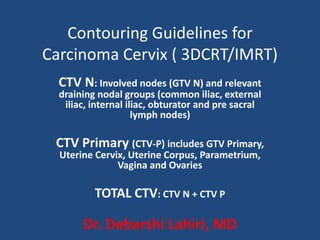
- 1. Contouring Guidelines for Carcinoma Cervix ( 3DCRT/IMRT) CTV N: Involved nodes (GTV N) and relevant draining nodal groups (common iliac, external iliac, internal iliac, obturator and pre sacral lymph nodes) CTV Primary (CTV-P) includes GTV Primary, Uterine Cervix, Uterine Corpus, Parametrium, Vagina and Ovaries TOTAL CTV: CTV N + CTV P Dr. Debarshi Lahiri, MD
- 6. Summary of Recommendations (LN CTV) CTV N includes involved nodes and relevant draining nodal groups (common iliac, external iliac, internal iliac, obturator and pre sacral lymph nodes). • Start contouring iliac vessels from aortic bifurcation down till the appearance of femoral head. • Uniformly, pelvic blood vessels are given a margin of 7mm. The upper border is maintained at aortic bifurcation. • The contour is extended around common iliac vessels posteriorly and laterally so as to include connective tissue between iliopsoas muscles and lateral surface of vertebral body. • All visible nodes (contoured as GTV node) are given a margin of 10mm to create CTV node. • Muscle and bone are excluded from CTV N.
- 8. Pre-sacral region is covered by connecting the volumes on each side of pelvis with a 10-mm strip over the anterior sacrum starting from aortic bifurcation till S2-S3 junction. Sacral foramina are not included in CTV N
- 9. To cover obturator nodes, a strip 17 mm wide is created medial to the pelvic sidewall, by joining the contour of external iliac vessels with internal iliac vessels. Contouring of obturator nodes with 17 mm brush is continued lower down along pelvic side wall, till superior part of obturator foramen The posterior margin of the contour over Internal Iliac vessels lies along anterior edge of pyriformis muscle.
- 10. The caudal margin of internal iliac nodes is at the level of Ischial spine. The caudal margin of external iliac nodes is till the appearance of femoral head. The caudal extent of obturator lymph node is till superior border of obturator foramen
- 11. CTV Primary (CTV-P) includes GTV Primary, Uterine Cervix, Uterine Corpus, Parametrium, Vagina and Ovaries The uterine corpus, entire cervix and the vagina are contoured along with the gross disease (GTV primary) as a single structure (CTV P1) VAGINA: paravaginal tissue is included along with the vaginal wall. A vaginal marker is placed at the lower extent of vaginal disease while taking CT and as per RTOG guidelines: Minimal or no vaginal wall involvement: The contouring is stopped few slices above the lower border of obturator foramen, so that when 1.5 cm ITV (internal target volume) margin is given over the uterus, the lower border does not extend beyond the lower border of obturator foramen. Upper vaginal involvement: Upper two-thirds Extensive vaginal involvement: Entire vagina
- 12. PARAMETRIUM (CTV P2) To delineate the parametrium , connective tissue extending from the cervix to the pelvic wall are included, along with the visible linear structures that run laterally (e.g. vessels, nerves and fibrous structures) Cranial border : defined at the level where the true pelvis begins. Contours should stop once loops of bowel are seen next to the uterus (Lim/Toita et al.) Anteriorly: contouring is done up to the level of posterior border of bladder in the central region, while, in periphery it extends till the anterior end of lateral pelvic bony wall. Posteriorly: parametrium is contoured only till the anterior part (semicircular) of mesorectal fascia. In case of significant parametrial invasion(IIIB)/uterosacral ligament involvement, include entire mesorectum.(Lim et al.(RTOG)/PGI Guidelines). Laterally, the parametrium is contoured till the lateral pelvic wall, upto the medial edge of internal obturator muscle. Caudal border of parametrium is taken at the pelvic floor.
- 14. Total CTV: CTV N and the CTV primary (CTVP1 & CTVP2) are combined and named as total CTV PTV: 10 mm over total CTV ITV Margin:The uterine motion is accounted for by giving an ITV margin on the uterus….??? An asymmetrical margin with ITV expansion of 15 mm antero-posteriorly, 15mm supero-inferiorly and 7 mm laterally, is taken from the uterus (CTV P1)
- 15. Final PTV: The ITV margin given over CTV P1 for uterine motion is added to the total PTV and this is taken as the total target volume (final PTV) to be treated.
- 16. Normal Tissue Delineation (RTOG) • Bowel: The small and large bowel can be contoured together as a Bowel- Bag. Inferiorly, the bowel bag should begin with the first small or large bowel loop or above the ano-rectum, whichever is most inferior. The contours should end 1 cm. above the PTV if coplanar beams are used. If non coplanar beams are chosen, the contours will need to extend further. • Ano-Rectum:Ano-Rectum should be contoured from the level of the anus to the sigmoid flexure. It should extend from the anal verge (marked by a radiopaque marker at simulation) to superiorly where it loses its round shape in the axial plane and connects anteriorly with the sigmoid. • Bladder: Contoured inferiorly from its base, and superiorly to the dome. • Femoral Heads & Pelvic Bones
- 17. Dose Prescription & Constraints • PTV: 50.4 Gy/ 28# • Bladder: V50 < 50% • Bowel Bag: V45 < 195 cc. • Femoral Head: V50 < 5% Mean < 45 Gy • Bone Marrow: V40 < 37% Mean < 34 Gy • Rectum: V50 < 35% Rectum Constraint not given ( IIIB disease/Risk of Uterosacral ligament infiltration) and included in PTV