Daniele Amato Poster - Cancer-associated Venous Thromboembolism
•Download as PPTX, PDF•
0 likes•11 views

Cancer-associated Venous Thromboembolism (VTE) is a critical complication in patients with cancer. However the pathological findings of VTE are limited. Here are investigated the histopathological features of cancer-associated VTE in human autopsy cases.
Recommended

Recommended
More Related Content
Similar to Daniele Amato Poster - Cancer-associated Venous Thromboembolism
Similar to Daniele Amato Poster - Cancer-associated Venous Thromboembolism (20)
More from Karolinska Institutet, University of Bergen, University of Oslo
More from Karolinska Institutet, University of Bergen, University of Oslo (10)
Recently uploaded
Recently uploaded (20)
Daniele Amato Poster - Cancer-associated Venous Thromboembolism
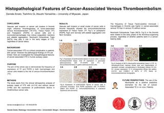
- 1. CONCLUSION Vascular wall invasion or cancer cell clusters in thrombi might influence thrombogenesis of tumor-associated venous thromboembolism (TA-VTE). Tissue Factor (TF) and Podoplanin (PDPN) in cancer cells and in monocyte/macrophages may induce coagulation reactions and platelet aggregation. Neutrophil Extracellular Traps (NETs) may play a role in the early stages of VTE, regardless of cancer status. PURPOSE The aim of this article was to demonstrate the frequency of expression of TF and PDPN in both macrophages and cancer cells related to the risk of venous thromboembolism (TE). Fig.1) Expression of prothrombotic factors of cancer cells in venous thromboembolism tissue. A-B) TF expression in pancreatic adenocarcinoma with TF+/PDPN- and TF+ (C, immunohistochemistry) in gastric adenocarcinoma cells. Histopathological Features of Cancer-Associated Venous Thromboembolism Daniele Amato, Toshihiro Gi, Atsushi Yamashita – University of Miyazaki, Japan FUTURE PERSPECTIVES: The use of the Neutrophils as a positive predictive biomarker for the risk of Cancer Associated TE in «in vivo» Patients. Daniele Amato, Radiology I Universitetet I Oslo – Universitetet i Bergen – Karolinska Institutet daniele.amato@stud.ki.se +39 3935236763 BACKGROUND Cancer-associated VTE is a critical complication in patients with cancer. However the pathological findings of VTE are limited. Here are investigated the histopathological features of cancer-associated VTE in human autopsy cases. 1-A 1-B 1-C 2-A 2-B 2-C Fig.2) Expression of prothrombotic factors of cancer cells in pulmonary thrombus. A-B) PDPN expression with TF-/PDPN+ pattern and PDPN+ (C, immunohistochemistry) in cutaneous squamous cell carcinoma. Fig.3) Analysis of NETs (Neutrophils extracellular traps) in VTE. A) Hematoxylin-Eosin staining shows lysis and fragmentation of neutrophil cromatin corresponding to citrullinated histone H3 (Cit- H3, arrows) expression; C) A corresponding diffuse lytic pattern (Cit-H3+ and CD66b+) of NETs in VTE tissue on immunohistochemical micrographs. METHODS The study starts from the clinical retrospective analysis of autopsy cases of VTE with (n=114) and without cancer (n=66) and the expression of prothrombotic factors in intrathrombus cancer cells. 3-A 3-B 3-C RESULTS Vascular wall invasion or small cluster of cancer cells in thrombi frequently showed the immunohistochemical expression of Tissue Factor (TF, Fig.1) or podoplanin (PDPN, Fig2) and necrosis with platelet aggregation and fibrin formation. The frequency of Tissue Factor-positive monocyte / macrophages in thrombi was higher in cancer-associated venous TE than in venous TE without cancer. Neutrophil Extracellular Traps (NETs, Fig.3) in the thrombi were related to the early phase of the thrombus-organizing process, regardless of whether patients were in a cancer- bearing state.