1. Special Article Journal of Veterinary Emergency and Critical Care 22(S1) 2012, pp S102–S131
doi: 10.1111/j.1476-4431.2012.00757.x
RECOVER evidence and knowledge gap
analysis on veterinary CPR.
Part 7: Clinical guidelines
Daniel J. Fletcher∗
, PhD, DVM, DACVECC; Manuel Boller∗
, Dr. med. vet., MTR, DACVECC;
Benjamin M. Brainard, VMD, DACVA, DACVECC; Steven C. Haskins, DVM, DACVA, DACVECC;
Kate Hopper, BVSc, PhD, DACVECC; Maureen A. McMichael, DVM, DACVECC; Elizabeth A.
Rozanski, DVM, DACVECC, DACVIM; John E. Rush, DVM, MS, DACVIM, DACVECC and Sean D.
Smarick, VMD, DACVECC
Abstract
Objective – To present a series of evidence-based, consensus guidelines for veterinary CPR in dogs and cats.
Design – Standardized, systematic evaluation of the literature, categorization of relevant articles according to
level of evidence and quality, and development of consensus on conclusions for application of the concepts to
clinical practice. Questions in five domains were examined: Preparedness and Prevention, Basic Life Support,
Advanced Life Support, Monitoring, and Post-Cardiac Arrest Care. Standardized worksheet templates were
used for each question, and the results reviewed by the domain members, by the RECOVER committee, and
opened for comments by veterinary professionals for 4 weeks. Clinical guidelines were devised from these
findings and again reviewed and commented on by the different entities within RECOVER as well as by
veterinary professionals.
Setting – Academia, referral practice and general practice.
Results – A total of 74 worksheets were prepared to evaluate questions across the five domains. A series of 101
individual clinical guidelines were generated. In addition, a CPR algorithm, resuscitation drug-dosing scheme,
and postcardiac arrest care algorithm were developed.
Conclusions – Although many knowledge gaps were identified, specific clinical guidelines for small animal
veterinary CPR were generated from this evidence-based process. Future work is needed to objectively evaluate
the effects of these new clinical guidelines on CPR outcome, and to address the knowledge gaps identified
through this process.
(J Vet Emerg Crit Care 2012; 22(S1): 102–131) doi: 10.1111/j.1476-4431.2012.00757.x
Keywords: canine, cardiac arrest, defibrillation, feline
From the Department of Clinical Sciences, College of Veterinary Medicine,
Cornell University, Ithaca, NY (Fletcher); Department of Emergency
Medicine, School of Medicine, Center for Resuscitation Science and the
Department of Clinical Studies, School of Veterinary Medicine, University
of Pennsylvania, Philadelphia, PA (Boller); Department of Small Animal
Medicine and Surgery, College of Veterinary Medicine, University of Geor-
gia, Athens, GA (Brainard); Department of Surgical and Radiological Sci-
ences, School of Veterinary Medicine, University of California at Davis, Davis,
CA (Haskins, Hopper); College of Veterinary Medicine, University of Illinois,
IL (McMichael); Cummings School of Veterinary Medicine, Tufts University,
North Grafton, MA (Rozanski, Rush); AVETS, Monroeville, PA (Smarick).
∗
These authors contributed equally.
The authors declare no conflict of interest.
Address correspondence and request for reprints to
Dr. Daniel Fletcher, Cornell University College of Veterinary Medicine, DCS
Box 31, Upper Tower Rd, Ithaca, NY 14853, USA.
E-mail: djf42@cornell.edu
Submitted March 29, 2012; Accepted March 29, 2012.
Abbreviations
ABC airway, breathing, circulation
ALS advanced life support
BLS basic life support
CPA cardiopulmonary arrest
CPR cardiopulmonary resuscitation
EtCO2 end tidal CO2
ETT endotracheal tube
ILCOR International Liaison Committee on Resus-
citation
LOE level of evidence
PEA pulseless electrical activity
PICO population, intervention, control group,
outcome
S102 C
" Veterinary Emergency and Critical Care Society 2012
2. RECOVER clinical guidelines
RECOVER Reassessment Campaign on Veterinary
Resuscitation
VF ventricular fibrillation
VT ventricular tachycardia
Introduction
The development of specific, evidence-based clini-
cal guidelines for human cardiopulmonary resuscita-
tion (CPR), based upon extensive surveys of the lit-
erature by the International Liaison Committee on
Resuscitation (ILCOR) has allowed consistent train-
ing for human healthcare professionals and the lay
public, leading directly to improved outcomes.1–3
No
comparable evidence-based guidelines have been avail-
able in veterinary medicine, although recommenda-
tions on practical execution of CPR in small animals
have been published.4–8
The absence of standardized,
comprehensive training coupled with a lack of con-
sensus on the content of the published recommen-
dations has led to significant variability in the ap-
proach to veterinary CPR, likely to the detriment of our
patients.9
The main goal of the Reassessment Campaign on
Veterinary Resuscitation (RECOVER) initiative was to
develop a set of clinical consensus guidelines for the
practice of CPR in dogs and cats based upon an ex-
tensive, systematic review of the literature in the con-
text of our target species. Although there is overlap be-
tween the literature examined by ILCOR and RECOVER,
the science was interpreted based upon applicability
to dogs and cats. This has led to conclusions that di-
verge, in some areas, from those reached by ILCOR.
Based upon the results of the evidence worksheet pro-
cess used in RECOVER,10
a total of 101 clinical guide-
lines were developed and made available for review
for a period of 4 weeks to members of the veterinary
community (see Appendix I). This feedback was used
to modify and refine the recommendations, yielding
the final set of consensus guidelines presented in this
manuscript.
In order to reflect the variability in the quality and
quantity of evidence examined, each guideline devel-
oped through the RECOVER consensus process has been
assigned two descriptors: (1) Class – this categorizes
the risk-benefit ratio of the intervention described in the
guideline, and (2) Level – this categorizes the strength of
the evidence available to support the recommendation.
This scheme was adapted from that used by ILCOR.11
The individual class and level categories are detailed
in Tables 1 and 2, and each guideline is labeled (Class-
Level).
Table 1: Class descriptors for the clinical guidelines, categorizing
the risk-benefit ratio associated with the intervention
Class Risk:benefit ratio Clinical recommendation
I Benefit >>> Risk Should be performed
IIa Benefit >> Risk Reasonable to perform
IIb Benefit ≥ Risk May be considered
III Risk > Benefit Should not be performed
Table 2: Level descriptors for the clinical guidelines, categorizing
the strength of the evidence available for the recommendation
Level
Populations
studied Criteria for recommendation
A Multiple populations Multiple high quality and/or high level of
evidence studies
B Limited populations Few to no high quality and/or high level
of evidence studies.
C Very limited
populations
Consensus opinion, expert opinion,
guideline based on physiologic/
anatomic principles, standard of care
Small Animal Veterinary CPR Algorithm
The guidelines presented in this document cover a wide
variety of CPR-related topics in 5 domains: Prepared-
ness and Prevention, Basic Life Support (BLS), Advanced
Life Support (ALS), Monitoring, and Post-Cardiac Arrest
Care. The main elements of CPR and their temporal se-
quence have been summarized in a CPR algorithm chart
(Figure 1). This algorithm was designed to deliver step-
by-step prompts to the veterinary rescuer engaged in
CPR and stresses the importance of early BLS inter-
ventions. The evidence reviewed strongly reinforced the
importance of early delivery of high-quality chest com-
pressions with minimal interruption. High-quality chest
compressions should be delivered in uninterrupted cy-
cles of 2 minutes with most patients in lateral recum-
bency, at a compression rate of 100–120/min and a com-
pression depth of 1/3–1/2 the width of the chest while
allowing for full elastic recoil of the chest between indi-
vidual compressions. In addition, it is likely that early
intubation and ventilation in veterinary CPR is highly
valuable, with a ventilation rate of approximately 10
breaths/min, a tidal volume of 10 mL/kg, and an in-
spiratory time of 1 second delivered simultaneously
with compressions. If intubation supplies are not avail-
able, mouth-to-snout ventilation is an acceptable alter-
native, and should be delivered in repeated rounds of
30 chest compressions followed by 2 rapid breaths in
cycles of 2 minutes. After each 2-minute cycle of BLS,
the compressor should be rotated to prevent fatigue,
which may decrease the quality of chest compressions.
Every effort should be made to minimize the duration
of chest compression interruptions between cycles. ALS
C
" Veterinary Emergency and Critical Care Society 2012, doi: 10.1111/j.1476-4431.2012.00757.x S103
3. D. J. Fletcher et al.
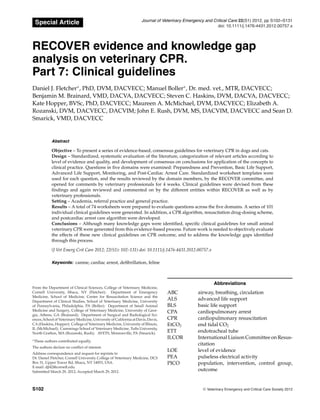
Figure 1: CPR algorithm chart. This chart summarizes the clinical guidelines most relevant to the patient presenting acutely in
CPA. The box surrounded by the grey dashed line contains, in order, the initial BLS and ALS actions to be taken when a patient is
diagnosed with CPA: (1) administration of chest compressions, (2) ventilation support, (3) initiation of ECG and EtCO2 monitoring,
(4) obtaining vascular access for drug administration, and (5) administration of reversal agents if any anesthetic/sedative agents have
been administered. The algorithm then enters a loop of 2-minute cycles of CPR with brief pauses between to rotate compressors, to
evaluate the patient for signs of ROSC, and to evaluate the ECG for a rhythm diagnosis. Patients in PEA or asystole should be treated
with vasopressors and, potentially, anticholinergic drugs. These drugs should be administered no more often than every other cycle of
CPR. Patients in VF or pulseless VT should be electrically defibrillated if a defibrillator is available, or mechanically defibrillated with
a precordial thump if an electrical defibrillator is not available. Immediately after defibrillation, another 2-minute cycle of BLS should
be started immediately. BLS, basic life support; CPA, cardiopulmonary arrest; CPR, cardiopulmomary resuscitation; C:V, compression
to ventilation ratio; EtCO2, end tidal CO2; PEA, pulseless electrical activity; ROSC, return of spontaneous circulation; VF, ventricular
fibrillation; VT, ventricular tachycardia.
S104 C
" Veterinary Emergency and Critical Care Society 2012, doi: 10.1111/j.1476-4431.2012.00757.x
4. RECOVER clinical guidelines
interventions, including initiation of monitoring, estab-
lishment of vascular access, administration of reversal
agents, vasopressor and vagolytic therapy, and defibril-
lation are also included in the algorithm. Recommended
dosing and indications for common CPR-related drugs
are included in Appendix II.
A post-cardiac arrest (PCA) algorithm chart, designed
to summarize the major interventions recommended in
the guidelines for patients that achieve return of sponta-
neous circulation (ROSC), is shown in Figure 2. The algo-
rithm is focused on initial respiratory optimization that
includes normalizing ventilation to achieve normocap-
nia and titration of oxygen supplementation to maintain
normoxemia while avoiding both hypoxemia and hyper-
oxemia. Once the patient’s respiratory status is assessed
and a treatment plan is initiated, cardiovascular concerns
are addressed. The hemodynamic optimization compo-
nent is based on the concept of early goal-directed ther-
apy, first described for patients in septic shock.12
Arterial
blood pressure is first assessed, and IV fluids, vasopres-
sors, and positive inotropes are administered as needed
to achieve normotension or mild hypertension. Severe
hypertension is addressed with adjustment of vasopres-
sors, pain management, and antihypertensives. Once ar-
terial blood pressure targets are met, central venous oxy-
gen saturation (ScvO2) or blood lactate concentration is
assessed to determine if oxygen delivery to tissues is
adequate. If a deficit in oxygen delivery is noted, hemo-
dynamic optimization is revisited and guided by oxy-
gen delivery targets rather than arterial blood pressure
targets. If oxygen delivery targets are still not met, red
blood cell transfusions are administered if indicated. A
PCV target of 25% is suggested, a departure from tra-
ditional early goal-directed therapy due to more recent
data in humans documenting improved outcomes with
more restrictive transfusion triggers.13
Once hemody-
namic optimization strategies have been initiated, neu-
roprotective interventions and intensive monitoring are
considered based on the neurologic status of the patient.
Recommended doses for common PCA-related drugs are
included in Appendix II. It should be noted that this
comprehensive treatment protocol is based in part on
evidence specific to the PCA condition and in part on
general critical care principles. Studies on the effects of
these types of optimization strategies during PCA care
are needed.
Preparedness and Prevention
The guidelines developed through the evidence col-
lected by this domain are based on the premise that
resuscitation attempts that are organized, cohesive, and
led by a well-functioning knowledgeable team adhering
to evidence-based CPR guidelines should improve sur-
vival from cardiopulmonary arrest (CPA). Strengthening
the links in the chain of survival, the time-sensitive, co-
ordinated actions necessary to maximize survival from
CPA, has the potential to lead to improved outcomes.4
The guidelines derived from this domain focus on inter-
ventions involving both environmental and personnel
factors that strengthen the chain of survival for dogs and
cats with CPA.
Equipment organization and cognitive aids
An organized and efficient response to an acute medical
or surgical crisis is crucial. The effects of ready access to
organized and consistently audited crash carts on out-
comes for patients receiving CPR have been well studied
in human medicine.14
Equipment and supply inaccessi-
bility or failure has been implicated in delays in initia-
tion of CPR in up to 18% of CPA cases.15
Therefore, it is
recommended that the location, storage, and content of
resuscitation equipment should be standardized and
regularly audited (I-A). In addition, the presence of cog-
nitive aids such as checklists, algorithm charts, and dos-
ing charts has been shown to improve compliance with
CPR guidelines.16
Formal training of personnel in the use
of these cognitive aids is also crucial to effective utiliza-
tion during a crisis.17
Figure 1 shows an example of a CPR
algorithm chart, and Figure 3 shows an example of an
emergency drug and dosing chart, containing only the
most commonly used drugs, separated into categories
based upon indication, and provided in volume of drug
to be administered by body weight to reduce dose cal-
culation errors. Availability and clear visibility of these
charts in areas in which CPA may occur, such as pro-
cedure areas, anesthesia induction rooms, and surgery
suites is recommended (I-B).
CPR training
Adherence to CPR guidelines can only be accomplished
if personnel receive effective, standardized training and
regular opportunities to refresh their skills. Because
high-quality CPR requires both cognitive skills to cor-
rectly perform all indicated steps in an orderly, rapid
fashion as well as psychomotor skills to provide effec-
tive manual interventions such as chest compressions
and ventilation, CPR training should include both didac-
tic components targeted at cognitive performance and
opportunities to practice hands-on skills with quality
feedback (I-A). Effective options for psychomotor skill
training include high-fidelity simulation technologies,
low-fidelity task trainers, and auditory and visual feed-
back devices.18–20
Regardless of the type of technology
used for initial training, refresher training at least every
6 months is recommended to reduce the risk of the de-
cay of skills (I-A). There is some evidence that the use
of simulation methodologies may be most beneficial for
C
" Veterinary Emergency and Critical Care Society 2012, doi: 10.1111/j.1476-4431.2012.00757.x S105
5. D. J. Fletcher et al.
Figure 2: Post-cardiac arrest (PCA) care algorithm. This chart summarizes a comprehensive treatment protocol for PCA care that
includes components of controlled ventilation and oxygenation, goal-directed hemodynamic optimization, and neuroprotective strate-
gies. The sequence shown reflects the order in which each component should be assessed and treatment initiated. Assessment and
initiation of treatment for the subsequent component will likely commence before the endpoints of the previous component have been
completely met. Thus respiratory, hemodynamic, and neuroprotective treatment strategies will be initiated in parallel in most cases.
CRT, capillary refill time; CVP, central venous pressure; EtCO2, end-tidal carbon dioxide; HTS, hypertonic saline; IPPV, intermittent
positive pressure ventilation; MAP, mean arterial pressure; MM, mucous membrane color; ROSC, return of spontaneous circulation;
SAP, systolic arterial pressure; ScvO2, central venous oxygen saturation.
S106 C
" Veterinary Emergency and Critical Care Society 2012, doi: 10.1111/j.1476-4431.2012.00757.x
7. D. J. Fletcher et al.
this booster training.21
Although high-fidelity simula-
tors may carry some advantage in this type of training,
simple mock codes run every 3–6 months on low-fidelity
manikins are likely to improve awareness of CPR guide-
lines and are achievable in most small animal practice
settings.
Improved learning outcomes have been documented
when CPR training culminates in performance testing.22
Therefore, regardless of the methods used for initial and
refresher training, structured assessment after CPR train-
ing is recommended (I-A). In addition to assessment af-
ter didactic and psychomotor skills training, structured
debriefing after a real resuscitation effort or simulated
CPR, allowing participants to review and critique their
performance and the performance of the team as a whole
is recommended (I-A). During the debriefing, the partic-
ipants should be encouraged to drive the discussion and
identify for themselves the strengths and weaknesses
of the team’s performance. Facilitation by a team mem-
ber trained in debriefing technique is useful, and care
must be taken to prevent focusing on blaming indi-
viduals for poor performance. Open, honest discussion
about opportunities for improvement immediately after
a CPR attempt can lead to significant enhancement in
CPR performance.23–25
Team dynamics
Several studies in human medicine have investigated the
effect of the presence of a physician on outcomes in out-
of-hospital CPA, and taken as a whole, there does not
appear to be a beneficial effect on outcome of CPR from
the presence of a physician acting as team leader.26,27
Although there have been no studies investigating this
question in veterinary medicine, based on the data avail-
able in human medicine, veterinarians or technicians
may be considered as leaders of a CPR team (IIb-B).
Regardless of the status of the team leader, there is
strong evidence in the literature that communication
and team skills training can improve the effectiveness
of a CPR attempt,28
and specific leadership training is
recommended for individuals who may need to lead in
a CPR attempt (I-A). Crucial roles of the team leader
include distributing tasks to other team members and
enforcing rules and procedures. Important leadership
behaviors that can improve CPR team performance in-
clude intermittently summarizing the code to ensure a
shared mental model among team members, actively so-
liciting input from team members to encourage situation
awareness and identify issues and ideas from all mem-
bers of the team, and assigning individual tasks to team
members rather than performing them personally to al-
low better attention to the global status of the code rather
than a specific task. Team performance can also be en-
hanced by using focused, clear communication directed
at individuals when tasks are assigned, and utilization
of closed loop communication.29
Closed loop communi-
cation is accomplished by a clear, directed order being
given to one team member by another, after which the
receiving team member repeats the order back to the
requestor to verify the accuracy of the receiver’s percep-
tion. This simple technique drastically reduces medical
errors, especially in an emergency situation, due to mis-
understanding of orders and prevents the possibility of
an order not being carried out because the receiver did
not hear the request.
BLS
In veterinary CPR, BLS includes the recognition of CPA,
administration of chest compressions, airway manage-
ment, and provision of ventilation. It is imperative that
BLS is provided immediately upon diagnosis or suspi-
cion of CPA, and lay rescuers and medical professionals
alike may accomplish most aspects. Numerous human
and animal experimental studies have shown that the
rapidity of initiation and quality of BLS performed is as-
sociated with ROSC and survival in victims of CPA.30–32
Although BLS is considered separately from ALS and
monitoring in this consensus statement, in clinical prac-
tice, the intent is that BLS will be performed simul-
taneously with ALS and monitoring, or that ALS and
monitoring will occur as soon after initiation of BLS as
possible.
Chest compressions
Chest compressions should be initiated as soon as possi-
ble upon recognition of CPA and if multiple rescuers are
present, airway and ventilation management should not
delay commencement of chest compressions.
Patient position and compressor hand placement
Due to experimental evidence suggesting higher left
ventricular pressures and aortic flow in dogs in lat-
eral recumbency compared to dorsal recumbency, and
clinical data in dogs and cats showing higher rates of
ROSC associated with compressions performed in lat-
eral recumbency,33,34
chest compressions should be done
in lateral recumbency in both dogs and cats (I-B). Either
left or right lateral recumbency is acceptable. However,
the profound variations in chest conformation among
dogs and cats suggest that a single, identical approach
to chest compressions is unlikely to be optimal in all pa-
tients with CPA. There are 2 main theories describing the
mechanism by which external chest compressions lead
to blood flow during CPR.35
The cardiac pump theory
postulates that the cardiac ventricles are directly com-
pressed between the sternum and the spine in patients
S108 C
" Veterinary Emergency and Critical Care Society 2012, doi: 10.1111/j.1476-4431.2012.00757.x
8. RECOVER clinical guidelines
in dorsal recumbency or between the ribs in patients
in lateral recumbency. The thoracic pump theory pro-
poses that chest compressions increase overall intratho-
racic pressure, secondarily compressing the aorta and
collapsing the vena cava leading to blood flow out of the
thorax. During elastic recoil of the chest, subatmospheric
intrathoracic pressure provides a pressure gradient that
favors the flow of blood from the periphery back into
the thorax and into the lungs where oxygen and carbon
dioxide exchange occurs. Although minimally studied,
it is believed that the predominant mechanism in any
patient will be dependent upon thoracic conformation,
and it is likely that both mechanisms contribute to blood
flow in most patients.
In the majority of medium, large, and giant breed dogs
with rounded chests, direct compression of the heart
with external chest compressions is unlikely. Therefore,
the thoracic pump mechanism is likely to predominate
in these patients, and chest compressions over the widest
portion of the chest will allow maximal increases in in-
trathoracic pressure (see Figure 4a). It is therefore reason-
able in most large and giant breed dogs, to deliver chest
compressions with the hands placed over the widest por-
tion of the chest (IIa-C). Conversely, in more keel-chested
(narrow, deep chested) dogs such as greyhounds, the
cardiac pump theory may be more easily employed with
external chest compressions in lateral recumbency; there-
fore, in dogs with this conformation, chest compressions
with the hands positioned directly over the heart is rea-
sonable (IIa-C). (Figure 4b). In dogs with barrel-chested
conformations, such as English bulldogs, sternal com-
pressions in dorsal recumbency, directed at the cardiac
pump theory, may be considered (IIb-C) (Figure 4c). Cats
and small dogs tend to have higher thoracic wall com-
pliance, and effective chest compressions using the car-
diac pump mechanism can likely be achieved with a 1-
hand technique with the compressor’s fingers wrapped
around the sternum at the level of the heart (see Fig-
ure 5a). Thus, circumferential compressions rather than
lateral compressions may be considered (IIb-C). How-
ever, if the compressor becomes fatigued or an individual
patient’s thoracic wall compliance is lower due to age,
obesity, or conformation, a 2-handed technique employ-
ing the cardiac pump mechanism can be used (Figure 5b).
Chest compression technique
There is strong evidence, including an experimental
study in dogs documenting increased rates of ROSC
and 24-hour survival, supporting a recommendation for
compression rates of 100–120/min in cats and dogs (I-
A).36
However, there is also some evidence that higher
compression rates of up to 150/min may be even more
advantageous, and further work in this area is needed.
Figure 4: Chest compression techniques for medium, large, and
giant breed dogs. (A) For most dogs, it is reasonable to do chest
compressions over the widest portion of the chest to maximally
employ the thoracic pump theory. Either left or right lateral re-
cumbency are acceptable. (B) In keel-chested (ie, deep, narrow
chested) dogs like greyhounds, it is reasonable to do chest com-
pressions with the hands directly over the heart to employ the
cardiac pump theory, again in either recumbency. (C) For barrel-
chested dogs like English Bulldogs, sternal compressions directly
over the heart with the patient in dorsal recumbency may be con-
sidered to employ the cardiac pump mechanism.
C
" Veterinary Emergency and Critical Care Society 2012, doi: 10.1111/j.1476-4431.2012.00757.x S109
9. D. J. Fletcher et al.
Figure 5: Chest compression techniques for small dogs and cats.
(A) For most cats and small dogs (<10 kg) with compliant chests,
the use of a 1-handed technique to accomplish circumferential
chest compressions with the hand wrapped around the sternum
directly over the heart may be considered. (B) An alternative chest
compression method for cats and small dogs is the 2-handed tech-
nique directly over the heart to employ the cardiac pump mech-
anism. This method may be considered in larger cats and small
dogs with lower thoracic compliance, or in situations in which
the compressor is becoming fatigued while doing 1-handed
compressions.
There is also good evidence to support deep chest com-
pressions of 1/3–1/2 the width of the thorax in most pa-
tients (IIa-A), with an experimental canine study show-
ing a linear relationship between compression depth
and mean arterial pressure, and multiple human clinical
trials and experimental animal studies supporting these
compression depths.37–39
Finally, experimental studies
in pigs have documented reduced coronary and cerebral
perfusion when full elastic recoil between chest compres-
sions is not permitted (ie, leaning). Observational stud-
ies in people have shown a high prevalence of leaning
during CPR. It is recommended that full chest wall recoil
is allowed between compressions (I-A).40,41
Ventilation
Both hypoxia and hypercapnia reduce the likelihood of
ROSC; therefore, securing a patent airway and providing
ventilation are essential during CPR.42,43
Although hu-
man CPR algorithms emphasize the importance of chest
compressions over ventilation in BLS, there is evidence in
human pediatric patients that ventilation is more impor-
tant in patients with CPA not of primary cardiac origin.44
Because the majority of canine and feline cardiac arrests
are due to noncardiac root causes, early endotracheal in-
tubation and provision of ventilation in CPR is likely to
be of benefit.
Ventilation technique for intubated patients
Given the documented detrimental effects of pauses in
chest compressions and the ease with which dogs and
cats can be intubated, if equipment and personnel are
available, rapid intubation of dogs and cats in CPA is
recommended. This should be accomplished with the
animal in lateral recumbency so that chest compressions
may be continued during the procedure. Once the endo-
tracheal tube (ETT) is in place, the cuff should be inflated
so that ventilation and chest compressions can occur si-
multaneously (I-A). The ETT should be secured to the
muzzle or mandible to prevent dislodgement. It may
be useful for veterinarians and technicians to practice
lateral endotracheal intubation in patients undergoing
routine anesthetic procedures to develop and maintain
these skills.
Although there are very limited data in dogs and
none in cats evaluating optimal ventilation strategies
for intubated patients during CPR, there are several
well-controlled experimental studies in pigs as well
as clinical studies in people supporting these recom-
mendations. Higher respiratory rates, longer inspiratory
times, and higher tidal volumes can lead to impaired
venous return due to increased mean intrathoracic pres-
sure as well as decreased cerebral and coronary per-
fusion due to vasoconstriction, and have been docu-
mented to lead to poorer outcomes in people during
CPR.45
Due to decreased pulmonary blood flow result-
ing from the reduced cardiac output achievable during
CPR (approximately 25–30% or normal), physiologically
“normal” ventilation rates are likely to lead to low ar-
terial CO2 tension. Lower respiratory rates are associ-
ated with elevated arterial CO2 tension and can cause
S110 C
" Veterinary Emergency and Critical Care Society 2012, doi: 10.1111/j.1476-4431.2012.00757.x
10. RECOVER clinical guidelines
peripheral vasodilation, worsening perfusion to the
core, and cerebral vasodilation, potentially increasing
intracranial pressure. Therefore, a ventilation rate of 10
breaths/min with a tidal volume of 10 mL/kg and a short
inspiratory time of 1 second are recommended (I-A).
Ventilation technique for nonintubated patients
There have been no studies examining the efficacy of
mouth-to-snout ventilation in dogs and cats, although
there is a case report describing successful application
of this technique in a dog with traumatic cervical spinal
cord injury during transport to a veterinary hospital,
suggesting that it can effectively maintain oxygenation
and ventilation in this species.46
In addition, there is
some evidence that effective ventilation can be accom-
plished in dogs using noninvasive techniques such as
tight-fitting masks, but obtaining an appropriate fit and
seal can be challenging.47,48
To accomplish mouth-to-
snout ventilation, the rescuer holds the patient’s mouth
tightly closed, places his or her mouth over the pa-
tient’s nares making a seal with the snout, and blows
into the nares (see Figure 6). There have been no studies
investigating the optimal compression-to-ventilation
(C:V) ratio during CPR in nonintubated dogs and cats
and the results of studies in other species are somewhat
conflicting. The preponderance of the evidence suggests
C:V ratios of at least 30:2 should be maintained. Until
further studies are done evaluating higher C:V ratios, a
C:V ratio of 30:2 in nonintubated dogs is recommended
(I-B). To accomplish this, a series of 30 chest compres-
sions at a rate of 100–120/min is performed, followed by
Figure 6: Mouth-to-snout breathing technique. The rescuer
holds the patient’s mouth closed with one hand, creates a seal
over the patient’s nares with his or her mouth, and blows into
both nares to achieve a normal chest rise.
a brief interruption of chest compressions during which
2 breaths are delivered quickly, after which another se-
ries of 30 chest compressions are delivered.
Cycles of CPR
Although there have been no studies in dogs and cats
evaluating the optimal timing of CPR cycles, there are
several high-quality prospective and retrospective stud-
ies in human medicine suggesting that uninterrupted
cycles of BLS lasting 2 minutes result in better sur-
vival and neurological outcomes than shorter cycles with
more frequent interruptions to chest compressions.49,50
Therefore, chest compressions should be performed in
2-minute cycles without interruption in intubated pa-
tients when several rescuers are present, or in 2-minute
cycles with brief interruptions after every 30 chest com-
pressions to allow 2 quick breaths to be delivered using
the mouth-to-snout technique if only 1 rescuer is present
or the animal is not intubated (I-A). After each 2-minute
cycle of compressions, the compressor should rotate to
reduce lean and compromise of compression efficacy due
to fatigue (I-B).
Delay in starting CPR
Rapid diagnosis of CPA is crucial because the deleteri-
ous effects of delaying the start of BLS are significant,
with reductions in survival to discharge and neurologic
status reported in numerous studies.51–53
Although not
examined in veterinary medicine, several human stud-
ies have documented the poor sensitivity of pulse palpa-
tion for diagnosis of CPA.54,55
In addition, it is common
for agonal breaths to be misidentified as spontaneous
breathing in people in CPA.56
There is also strong evi-
dence in the human literature that less than 2% of pa-
tients in CPA experience any serious harm when BLS is
started, likely because patients will commonly respond
to the stimulation associated with CPR.57
Therefore, ag-
gressive administration of CPR in patients suspected of
being in CPA is recommended, as the risk of injury due
to CPR in patients not in CPA is low (I-B). When assess-
ing patients that are apneic and unresponsive, a rapid
airway, breathing, circulation (ABC) assessment lasting
no more than 5–10 seconds is recommended. If there
is any doubt as to whether the patient has experienced
CPA, CPR should be initiated immediately while further
assessment to support the diagnosis of CPA is accom-
plished simultaneously by other personnel or after an
initial cycle (2 min) of CPR.
Interposed abdominal compressions
To facilitate venous return from the abdomen and im-
prove cardiac output, the use of abdominal compressions
C
" Veterinary Emergency and Critical Care Society 2012, doi: 10.1111/j.1476-4431.2012.00757.x S111
11. D. J. Fletcher et al.
interposed with chest compressions has been extensively
studied in experimental canine and porcine models as
well as in human clinical trials.58,59
There is minimal evi-
dence of abdominal trauma due to the use of interposed
abdominal compressions when rescuers are trained in
the technique. Therefore, the use of interposed abdomi-
nal compressions in dogs and cats with CPA is reasonable
when sufficient personnel trained in its use are available
(IIa-B).
ALS
ALS encompasses the components of veterinary CPR
performed after BLS has been initiated and until ROSC is
achieved. ALS includes therapy with vasopressors, pos-
itive inotropes, and anticholinergics, correction of elec-
trolyte and acid-base disturbances and volume deficits,
and prompt defibrillation. If BLS and ALS are performed
promptly, initial ROSC rates may be as high as 50% in
dogs and cats.34
Vasopressor and vagolytic therapy
Because only 25–30% of a normal cardiac output is
achieved with even high-quality external chest compres-
sions, generation of adequate coronary and cerebral per-
fusion pressures during CPR requires high peripheral
vascular resistance, directing more of the circulating vol-
ume to the central circulation. Vasopressors are therefore
an essential component of ALS drug therapy.
Epinephrine
Epinephrine, a catecholamine that acts as a nonspecific
adrenergic agonist, has been widely used for its vaso-
pressor (!1) activity during CPR for decades. It also has
"1 adrenergic activity, the inotropic and chronotropic ef-
fects of which are likely less crucial, and may be harmful
when treating CPA due to increased myocardial oxy-
gen demand, exacerbating myocardial ischemia, and
predisposing to arrhythmias once ROSC is achieved.60
Although higher doses (0.1 mg/kg IV) of epinephrine
have been associated with increased rates of ROSC,
they have not been associated with increased survival
to discharge, possibly due to the exaggerated adrener-
gic effects.61
Therefore, the use of low-dose (0.01 mg/kg
IV) epinephrine administered every 3–5 minutes early
in CPR is recommended (I-B), but high-dose (0.1 mg/kg
IV) epinephrine may be considered after prolonged CPR
(IIb-B). In order to minimize underdosing or overdosing
during CPR, this drug should be administered during
every other cycle of BLS.
Vasopressin
The vasopressor effects of vasopressin are mediated
through the peripheral V1 receptor located on vascular
smooth muscle. This mechanism of action is completely
independent of the !1 effects of epinephrine. Unlike !1
receptors, V1 receptors remain responsive in the face
of an acidic pH, and vasopressin has no inotropic or
chronotropic effects that could worsen myocardial is-
chemia. Therefore, it has been studied as an alternative
to epinephrine during CPR. Evidence of the efficacy of
vasopressin compared to epinephrine in dogs and cats
during CPR is limited, with 1 prospective observational
study suggesting a beneficial effect of vasopressin34
while a prospective trial in dogs found equivalent sur-
vival rates.62
The human literature is mixed, with va-
sopressin potentially associated with increased survival
in human patients with asystole, prolonged CPA, or
hypovolemia,63,64
but large meta-analyses have failed
to show any benefit (or detriment) to the use of vaso-
pressin over epinephrine in CPR.65,66
Although further
study is needed, the use of vasopressin (0.8 U/kg IV) as
a substitute or in combination with epinephrine every
3–5 minutes may be considered (IIb-B).
Atropine
Atropine is a parasympatholytic agent that has been used
widely in patients with CPR. Many studies have evalu-
ated the use of atropine during CPR, and have largely
shown no beneficial or detrimental effect of its use at
standard dosing (0.04 mg/kg). Higher doses (0.1, 0.2, 0.4
mg/kg) have been associated with worse outcomes in an
experimental study in dogs.67
However, an experimental
study showed that dogs with asphyxia-induced pulse-
less electrical activity (PEA) were more likely to be resus-
citated when administered a combination of epinephrine
and atropine than dogs administered epinephrine and
5% dextrose.68
Although not strongly supported by the
literature, atropine is most likely to be of use in dogs
and cats with asystole or PEA associated with high va-
gal tone, and use of standard dose (0.04 mg/kg) at-
ropine in these cases is reasonable (IIa-B). Due to the lack
of any clear detrimental effect, routine use of atropine
(0.04 mg/kg IV) during CPR in dogs and cats may be
considered (IIb-C).
Defibrillation
Sudden cardiac arrest due to ventricular fibrillation (VF)
is common in people, and a large body of literature
suggests that electrical defibrillation is the most effec-
tive therapy. Widespread implementation of by-stander-
operated electrical defibrillators has been associated
with marked improvement in survival in people. In a
S112 C
" Veterinary Emergency and Critical Care Society 2012, doi: 10.1111/j.1476-4431.2012.00757.x
12. RECOVER clinical guidelines
hospital setting, current guidelines in human medicine
recommend that “shockable” rhythms (VF and pulseless
VT) be promptly treated with electrical defibrillation if
available. Because VF and VT are the result of abnormal
pacing of groups of ventricular myocardial cells by the
myocardial cells themselves rather than the pacemak-
ers, the goal of electrical defibrillation is to depolarize
as many of these cells as possible, driving them into
their refractory period, and stopping the random elec-
trical and uncoordinated mechanical activity, that is, to
stop the ventricles from fibrillating. If this is successful,
the pacemakers may then begin driving the myocardial
cells (establishing a sinus rhythm), or the patient may
develop asystole. Note that either of these outcomes is
considered a successful defibrillation. In the absence of
an electrical defibrillator, mechanical defibrillation may
be accomplished with a precordial thump, but the effi-
cacy of this intervention is likely poor.
Electrical defibrillation technique
Modern defibrillators use one of two main technologies:
(1) monophasic, in which a unidirectional current flows
from one electrode to the other, and (2) biphasic, in which
current initially flows in one direction, then reverses and
flows in the other direction. Biphasic defibrillators have
been shown to more effectively terminate VF at lower
defibrillation energy than monophasic defibrillators, in
turn leading to less myocardial injury.69
Therefore, the
use of a biphasic defibrillator is recommended over a
monophasic defibrillator (I-A), at a dose of 4–6 J/kg with
a monophasic defibrillator or 2–4 J/kg with a bipha-
sic defibrillator (IIa-B). If the first shock is unsuccessful,
there is some evidence from experimental and clinical
human studies that increasing the defibrillation energy
may increase the rate of success.70,71
Although no studies
have shown a direct detrimental effect of dose escalation,
there is a risk of increased myocardial damage with in-
creasing defibrillation dose. However, in dogs and cats
with VF/pulseless VT, defibrillation energy escalation
(eg, 50% dose increase) is reasonable if the first counter-
shock is unsuccessful (IIa-B).
To maximize current through the ventricles, the pad-
dles should be placed on opposite sides of the thorax
approximately over the costochondral junction directly
over the heart. To facilitate this, the patient will likely
have to be placed in dorsal recumbency. The use of a
plastic trough may facilitate this. Defibrillator paste or
gel should be liberally applied to the paddles, which
must be pressed firmly against the chest to establish con-
tact with the skin. If defibrillation patches are used, the
fur must be shaved to facilitate contact, which will re-
sult in a longer pause in chest compressions. Once the
defibrillator is charged, the operator must ensure that
Figure 7: Posterior paddle assembly. The black arrow indicates
the posterior paddle. The dog is laid on the posterior paddle, and
when defibrillation is required, the hand paddle is placed on the
opposite side of the chest directly over the heart to defibrillate.
Chest compressions can then be immediately continued with the
posterior paddle in place.
no personnel are making any contact with the patient or
the table to prevent injury by announcing the intent to
defibrillate with a term such as “Clear” and visually con-
firming that all personnel are clear before discharging the
defibrillator. The person discharging the defibrillator is
also at risk, and must ensure that he or she is not touch-
ing the patient or the table; the use of exam gloves can
reduce the risk of contact, but he or she must ensure that
no fluid, gel, or paste is bridging the cuff of the glove
and allowing contact with the skin. In addition, electri-
cal defibrillation should not be attempted if alcohol is on
the fur due to the high risk of fire. The use of a posterior
paddle assembly, a flat paddle replacement, can improve
the efficiency and safety of defibrillation, minimizing the
interruption to compressions, and eliminating the need
to place the patient in dorsal recumbency. The flat paddle
is coated with gel or paste and placed under the patient’s
thorax. Defibrillation is then accomplished using a stan-
dard hand paddle on the upward facing chest wall, and
chest compressions can resume immediately while the
posterior paddle is still in place (see Figure 7).
Timing of electrical defibrillation
It is generally accepted that after a loss of perfusion,
the ischemic heart passes through 3 phases: (1) the elec-
trical phase during which minimal ischemic damage
C
" Veterinary Emergency and Critical Care Society 2012, doi: 10.1111/j.1476-4431.2012.00757.x S113
13. D. J. Fletcher et al.
occurs, lasting 4 minutes; (2) the circulatory phase dur-
ing which reversible ischemic damage occurs, lasting 6
minutes; (3) the metabolic phase during which poten-
tially irreversible ischemic damage begins to occur, and
which may necessitate more advanced techniques such
as therapeutic hypothermia and cardiopulmonary by-
pass to reverse.72
Therefore, immediate defibrillation is
recommended in cases of CPA due to VF/pulseless VT
of duration of 4 minutes or less (I-B), or if VF is diag-
nosed during a rhythm check between cycles of CPR
(IIb-B). If the patient is known or suspected to have been
in VF/pulseless VT for greater than 4 minutes and is
beyond the electrical phase, energy substrates are likely
depleted, and the patient will most likely benefit from a
2-minute cycle of BLS before defibrillation (I-B).
Although older CPR algorithms recommended the
use of 3 stacked shocks for patients with refractory
VF/pulseless VT, compelling experimental data in pigs
and clinical data in people showed better outcomes when
a single shock was followed by a full 2-minute cycle
of CPR before re-evaluating the ECG and defibrillating
again.73–75
Therefore, administration of a single shock as
opposed to 3 stacked shocks is recommended, with im-
mediate resumption of CPR in the case of nonsuccessful
defibrillation (I-B).
Precordial thump
The precordial thump was first described as a treatment
option for VF in a case report in 1969 and a case se-
ries in 1970.76,77
Briefly, this is a method of mechanical
defibrillation, accomplished by striking the patient with
the heel of the hand directly over the heart. Unfortu-
nately, more recent studies have documented minimal
efficacy of this technique for treatment of VF.78–80
Al-
though a worksheet was not completed on this topic,
given that there is some limited evidence that a precor-
dial thump may have some efficacy for the treatment of
VF/pulseless VT, this intervention may be considered.
However, given the overwhelming evidence of the su-
periority of electrical defibrillation for the treatment of
VF/pulseless VT, a precordial thump should only be
considered if an electrical defibrillator is not available.
Antiarrhythmic drug therapy
The utility of antiarrhythmic agents such as amiodarone,
lidocaine, and magnesium for patients with CPA due
to VF/pulseless VT has been extensively studied in ex-
perimental models and clinical trials in people, and the
data have been summarized in a recent meta-analysis.81
Of the agents studied, only amiodarone has shown
consistent benefit and may be considered in cases of
VF/pulseless VT resistant to electrical defibrillation (IIb-
B). Some studies have also shown a beneficial effect of
lidocaine in patients with refractory VF/pulseless VT,
although one experimental study showed an increase
in the energy required to successful defibrillate dogs
with induced VF.82
However, more recent data in pigs
suggested that this phenomenon occurs when using
monophasic defibrillators, but not when using bipha-
sic defibrillators.83
Given the uniformly grave prognosis
for patients in refractory VF/pulseless VT, when amio-
darone is not available, lidocaine may be considered in
cases of pulseless VT/VF resistant to defibrillation (IIb-
B), especially when a biphasic defibrillator is used. Data
on the use of magnesium are less compelling, and routine
use of magnesium sulfate is not recommended for car-
diac arrhythmias during CPR, although it may be consid-
ered for treatment of torsades de pointes (IIb-B). It should
be recognized that the use of antiarrhythmic agents may
be considered as adjunctive therapy in refractory cases,
but electrical defibrillation is the recommended primary
treatment for VF/pulseless VT (I-B).
Reversal agents
Of the reversal agents available, only naloxone has been
evaluated for use in patients in CPA. Although evidence
of a beneficial effect is limited, in cases of opioid toxicity,
naloxone should be used during CPR (I-B).84
Even in the
absence of opioid toxicity, the data available suggest that
in cases of recent opioid administration, the use of nalox-
one during CPR may be considered (IIb-B). Although
no specific studies have evaluated the use of other re-
versal agents, in dogs and cats that have received re-
versible anesthetic/sedative medication, administering
reversal agents during CPR may beconsidered (IIb-C),
as the potential risks associated with administration of
these drugs are low. The drug and dosing chart in Ap-
pendix II contains recommended doses during CPR for
naloxone (to reverse opioids), flumazenil (to reverse ben-
zodiazepines), and atipamezole (to reverse !2 agonists).
Electrolyte therapy
Calcium
Calcium is vital for many cellular processes, includ-
ing cellular communication and muscle contraction. Al-
though hypocalcemia commonly develops in patients
with prolonged CPA, the majority of studies investigat-
ing the utility of routine calcium administration during
CPR demonstrated no effect on outcome or worse out-
comes, suggesting that IV calcium should not be used
routinely during CPR (III-B). No studies investigating
the use of calcium in patients with documented hypocal-
cemia during CPR were identified. Given the importance
of calcium for skeletal and smooth muscle contraction,
intravenous calcium may be considered in dogs and
S114 C
" Veterinary Emergency and Critical Care Society 2012, doi: 10.1111/j.1476-4431.2012.00757.x
14. RECOVER clinical guidelines
cats with documented moderate to severe hypocalcemia
during CPR (IIb-C), but studies directly addressing this
question are needed.
Potassium
Hyperkalemia develops commonly in patients with pro-
longed CPA, and treatment of hyperkalemia during
CPR using hemodialysis is associated with improved
outcomes.85
Given this evidence, documented hyper-
kalemia should be treated during CPR (I-B). Although
hemodialysis is rarely available in veterinary clinical
practice, administration of medical therapies directed at
treating hyperkalemia would be reasonable.86
Although
hypokalemia has been associated with CPA in people,
no studies of the efficacy of treatment of hypokalemia
during CPR have been done.87
Therefore, treatment of
documented hypokalemia during CPR may be consid-
ered (IIb-C), but there is no evidence to support or refute
this treatment.
Other therapies
Corticosteroids
Several case series and experimental studies have exam-
ined the utility of corticosteroids in CPR with mixed re-
sults, most involving multiple treatment variables other
than steroids. Only one placebo-controlled randomized
trial specifically investigated the efficacy of cortico-
steroids (dexamethasone) in people during out of hos-
pital CPR, which showed no benefit with the use of
steroids.88
Given the lack of compelling evidence of a
beneficial effect and the potential for deleterious side ef-
fects from corticosteroids,89,90
especially in animals with
poor perfusion,91
the routine use of corticosteroids dur-
ing CPR is not recommended (III-C).
Impedance threshold device
Impedance threshold devices (ITD) have been shown
to improve hemodynamics in anesthetized dogs by in-
creasing venous return due to decreased intrathoracic
pressure.92
While some experimental studies in non-
target species have demonstrated a benefit of these
devices during CPR, the largest clinical trial to date
failed to demonstrate any improvement in ROSC or sur-
vival to discharge in people in CPA with the use of
an ITD.93
In addition, because the device requires chest
wall recoil to generate a “cracking pressure” of at least
−12 cm H2O, use is not feasible in small dogs or cats
weighing less than 10 kg because they are unlikely to be
capable of generating those types of pressures from elas-
tic recoil alone. Therefore, the use of an ITD to enhance
circulation is reasonable in animals > 10 kg (IIa-B), but
studies to date have not demonstrated a survival advan-
tage with their use.
Alkalinization therapy
Severe acidemia due to metabolic acidosis is common
in patients with CPA, and this acid-base disturbance
can lead to detrimental metabolic dysfunction. Sev-
eral experimental studies in dogs have documented
improved survival with bicarbonate therapy with pro-
longed (>10 min) duration of CPA.94,95
However, other
experimental studies in dogs have demonstrated worse
outcomes and metabolic derangements with bicarbonate
therapy, especially when given early in CPR.96
Given the
evidence available, bicarbonate therapy after prolonged
CPA of greater than 10–15 minutes with administration
of 1 mEq/kg of sodium bicarbonate may be considered
(IIb-B).
Intratracheal drug administration
When available, intravenous or intraosseous adminis-
tration of resuscitation drugs is preferred over intratra-
cheal administration, and is associated with improved
survival from CPA.97
However, in animals in which in-
travenous or intraosseous access is not possible, the use
of the intratracheal route for epinephrine, vasopressin, or
atropine may be considered (IIb-B). The optimal location
within the respiratory tract for administration of these
drugs is not fully understood, nor is the optimal drug
dose, or volume and type of diluent. There is some evi-
dence that use of a long catheter advanced to or beyond
the level of the carina results in higher plasma concentra-
tions of drug than shorter catheters or direct instillation
of drug into the ETT.98
If the intratracheal route is used
for drug administration during CPR, drugs should be di-
luted with saline or sterile water and administered via a
catheter longer than the ETT (I-B). Increased doses of up
to 10× standard doses (in the case of epinephrine) have
been recommended, but data regarding optimal dosing
are lacking.
Supplemental oxygen administration
The use of a fraction of inspired oxygen (FiO2) of 100%
during CPR has been justified as a means to maxi-
mize arterial oxygen content in an effort to compen-
sate for the decreased cardiac output (25–30% of nor-
mal) during external chest compressions. However, the
presence of hyperoxia may predispose patients to in-
creased concentrations of reactive oxygen species, wors-
ening tissue damage during CPR. There is limited evi-
dence in experimental animals, but the preponderance of
the evidence suggests decreased neurologic injury when
C
" Veterinary Emergency and Critical Care Society 2012, doi: 10.1111/j.1476-4431.2012.00757.x S115
15. D. J. Fletcher et al.
oxygen supplementation is titrated to achieve normox-
emia (PaO2 of 80–105 mm Hg) compared to animals that
are hyperoxemic.99,100
Given this evidence, during CPR
in dogs and cats, the use of an FiO2 of 21% (room air)
may be considered (IIb-B). However, this approach is
best used in circumstances in which arterial blood gas
analysis during CPR is possible so that the FiO2 can be
titrated to maintain normoxemia. In the absence of ar-
terial blood gas data, the risks of hypoxemia likely out-
weigh the risks of hyperoxemia, and the use of an FiO2
of 100% is reasonable (IIa-B).
IV fluid administration
A worksheet on IV fluid administration during CPR was
not completed as part of the RECOVER initiative. How-
ever, the ILCOR fluid therapy worksheet (ALS-D-016A)
was evaluated and guidelines extracted from evalua-
tion of that data.101
Multiple experimental studies in
animals have shown that fluid administration during
CPR in animals that are euvolemic is associated with de-
creased coronary perfusion pressure.102,103
This is likely
due to the fact that the administration of IV fluids pre-
dominantly increases central venous pressure, oppos-
ing blood flow to the coronary and cerebral circulation.
Therefore, during CPR in euvolemic or hypervolemic
dogs and cats, routine administration of intravenous flu-
ids is not recommended (III-B). Although no specific ev-
idence was identified, patients with preexisting hypo-
volemia are likely to benefit from increased circulating
volume during CPR, and administration of intravenous
fluids in these patients is reasonable (IIa-C).
Open-chest CPR
Open-chest CPR is more effective than closed-chest CPR
in restoring ROSC and promoting a good outcome in
canine models of VF. In practice, open-chest CPR re-
quires significant resources, is a procedure that requires
a skillful veterinary team, and demands advanced PCA
supportive care. Although studies investigating the util-
ity of open-chest CPR in veterinary medicine are lack-
ing, in cases of significant intrathoracic disease, such as
tension pneumothorax or pericardial effusion, promptly
performing open-chest CPR may be considered (IIb-C).
Monitoring
Two overarching clinical goals of RECOVER led to the
development of a domain devoted exclusively to mon-
itoring. First, special considerations apply to the use
of familiar hemodynamic monitoring technology dur-
ing CPR due to significant alterations in cardiovascu-
lar and respiratory physiology that occur under these
conditions. Second, specific recommendations regarding
monitoring equipment and techniques necessary for the
performance of high-quality CPR are provided for prac-
titioners aiming to update clinical CPR practices and pre-
paredness.
Four important aspects of veterinary CPR are ad-
dressed in these monitoring guidelines. The first is fo-
cused on methods to confirm a diagnosis of CPA and
endotracheal intubation. The second section, and the
bulk of this domain, evaluates monitoring options dur-
ing CPR, covering both commonly used monitoring pro-
tocols as well as newer options for assessing adequacy
of CPR and ROSC. The third examines monitoring ap-
proaches that may be useful in patients at risk of CPA.
The final section of this domain is concerned with sug-
gested monitoring protocols for small animal patients
following ROSC.
Diagnosing CPA
Early initiation of CPR in patients that have experienced
CPA is crucial for a successful outcome; therefore, a rapid
initial airway, breathing, and circulation (ABC) assess-
ment of any unresponsive, apneic patient to rule out CPA
is essential. Several monitoring techniques have been
proposed to aid in this diagnostic assessment. Pulse pal-
pation is widely employed by veterinary practitioners
as part of their initial assessment of any acutely present-
ing patient. Although no clinical research was identi-
fied in veterinary medicine, many human studies have
shown that pulse palpation is an unreliable technique
to confirm CPA, and that only 2% of rescuers correctly
recognize the lack of a pulse within 10 seconds.54
The
specificity of pulse palpation for diagnosis of CPA is ap-
proximately 65%, meaning that in 35% of cases, rescuers
believed a pulse was present when one was not. Until
studies in veterinary medicine in unresponsive, apneic
dogs and cats are done, the use of pulse palpation to
support a diagnosis of CPA before initiating CPR is not
recommended (III-B). It may be challenging for many
practitioners to begin CPR without attempting to iden-
tify a pulse. However, the data suggest that prolonged
pulse palpation to refute an initial diagnosis of CPA is
not beneficial, and CPR should be started immediately
in any patient in which a pulse cannot be readily identi-
fied during an initial ABC assessment. Although there is
evidence that Doppler blood pressure monitoring may
be useful for early recognition of CPA in patients at risk
of arrest, no studies investigated the ease of placement
of a Doppler flow probe in patients suspected of being in
CPA. Given the time associated with placing a Doppler
sensor and acquiring a signal, in unresponsive, apneic
dogs and cats, the use of Doppler to support a diagnosis
of CPA before initiating CPR is not recommended (III-C),
S116 C
" Veterinary Emergency and Critical Care Society 2012, doi: 10.1111/j.1476-4431.2012.00757.x
16. RECOVER clinical guidelines
unless the probe had been placed prior to CPR (eg, as part
of anesthetic monitoring). Although ECG monitoring is
useful during CPR to identify specific arrest rhythms
that may guide ALS therapy, some rhythms (eg, PEA,
pulseless VT) may appear as perfusing rhythms despite
the presence of CPA, and thus have the potential to delay
the start of BLS. Therefore, in unresponsive, apneic dogs
and cats, the use of ECG as the sole parameter to ac-
cept or reject a diagnosis of CPA before initiating CPR is
not recommended (III-B). Finally, EtCO2 monitoring has
been investigated as a tool for diagnosing CPA. Because
of decreased pulmonary blood flow, a low EtCO2 is ex-
pected in the presence of CPA. However, initial EtCO2
values (ie, the first values obtained after endotracheal in-
tubation) have been shown to be unreliable for this task
in dogs, pigs, and humans. In dogs with asphyxial car-
diac arrest, initial EtCO2 can be higher than the prearrest
mean value.104,105
Therefore, the immediate postintuba-
tion EtCO2 value should not be used for diagnosis of
CPA in dogs and cats (III-B), although subsequent val-
ues may be associated with pulmonary perfusion.
Monitoring patients during CPA
A large part of the monitoring domain focused on recom-
mendations for assessments that should be performed
during CPR, as well as for the appropriate application of
these techniques. The following guidelines are the result
of an analysis of the monitoring domain worksheets as
well as worksheets from the other RECOVER domains.
Of the monitoring devices evaluated, there is strong evi-
dence to support the use of ECG and EtCO2 monitoring
in dogs and cats with CPA, and if they are available,
these devices should be used early in any CPR attempt.
Verification of endotracheal intubation
In contrast to the American Heart Association (AHA)
guidelines for CPR in people, the RECOVER guidelines
recommend early intubation and ventilation in dogs and
cats in CPA because of the ease with which most dogs
and cats may be intubated and the higher prevalence
of asphyxial arrest in these species. Verification that the
ETT is correctly placed into the trachea as opposed to
the esophagus is crucial, and EtCO2 monitoring has been
used to assist in this verification process because CO2 will
not be consistently measured if the esophagus has been
intubated. Based on the evidence evaluated, EtCO2 mon-
itoring is likely a valuable adjunct for verification of cor-
rect ETT placement in conjunction with direct visualiza-
tion, auscultation, or observation of chest excursions in
dogs and cats with CPA to verify correct ETT placement
(IIa-B), but should not be used as a sole measure of correct
placement (III-B). The majority of the assessed studies
found that in patients with primary cardiac arrest, a low
EtCO2 value may be obtained despite correct ETT place-
ment, and that more accurate evaluation of ETT place-
ment requires other assessments as described above.106
Electrocardiogram
The ECG is a valuable monitor during CPR. Although
it is susceptible to artifact during chest compressions,
evaluation of the ECG during intercycle pauses is recom-
mended to obtain an accurate rhythm diagnosis and to
guide ALS therapy (I-C). However, the ECG evaluation
must be done rapidly, and should not significantly delay
resumption of chest compressions. Chest compressions
should not be stopped during a complete 2-minute cycle
of CPR to allow ECG interpretation (III-B).107
Similarly,
for patients in VF, rapid assessment of the ECG to deter-
mine if VF has resolved immediately after defibrillation
is reasonable, but should minimally delay resumption
of chest compressions for another cycle (IIa-B). Several
studies have demonstrated no harm in these short delays
in chest compressions (eg,108
), but there is also evidence
that 72% of patients will develop recurrent VF within 60
seconds of defibrillation while only 20% have evidence
of recurrence within 6 seconds, suggesting that an ECG
rhythm diagnosis immediately after defibrillation may
not be an accurate reflection of sustained defibrillation
success.109
End tidal CO2
There is strong evidence supporting the use of EtCO2
monitoring during CPR as an early indicator of ROSC
(I-A) and as a measure of efficacy of CPR (IIa-B), poten-
tially allowing rescuers to adjust their treatment to max-
imize perfusion during CPR. Because EtCO2 is affected
by both pulmonary perfusion and minute ventilation,
rescuers should be cautious to maintain constant minute
ventilation when using EtCO2 measurement for these
purposes. Multiple high-quality studies support the con-
clusion that sudden increases in EtCO2 occur rapidly
with ROSC due to an increase in pulmonary blood flow.
There is limited data in dogs and cats suggesting that
higher EtCO2 values during CPR (>15 mm Hg in dogs,
> 20 mm Hg in cats) may be associated with an increased
rate of ROSC, although a statistically significant differ-
ence was only noted in dogs.34
Other monitoring approaches during CPR
The evidence supporting the use of other monitoring
approaches during CPR is less compelling. As described
previously, although not studied in veterinary medicine,
pulse palpation is not a reliable diagnostic tool for CPA
C
" Veterinary Emergency and Critical Care Society 2012, doi: 10.1111/j.1476-4431.2012.00757.x S117
17. D. J. Fletcher et al.
in people, and interruption of chest compressions during
CPR specifically to palpate the pulse is not recommended
(III-B). However, palpation of the pulse to identify ROSC
during intercycle pauses in CPR is reasonable as long as it
does not delay resumption of compressions (IIb-C). Pulse
palpation during chest compressions is also reasonable,
but should be interpreted cautiously, as retrograde flow
through the venous system may be mistakenly inter-
preted as an arterial pulse.
Although no published data are available, the use of
a Doppler flow probe during CPR has been described
anecdotally as a measure of CPR quality and ROSC. Due
to the lack of evidence at this time, no recommenda-
tion on the utility of this approach can be made, but
Doppler signals should be interpreted with caution in
patients undergoing chest compressions due to the possi-
bility of motion artifact or detection of retrograde venous
blood flow. During intercycle pauses in chest compres-
sions, Doppler flow probe assessment may be useful,
but should not delay resumption of chest compressions.
Further studies are needed to assess the utility of this
monitoring technology.
Audiovisual prompting and feedback devices have
been shown to improve adherence to guidelines during
CPR in people, but have not directly been shown to im-
prove outcomes.110
No studies in veterinary medicine
have evaluated the use of these devices, but it is reason-
able to use such devices to improve the quality of CPR
(IIa-C) if they may be modified for veterinary patients
and veterinary CPR goals.
Electrolyte disturbances such as hyperkalemia and
hypocalcemia commonly develop with prolonged CPR,
and routine monitoring of electrolytes, especially dur-
ing prolonged CPR, may be considered (IIb-B).111
In
cases of CPA that are known or suspected to be due
to electrolyte derangements, monitoring of electrolytes
will help guide therapy and is recommended (I-C). The
use of blood gases during CPR is controversial, but the
available data generally support that central or mixed ve-
nous blood gases more accurately reflect ventilation and
perfusion deficits than arterial blood gases. This sug-
gests that central or mixed venous blood gas analysis
to evaluate the effectiveness of CPR may be considered
(IIb-B), but that arterial blood gas analysis during CPR is
not recommended (III-A).112
Quantitative VF waveform
analysis using wavelet analysis has been evaluated in
experimental dog and pig models, but data on its util-
ity are limited. The major conclusions from this work
are that coarse (high amplitude, low frequency) VF ap-
pears to be associated with a higher likelihood of ROSC
than fine (low amplitude, high frequency) VF. This type
of analysis may be considered during intercycle pauses
in chest compressions (IIb-B), but more studies are
needed.
Monitoring patients at risk of CPA
Given the grave prognosis associated with CPA in dogs
and cats, early identification of at risk patients and early
diagnosis of CPA are crucial for improving outcomes.
Therefore, critically ill patients at risk of CPA must be
vigilantly monitored. Although no specific studies in-
vestigated the effect of pre-CPA monitoring on outcome,
the risk:benefit ratios of these types of monitoring ap-
proaches are highly favorable. Therefore, it is reasonable
to utilize continuous ECG monitoring (IIa-C) and con-
tinuous Doppler monitoring of arterial blood flow or
direct arterial pressure monitoring (IIa-C) in patients at
risk of CPA. In addition, because of the close association
between cardiac output and EtCO2 in patients with con-
stant minute ventilation, continuous EtCO2 monitoring
is recommended in intubated and ventilated patients at
risk of CPA (I-A).
Monitoring patients after ROSC
There are limited data available to provide guidelines
for monitoring of patients after ROSC; therefore, basic
principles of monitoring critically ill patients should be
applied. Because of the high risk of recurrence in pa-
tients with ROSC after CPA, postresuscitation monitor-
ing should be sufficient to detect impending reoccur-
rence of CPA (I-C) and should be sufficient to guide
therapy appropriate for the patient’s condition (I-C).
Based on the evidence presented above for monitor-
ing patients at risk of CPA, minimum postresuscitation
monitoring should include continuous ECG, intermit-
tent arterial blood pressure monitoring, and assessment
of oxygenation and ventilation (I-B). Other parameters
that might be abnormal in patients at risk for reoccur-
rence of CPA include blood glucose and lactate concen-
trations and body temperature; PCA monitoring of these
parameters may be considered depending on the patient
and any underlying diseases (IIb-B). Serial body temper-
ature measurements are also recommended in order to
avoid high rewarming rates and hyperthermia.
PCA Care
Many animals will ultimately die despite initial success-
ful resuscitation, leading to the conclusion that ROSC
is only an intermediate endpoint in CPR. Between 60%
and 70% of human CPA victims achieving ROSC will
not survive to hospital discharge,113,114
and survival to
discharge rates range from 2 to 10% for dogs and cats,
despite initial ROSC in 35 to 45% of the animals.34,115
Optimizing care after ROSC can, perhaps, positively im-
pact outcome. Thus PCA care is an essential portion of
CPA management and may be the missing link of suc-
cessful CPR.4,116
A PCA syndrome, characterized by a
S118 C
" Veterinary Emergency and Critical Care Society 2012, doi: 10.1111/j.1476-4431.2012.00757.x
18. RECOVER clinical guidelines
combination of multiorgan failure, cardiogenic shock,
anoxic brain injury, and the sequelae of preexisting dis-
eases, is believed to be the cause of this high mortality.117
The veterinary clinician faces the challenge of providing
care to a highly heterogeneous population of patients. In
addition, the goals of PCA care change over the course
of the post-ROSC phase, initially focused on prevention
of rearrest, and later targeting prevention of further or-
gan injury and rehabilitation care.117
General veterinary
critical care considerations build the foundation of care
for all of these patients; treatment principles specific to
the PCA phase have been described. It was the focus of
this RECOVER domain to examine the evidence and pro-
vide treatment guidelines for dogs and cats in the PCA
phase.
The clinically relevant questions asked in the RE-
COVER PCA care domain centered on hemodynamic
optimization strategies, control of respiratory function,
mild hypothermia, and rewarming rates. Drug therapies
including corticosteroids, seizure prophylaxis, hyperos-
molar therapy, and metabolic protection were also exam-
ined. Finally, outcome benefits associated with combina-
tion (bundle) therapies to achieve additive or synergistic
effects and referral center management of the PCA pa-
tient were investigated.
Hemodynamic optimization strategies
There is some limited evidence in humans that strate-
gies targeted at ensuring adequate oxygen delivery to
tissues with hemodynamic optimization algorithms may
improve survival in the PCA period.118
Use of these types
of hemodynamic optimization strategies, including pri-
mary resuscitation endpoints of central venous O2 sat-
uration (ScvO2) or lactate, and secondary endpoints in-
cluding arterial blood pressure, central venous pressure,
PCV, and arterial oxygen saturation may be considered
(IIb-B) in dogs and cats in the PCA period (see Figure 2).
Use of such an endpoint-driven approach to the provi-
sion of cardiovascular support allows titration of therapy
to the individual needs of the patient, an important as-
pect given the inherent heterogeneity of PCA patients.
To reach these hemodynamic goals, IV fluid therapy is
often indicated. However, the routine use of large vol-
umes of IV fluids postarrest is not recommended except
in the case of strongly suspected or confirmed hypo-
volemia; fluid therapy should instead be adjusted ac-
cording to criteria customary to veterinary small animal
emergency and critical care, and should be avoided in
patients with evidence of congestive heart failure (III-
C). Measurement of central venous pressure in patients
at increased risk of pulmonary edema may be used
as an integrated part of the RECOVER PCA algorithm
(Figure 2). In the PCA period, the evidence for the use of
vasopressor and/or positive inotropic support to reach
hemodynamic goals is generally supportive or neutral,
suggesting that the use of these drugs in dogs and cats
with persistent hypotension and/or cardiovascular in-
stability is reasonable (IIa-B). In addition, there is ex-
perimental evidence in dogs that after prolonged CPA,
hypertension (mean arterial pressure [MAP] > 150 mm
Hg) during reperfusion and the first few hours of PCA
may be associated with improved survival and neuro-
logic outcomes.119
Therefore, it is reasonable to assume
that hypertension in the immediate PCA period in dogs
and cats is beneficial (IIa-B).
Control of respiratory function
Ventilation impacts a number of important physiologic
processes that deserve special consideration in the con-
text of PCA care, although data describing the effect of
ventilatory disturbances during the PCA period on out-
come are sparse. First, ventilation is the main pathway
for elimination of CO2 from the body. Since the cere-
brovascular reactivity to CO2 is maintained after resus-
citation from CPA, at least in humans, control of CO2
should have an impact on cerebral blood flow.120–122
Hypocapnia can lead to decreased cerebral blood flow
potentially causing cerebral hypoxia, while hypercap-
nia may increase cerebral blood flow and blood volume,
potentially causing increased intracranial pressure. Sec-
ond, positive pressure ventilation may lead to increased
intrathoracic pressure, decreasing venous return to the
heart, and compromising cardiac output. The use of high
tidal volumes will worsen this effect and contribute to
lung injury.123
Third, insufficient tidal volume and res-
piratory rate may lead to pulmonary atelectasis and hy-
poxemia. However, direct evidence to suggest a gener-
ally applicable ventilation strategy in PCA patients is not
available. One experimental study in dogs demonstrated
improved cerebral blood flow, neurologic function, and
histopathologic evidence of neuronal damage in animals
that were normocapnic, although multiple simultaneous
interventions were employed.124
It is reasonable to target
normocapnia (PaCO2 of 32–43 mm Hg in dogs and 26–
36 mm Hg in cats) in the PCA period (IIa-B), and serial
monitoring of EtCO2 or arterial blood gases is necessary
to assure adequacy of ventilation. Mechanical ventila-
tion, ie, intermittent positive pressure ventilation (IPPV)
may be necessary to achieve and maintain normocapnia
and normoxia in some patients. One veterinary study
found that IPPV in the PCA period is associated with
poor survival.125
This finding was likely confounded by
the fact that the lung disease was severe in animals re-
ceiving IPPV, rather than documenting a detrimental ef-
fect of IPPV. Cost and case management considerations
may further limit routine application of IPPV for PCA
C
" Veterinary Emergency and Critical Care Society 2012, doi: 10.1111/j.1476-4431.2012.00757.x S119
19. D. J. Fletcher et al.
care. It is reasonable to employ manual or mechanical
ventilation in patients that are hypoventilating in the
PCA period, are hypoxemic or require high inspiratory
oxygen concentrations (FiO2 ≥ 0.60) to maintain nor-
moxemia, or are at risk of respiratory arrest (IIa-C), but
routine mechanical ventilation in all PCA patients is not
recommended (III-B).
Although hypoxemia is harmful in critically ill pa-
tients, there is good evidence from multiple studies
in various species, including dogs, that normoxia/
normoxemia is preferable to hyperoxia/hyperoxemia
in the early PCA period, probably due to the injuri-
ous effects of reactive oxygen species that are elabo-
rated in high concentrations during reoxygenation of
ischemic tissue.126
Therefore, oxygen supplementation
should be titrated to maintain normoxemia (PaO2 = 80–
100 mm Hg, SpO2 = 94–98%) especially early after re-
suscitation. Both hypoxemia and hyperoxemia should
be avoided (I-A).
Hypothermia and rewarming
The increasingly widespread use of mild therapeutic hy-
pothermia (MTH; core body temperature of 32–34◦
C)
in human PCA care seen today originated from two suc-
cessful landmark randomized controlled trials.127,128
The
preponderance of evidence suggests that MTH has organ
protective effects in PCA patients, leading to improved
cardiac, and most importantly, neurologic outcomes.129
Of key importance is that MTH is one of the few treat-
ments that is effective when administered after reper-
fusion, in contrast to many other interventions that are
efficacious only with pretreatment. Questions on the op-
timal onset and duration of hypothermia, as well as
which subpopulations benefit most, still need to be ad-
dressed in human medicine. Much experimental data
support the efficacy of MTH in dogs, but its benefit in
clinical veterinary medicine is undocumented.130
Safe
application of MTH principles requires advanced criti-
cal care capabilities and mechanical ventilation, although
it was found to be feasible in at least 1 veterinary case
report.131
Based on strong evidence from experimental
studies in dogs and human clinical trials, MTH should
be initiated in dogs and cats that remain comatose as
soon as possible after ROSC and maintained for 24–
48 hours if mechanical ventilation and advanced crit-
ical care capabilities are available (I-A). If advanced
critical care capabilities including mechanical ventila-
tion are not available, MTH should not be initiated
(III-C). However, if mild accidental hypothermia is
present in these cases, it is reasonable to not rapidly re-
warm these patients. This is clinically relevant even with-
out use of MTH, as many patients that experience CPA
and subsequently achieve ROSC develop unintended
hypothermia. Although not investigated in clinical stud-
ies targeted at rewarming in the PCA period, there are
several good quality experimental studies in dogs that
suggest a slow rewarming rate of 0.25–0.5◦
C/h is reason-
able (IIa-A), and that rewarming rates of >1◦
C/h should
be avoided (III-A).132–136
Drug therapies
The utility of other select neuroprotective and metabolic
drug therapies during CPA care have been investigated.
There have been no clinical studies conducted in veteri-
nary species investigating these therapies, but some data
from human clinical trials and experimental studies are
available.
Corticosteroids
There is conflicting evidence in the literature regard-
ing the utility of corticosteroids for neuroprotection in
the PCA period. Although a few experimental stud-
ies have shown some benefit to their use, human clin-
ical trials have failed to demonstrate any positive ef-
fect. Clinical trials in veterinary species have not been
done. Given the limited evidence in support of a bene-
ficial effect and the potential for severe adverse events
from corticosteroids,89,90
especially in animals with poor
perfusion,91
routine administration of corticosteroids
during PCA care is not recommended (III-C). However,
there is evidence that people experiencing PCA shock
may have improved global hemodynamics, ScvO2, and
survival to discharge when treated with low-dose hydro-
cortisone for relative adrenal insufficiency.137
Therefore,
administration of hydrocortisone (1 mg/kg followed by
either 1 mg/kg q 6 h or an infusion of 0.15 mg/kg/h and
then tapered as the patient’s condition allows) to cats
or dogs that remain hemodynamically unstable despite
administration of fluids and inotropes/pressors during
PCA care may be considered (IIb-C).
Hyperosmotic therapy
Cerebral edema has been identified in people in the PCA
period and is associated with poor outcome.138
Although
there is evidence of improved survival from CPA with
administration of hypertonic saline or mannitol when
administered during CPR, there have been no studies
investigating the utility of such therapy during PCA
care.139–142
Given the documented utility of both hyper-
tonic saline and mannitol for treating cerebral edema,
the use of these drugs in dogs and cats with neurologic
signs consistent with cerebral edema (eg, coma, cranial
nerve deficits, decerebrate postures, abnormal menta-
tion) may be considered (IIb-C), but the diuretic effects of
S120 C
" Veterinary Emergency and Critical Care Society 2012, doi: 10.1111/j.1476-4431.2012.00757.x
20. RECOVER clinical guidelines
mannitol should be recognized and fluid therapy titrated
to prevent development of hypovolemia.
Seizure prophylaxis
Seizures and myoclonus occur in 5–15% of adult human
patients in the PCA period and in 40% of human pa-
tients that remain comatose after ROSC.143
Many of these
seizures are nonconvulsive, and can therefore only be de-
tected by EEG monitoring.144
The presence of seizures is
associated with poor outcomes in people, but the inci-
dence and prognostic significance of these abnormalilties
in dogs and cats in the PCA period is unknown. Prophy-
lactic anticonvulsant therapy in the PCA period in peo-
ple has been associated with reduced seizure frequency
and improved outcomes in some studies, but no effect
has been found in others.117
One experimental VF study
in cats showed reduced EEG evidence of seizures in the
PCA period in animals treated with thiopental, but no
difference in neurologic outcome.145
Seizure prophylaxis
with barbiturates (eg, phenobarbital) may be considered
in dogs and cats during the PCA period (IIb-B).
Metabolic protection
Although there is much preclinical evidence that
metabolic protectants such as poly-ADP-ribose poly-
merase inhibitors (to prevent DNA damage), mitochon-
drial protectants, and antioxidants show benefit in PCA
care, their clinical efficacy remains to be demonstrated.117
The evidence, to date, can only be described as sugges-
tive and promising, and no clinical guidelines can be
developed at this time.
Bundle therapies for PCA care
Administration of several interventions in combina-
tion has been used in treatment of complex disease
states, such as cancer or sepsis, and such a bun-
dle of therapies may also be required to address the
PCA syndrome.146–148
The concept of using a bundle
of individual treatment components for PCA care has
found more attention recently, as a multisystem ap-
proach is currently considered a promising strategy
for PCA care in people and may include MTH, goal-
directed hemodynamic optimization, controlled reoxy-
genation, early percutaneous coronary intervention, and
glycemic control.114,118,149–151
Although these studies
demonstrated feasibility and promising trends toward
benefit, no conclusive superiority of such an approach to
PCA care has been demonstrated in humans compared to
historic controls, and there have been no clinical studies
of bundle therapies in veterinary medicine. In 1 canine
cardiac arrest study, the combination of MTH (34.2◦
C
compared to 37.6◦
C), hemodilution (PCV 31% versus
41%), and normocapnia (36 versus 30 mm Hg) was as-
sociated with significantly reduced neurologic deficits
and histopathologic evidence of neuronal injury.124
Ap-
plying such a bundle to veterinary PCA care is reason-
able (IIa-B). In addition, controlled reoxygenation and
goal-oriented hemodynamic optimization with possible
inclusion of early hypertension, could be considered as
additional bundle components (IIb-B). The RECOVER
PCA care algorithm (Figure 2) suggests such a bundle of
care, including respiratory optimization, hemodynamic
optimization, and neuroprotective interventions, but its
efficacy remains to be demonstrated.
Referral center care
There is some evidence in the human literature sug-
gesting that intensivist-led human ICUs achieve better
outcomes.152
However, there have been no clinical trials
comparing outcomes during the PCA period in higher
level care centers to those from centers lacking advanced
care capabilities. Nevertheless, given the higher likeli-
hood of availability of 24-hour care, intensive monitor-
ing, and advanced therapeutics as described above in a
specialty facility, referral of critically ill dogs and cats for
PCA care to such facilities is reasonable (IIa-B).
Discussion
This manuscript represents the culmination of the efforts
of over 100 veterinary specialists tasked with developing
a set of evidence-based, consensus guidelines for the clin-
ical practice of CPR in dogs and cats. While this review
resulted in the development of 101 individual clinical
recommendations (see Appendix I for a complete list),
careful review of the class and level descriptors for these
recommendations makes it clear that the veterinary pro-
fession has much work to do. This should not be viewed
as a failing of this endeavor, as identification of knowl-
edge gaps was one of the primary goals of the RECOVER
initiative, and it is our hope and expectation that the vet-
erinary community will rise to this call to address these
gaps.
The guidelines contained in this summary are the re-
sult of a consensus process. They were developed in the
Fall of 2011 after completion of an evidence worksheet
process, were announced and introduced at the 2011 In-
ternational Veterinary Emergency and Critical Care Sym-
posium (IVECCS) meeting, and published on the internet
for public comment for a period of 4 weeks.10
A few of
the guidelines were clarified as a result of this process,
but they remained largely unchanged, a testament to
the thorough work and dedication of the worksheet au-
thors. It should be noted that consensus does not imply
that all evaluators fully agreed with the final guidelines,
but that these guidelines and their assigned classes and
levels represent compromise positions that all interested
C
" Veterinary Emergency and Critical Care Society 2012, doi: 10.1111/j.1476-4431.2012.00757.x S121
21. D. J. Fletcher et al.
parties could “live with.” As such, we believe strongly
that these guidelines represent a standard for veterinary
CPR that all veterinary health care professionals should
strive to master, much as the AHA Guidelines serve as
the standard in human CPR. We are optimistic that, as
with the development of the AHA Guidelines, provid-
ing a standard for veterinary CPR practice will lead to
improved outcomes for our patients. However, the avail-
ability of guidelines is only the first step. Development of
standardized training tools, evaluation of outcomes, and
scientific investigation to address the many knowledge
gaps identified are essential as well. As a profession, it is
incumbent upon us to strive to monitor the effectiveness
of this approach and to continue to refine it.
This set of guidelines should be viewed as a first step
in a continuing endeavor to improve and extend our ap-
proach to CPR in veterinary medicine. It is the intention
of the organizers of the RECOVER initiative that this
serve as an initial foundation for veterinary CPR practice
and training. A thorough review of the guidelines and
the 5 domain evidence reviews in this issue of JVECC
will make it clear that, like all foundations, it will require
much work to maintain, strengthen, and replace the
occasional misplaced or defective components. But it
is our hope that it provides a solid base on which we,
as a profession, can continue to build, improve, and
refine our approach to CPR to better serve our patients
and clients. Continued meticulous inspection of this
product, vigorous debate about the conclusions drawn,
and scientific investigation to identify its weaknesses
and limitations are our greatest hope. We look forward
to RECOVER 2017 and the opportunity to recognize the
progress we are confident will be made in the next 5
years. We hope that this initial step will serve as a guide
map to future research and as a means of documenting
the progress to come.
Acknowledgments
The authors would like to thank the American College
of Veterinary Emergency and Critical Care (ACVECC)
and the Veterinary Emergency and Critical Care Soci-
ety for their financial and scientific support, as well as
Armelle deLaforcade, the ACVECC Executive Secretary
and Kathleen Liard, ACVECC Staff Assistant for their
administrative and organizational support. This work
would have been impossible without the tireless efforts
of the worksheet authors in the 5 RECOVER domains.
Their contribution to this product cannot be overstated,
and their dedication to this challenging task serves as
an inspiration to the veterinary profession. Also, we
would like to thank the RECOVER Advisory Board for
their guidance and invaluable input during the planning
and execution of this initative: Dennis Burkett, ACVECC
Past-President; Gary Stamp, VECCS Executive Director;
Daniel Chan, JVECC Liason; Elisa Mazzaferro, Private
Practice Liaison; Vinay Nadkarni, ILCOR Liason; Erika
Pratt, Industry Liaison; Andrea Steele, AVECCT Liai-
son; Janet Olson, Animal Rescue Liaison; Joris Robben,
EVECCS Liaison; Kenneth Drobatz, ACVECC Expert;
William W. Muir, ACVECC and ACVA Expert; Erik
Hofmeister, ACVA Expert. Finally, we would like to
thank the many members of the veterinary community
who provided input on the RECOVER guidelines at the
IVECCS 2011 session and during the open comment pe-
riod via the RECOVER website.
Appendix I
RECOVER clinical guidelines. Class and level definitions are contained in Tables 1 and 2, respectively. BLS, basic life support; CPA,
cardiopulmonary arrest; IV, intravenous; IO, intraosseus; IT, intratracheal; PCA, postcardiac arrest; PEA, pulseless electrical activity;
VF, ventricular fibrillation; VT, ventricular tachycardia.
Issue Guideline Class-level Worksheets
Preparedness and prevention
Crash cart Standardization and regular audit of the location, storage, and content of
resuscitation equipment is recommended.
I-A PRE01
Cognitive aids Immediate availability of cognitive aids describing the standard CPR algorithms
(eg, display of algorithm and dosing charts, carrying procedural CPR checklists)
is recommended.
I-B PRE01
Anesthesia-related
arrests
The evidence supports that in anesthetized patients with CPA, prompt CPR should
be attempted considering that these patients have a better prognosis for
survival (47%) and discharge from a hospital than the overall CPR survival rate
(4–9.6%).
I-B PRE02
CPR training CPR training should include both didactic components to teach I-A PRE03
cognitive skills and high-fidelity simulation technologies that provide immediate
feedback to teach psychomotor skills.
PRE07
S122 C
" Veterinary Emergency and Critical Care Society 2012, doi: 10.1111/j.1476-4431.2012.00757.x
22. RECOVER clinical guidelines
Issue Guideline Class-level Worksheets
Regardless of initial training technology, refresher training every 6 months is
recommended due to decay of skills.
I-A PRE03
PRE07
Structured assessment after CPR training is recommended. I-A PRE10
Leadership Veterinarians or technicians may be considered as leaders of a CPR team. IIb-B PRE04
PRE05
Specific leadership training is recommended for individuals who may need to lead in I-A PRE04
a CPA situation. PRE05
Debriefing Debriefing after a resuscitation effort to review and critique the procedure is
recommended.
I-A PRE09
Basic life support
Chest compressions In dogs and cats, chest compressions should be done in lateral recumbency. I-B BLS02
BLS06
In dogs and cats, chest compression depth of between 1/3 and 1/2 the width of the
chest is reasonable.
IIa-A BLS02
In large and giant breed dogs, chest compressions with the hands placed over the
widest portion of the chest is reasonable.
IIa-C BLS05B
BLS06
In keel-chested dogs, performing chest compressions with the hands directly over
the heart is reasonable.
IIa-C BLS05B
BLS06
In barrel-chested dogs, sternal chest compressions in dorsal recumbency may be
considered.
IIb-C BLS05B
BLS06
In cats and small dogs, circumferential compressions rather than lateral
compressions may be considered.
IIb-C BLS05A
Chest compression rate of 100–120 compressions/min are recommended for both
dogs and cats, independent of size.
I-A BLS07
Allowing full chest wall recoil between compressions and avoiding leaning on the
chest during recoil are recommended.
I-A BLS08
Ventilation In nonintubated dogs and cats or single-rescuer CPR, a C:V ratio of 30:2 is
recommended.
I-B BLS03
In intubated, multiple-rescuer CPR, continuous chest compressions with
simultaneous ventilation are recommended.
I-A BLS03
Ventilation of dogs and cats with CPA at a rate of 10 breaths per minute with a tidal I-A BLS14
volume of 10 mL/kg and an inspiratory time of 1 sec is recommended. BLS15
Cycles of CPR Rotation of chest compressors every 2 minutes is recommended to reduce lean
and compromise of compression efficacy due to fatigue.
I-B BLS12
BLS18
It is recommended that CPR be performed in 2-minute cycles without interruption,
and duration of pauses between cycles minimized.
I-A BLS12
Delay in starting CPR Aggressive administration of CPR in patients suspected of being in CPA is
recommended, as the risk of injury due to CPR in patients not in CPA is low.
I-B BLS11
Interposed abdominal
compressions
The use of interposed abdominal compressions in dogs and cats with CPA is
reasonable when sufficient personnel trained in its use are available.
IIa-B BLS09
Advanced life support
Epinephrine The use of low dose (0.01 mg/kg) epinephrine administered every 3–5 minutes
early in CPR is recommended.
I-B ALS01
The use of high dose (0.1 mg/kg) epinephrine may be considered after prolonged
CPR.
IIb-B ALS01
Atropine In dogs and cats with asystole or PEA potentially associated with increased vagal
tone, use of atropine is reasonable.
IIa-B ALS02
In dogs and cats, routine use of atropine during CPR may be considered. IIb-C ALS02
Vasopressin The use of vasopressin (0.8 U/kg) as a substitute or in combination with
epinephrine every 3–5 minutes may be considered.
IIb-B ALS03
Defibrillation The use of a biphasic defibrillator is recommended over a monophasic defibrillator. I-A ALS05
C
" Veterinary Emergency and Critical Care Society 2012, doi: 10.1111/j.1476-4431.2012.00757.x S123
23. D. J. Fletcher et al.
Issue Guideline Class-level Worksheets
External defibrillation dosing should start at 4–6 J/kg with a monophasic
defibrillator and 2–4 J/kg with a biphasic defibrillator.
IIa-B ALS05
Administration of a single shock as opposed to 3 stacked shocks is recommended,
with immediate resumption of CPR in the case of nonsuccessful defibrillation.
I-B ALS05
Defibrillation for treatment of VF/pulseless VT is recommended over routine use of
antiarrhythmic drugs.
I-B ALS07
Immediate defibrillation is recommended in cases of CPA due to VF of duration of
4 minutes or less.
I-B ALS08
Immediate defibrillation may be considered if VF is diagnosed during a rhythm
check between cycles of CPR.
IIb-B ALS08
A 2-minute cycle of CPR should precede defibrillation in cases of CPA due to VF of
known or suspected duration of greater than 4 minutes.
I-B ALS08
In dogs and cats with VF, defibrillation energy escalation is reasonable if the first
countershock is unsuccessful.
IIa-B ALS15
Amiodarone Amiodarone may be considered in cases of pulseless VT/VF resistant to
defibrillation.
IIb-B ALS07
When amiodarone is not available, lidocaine may be considered in cases of
pulseless VT/VF resistant to defibrillation.
IIb-B ALS07
Magnesium Routine use of MgSO4 is not recommended for cardiac arrythmias, but may be
considered for treatment of torsades de pointes.
IIb-B ALS07
Impedance threshold
device
Use of an impedance threshold device to enhance circulation is reasonable in
animals >10 kg.
IIa-B ALS10
Corticosteroids The routine use of corticosteroids during CPR is not recommended. III-C ALS11
Reversal agents In dogs and cats that have received reversible anesthetic/sedative medication,
administering reversal agents during CPR may be considered.
IIb-C ALS13
Naloxone In cases of opioid toxicity, naloxone should be used during CPR. I-B ALS13
In cases of recent opioid administration, the use of naloxone during CPR may be
considered.
IIb-B ALS13
Calcium The routine use of intravenous calcium in dogs and cats during CPR is not III-B ALS12
recommended. ALS14
Intravenous calcium may be considered in dogs and cats with documented IIb-C ALS12
moderate hypocalcemia during CPR. ALS14
Potassium Documented hyperkalemia should be treated during CPR. I-B ALS12
Treatment of documented hypokalemia during CPR may be considered. IIb-C ALS12
Intratracheal
administration
In animals in which intravenous or intraosseous access is not possible, the use of
the intratracheal route for epinephrine, vasopressin, or atropine may be
considered.
IIb-B ALS09
If the intratracheal route is used for drug administration during CPR, drugs should
be diluted with saline and administered via a catheter longer than the
endotracheal tube.
I-B ALS09
Supplemental oxygen
administration
During CPR in dogs and cats, the use of an FiO2 of 100% is reasonable. IIa-B ALS-CPR-A-011A
(ILCOR) PA08
During CPR in dogs and cats, the use of an FiO2 of 21% (room air) may be IIb-B ALS-CPR-A-011A
considered. (ILCOR) PA08
IV fluid administration During CPR in euvolemic or hypervolemic dogs and cats, routine administration of
intravenous fluids is not recommended.
III-B ALS-D-016A
(ILCOR)
During CPR in dogs and cats with documented or suspected preexisting
hypovolemia, administration of intravenous fluids is reasonable.
IIa-C ALS-D-016A
(ILCOR)
Alkalinization therapy Alkalinization therapy after prolonged CPA of greater than 10–15 minutes with
administration of 1 mEq/kg of sodium bicarbonate may be considered.
IIb-B ALS16
Open-chest CPR In cases of significant intrathoracic disease, such as tension pneumothorax or
pericardial effusion, promptly performing open-chest CPR may be considered.
IIb-C ALS06
S124 C
" Veterinary Emergency and Critical Care Society 2012, doi: 10.1111/j.1476-4431.2012.00757.x