SlideShare a Scribd company logo
1 of 106
Download to read offline
Figure 1 >1 Cleft Infant with bilateral Cleft of the upper lip.
(Courtesy of Dr, William Bruceā€˜)
FlSlllc I 7 Paramedian lip pits. Bilateral piis (arrows) on lhe
lower lip in a patient with van der Woude syndrome.
m
Double lip. Redundant fold of (issue on the upper lip
in a patient with Ascher syndrome. (Courtesy of RC. Zeigā€˜er.)
Figure Iā€™ll Fordyce granules. Lesions at the commissure
Fxgmv l H,Ā» Microglossia. Abnormally small tongue associated
with constricted mandibular arch.
m.ā€˜ .mmmā€˜
Flā€˜hā€™ullā€˜ I I Microglossia. Associated constriction of the maxi
Iary arch in he same patient shown in Figure 1716.
Figure 1-19 ' Ankyloglossia. Abnormal attachment of the lingual
frenum, limitlng tongue mobility.
w
Fugum I ~22 Fissured tongue. Moderate ļ¬ssuring of the dorsal
tongue. (From Allen CM, Camisa C: Diseases of the mouth and
lipsā€˜ In Sams WM, Lynch P, editors: Principles ofdermatology, New
York, 1990, Churchill Livingstone.)
Figure 1724 H tongue. Eiongated. black-ļ¬‚aming ļ¬hform
papillae on the posterior dorsai tongue.
figure lĀ»27 >Van'cosities. Multiple purple dilated veins on the
ventval and lateral surfam of the tongue.
Figure 1735 Exostoses. Multiple buccal exostoses of thc maxilr
law and mandlbular alveolar ridges.
Figure lĀ»38 Torus palatinus. Midline bony nodule of the
palatal vault.
Figure 1-39 Torus palatinus. Large, lobulated palatal mass.
Figure 1-40 Torus palatinus. Asymmetric. lobulated bony mass.
Figure 1741 Torus mandibularis. Bllulmdl Iobulatod bony
protuberames of the mandibular lingual alveolav ridge.
Figure 1742 Torus mandibularis. Masswe "kissmg" (on meet in
the midline.
Figure 1 -48 ~ Stafne defect. Anterior radiolucent lesion ofthe
body of the mandible associated with the sublingual gland.
Figurel-67 ā€˜ Dermoid cyst. Fluctuant midline swelling in the
ļ¬‚oor ofthe mouth (From Budnick SD: Handbook afpediatrir oral
pathology. Chicago, 1981, Year Book Medical.)
Figure 1-69 Thyroglossal duct cyst. Swelling (arrow) of the
anterior midline ofthe neckā€˜ (Courtesy of Drl Philip Sprinkle.)
Figure L77 Hemihyperplasia. Enlargement of the right sbde of
the face. (Courtesy of Dr George Blazis)
Figure 1-78 Hemihyperplasia. Same patient as depicted in
Figure 1777, with associated enlargement of the right half of the
tongue. (Courtesy of Dr. George Blozis.)
Figure 2-1 > Environmental enamel hypoplas . Bilaterally
symmetric pattern of horizontal enamel hypoplasia of the anterior
dentition. Maxillary central incisors have been restored previously
(From Neville BW, Damm DD, White DK: Color atlas ofclinicul oral
pathology. ed 2, Baltimore, 1999. Williams & Wi ins.)
Figure 2-2 Environmental enamel hypoplasia. Same patient
as depicted in Figure 271ā€˜ Note the lack of enamel damage on
bicuspids, (From Neville BW, Damm DD, White DK: Color atlas of
clinical oral pathology, ed 2, Baltimore, 1999, Williams & Wilkins.)
Turnerā€™s hypoplasia. Extensive enamel hypoplasia
of mandibular ļ¬rst bicuspid secondary to previous inļ¬‚ammatory
process associated with overlying ļ¬rst deciduous molar. (From
Halstead CL, Blozis CG, Drinnan AJ, et al: Physical evaluation ofthe
dental patient, St Louis, 1982, Mosbyt)
F ure 2-6 - Turner's hypopla 3. Extensive corona! hypopfasia
of permanent maxillary left (entral incisor secondary to previous
trauma to deciduous ce (ral incisor,
Figure 2A8 Dental ļ¬‚uorosis. Dentition exhibiting lusterless,
white, and opaque enamel.
Figure 2-9 ' Dental ļ¬‚uoros White opaque alteration of the
bicuspids and second molars in a patient who also exhibits
discoloration of the teeth secondary to (etracycline use. Patient
moved to area of endemic ļ¬‚uorosis at 3 years of age.
Figure 2-10 ~ Attrition. Extensive loss of coronal tooth height
WIthout pulp exposure in patient with anterior edge-to-edge
occlusionv
Figure 2-11 * Abrasion Horizontal cervical nokhes on the
anterior mandibular dentition
Figure 2-12 Abrasion. Extensive recession and loss of buccal
radlculardentin. Note visible pulp Canals that have been ļ¬lled
wllh lertiary dentin.
Abrasion. Notching of the right central incisor
caused by improper use of bobby pins. The patient aiso exhibits
environmental enamel hypopiasia of the anterior dentition.
(Courtesy of Dr. Robert]. Corlin.)
Figure 2 14 Abra on. Notching ofthe ante or dentition on
the right side caused by long-term use of tobacco pipe.
Erosion Extensive loss of buccal and occlusalh 'rnz
tooth structure. Note that the amalgam margins are above the
surface of the dentin.
mum
Figure 2716 Erosion. Occlusal surface of the mandibular
dentition exhibiting concave dentin depressions surrounded by
eievated rims of enamel.
Erosion. Extensive loss of enamel and dentin on
the buccal surface of the maxillary bicuspids. The patient had
sucked chronically on tamarinds (an acidic fruit).
Figure 2-18 Erosion. Palatal surfaces of the maxillary dentition
in which the exposed dentin exhibits a concave surface and a
peripheral white line ofenamel. The patient suffered from bulimia
Figure 2-19 Abfraction. Deep and rrow enamel cervncal
defects on the facial surface of the mandibular dentition (From
Neville BW. Damm DD. White DK: Coloratlus of clinical omā€˜l
pathology. ed 2, Baltimore, 1999, Williams & Wilkinsl)
m mĀ»
, Internal resorption (pink tooth of Mummery).
Pmk disrolomh'on of H19 maxillary (Pntml indsor,
Exgm'e 1 21 Internal resorption. Same patient as depicted in
Figure 2-21ā€˜ Note extensive resorption of both maxil|ary central
incisors,
Internal resorption. Balloonlike enlargement of
the root canal,
Flgure 2-24 0 External resorption. Extensive Irregular destrucā€”
tion of both roots of1he mandibular second molar associated
wixh chronic periodontitis. (Courtesy of Dr. Tommy Shimer)
w
Figure 2<25 ā€˜ External resorp Ion. ā€œMotheatenā€ radiolucent
alteration of the maxillary left central incisor The tooth had been
reimplanted after traumatic avulsion. (Courtesy of Drā€˜ Harry Meyers)
Figure 227 ~ External resorption. Diffuse external resorption of
radicular dentin of maxillary dentition. This prOCESS arose after
initiation of orthodontics.
Figure 2-40 Hypodont . MultIple developmentally mlssmgpel
manent teeth and several retained deciduous teeth in a female adult
Figure 2742 Hypodontia, Developmentally missing maxillary
lateral incisors. Radiographs revealed no underlying teeth, and
there was no history of trauma or extraction.
Figure 2-43 * Hyperdon a (mesiodens). Erupted superr
numerary, rudl'memary tooth of the anterior maxilla
Figure 2-44 Hyperdon a (mesiodens). U teral :uper-
numerary tooth of the anterior maxilla, which has altered the
eruption path of the maxil|ary right permanent central incisor.
supernumerary teeth of the anterior maxilla.
Figure 2ā€”45 % Hyperdontia (meslodens). Bulateral Inverted
Figure 2ā€”46 '-Ā» Hyperdontia. Right mandibular dentition
exhibiting four erupted bicuspids.
Figure 2ā€”47 v> Paramolar. Rudimentary tooth situated palatal to a
maxiilary meter in a patient who also exhibits hypodontia.
Figure 2-48 + Paramolar. Radiograph of the same patient
depicted in Figure 2-4.7 Note the fully formed tooth overlying
crown of the adjacent molar.
Flgure 2749 Natal teeth. Mandihu|ar rentral I'n(i<ors (hat were
erupted at birth.
Figure Z~50 ' Diffuse microdontla. Dentition in which the teeth
are Smaller than normal and widely spaced within the arch
Figure 2ā€”51 Ā» Isolated microdon Ia (peg lateral). Small, cone,
shaped right maxillary lateral incisor.
Figure 2-52 ā€™v Bilateral gemination. Two double teeth, The
moth count was normal when each anomalous tooth was counted
axoneā€˜
Figure 2-53 Gemina n. Mandibular bicuspid exhibiting biļ¬d
crowni
Figme 2 SC Fusion. Double tooth in the place of the
mandibular right lateral incisor and cuspid.
Figure 2ā€”57 "v Fusnon. Bilateral double teeth in the place of the
mandibular lateral incisors and cuspids.
Figure 2-59 Concrescence. Union by cementum of adjacent
maxillary molars.
#
Figure 2761 Concrescence. Cross photograph of the same
teeth depicted in Figure 260. Histopathologir examination
revealed that union occurred in the area of cemental repair
previously damaged by a periapical inļ¬‚ammatory lesion.
Figure 2 3 Talon cusp. Acce ory cusp present on the palatal
surface of H19 maxill y loft rpntral incisor: Note the (hreer
pronged pattern, which rezemlļ¬e: an eagle talon.
Figure 2-70 Ā« Coronal den vag atus, type II. Mandibular
lateral incisor exhibiting lingual bulbous enlargement at the site of
(clonal opening of enamel invagination.
F gure 2-7] x Coronal dens vag natus, type II. Radiograph of
the mandibular lateral incisor depicted in Figure 2-70. Note the
radiopaque and enamel-lined invagination extending below the
level of the cementoenamel junction.
Figure 2ā€”72 Coronal dens invaginatus, type M. Cross photo-
graph ofa sectioned tooth. Note the dilated invagination with
apical accumulation of dystrophic enamel.
Figure 2773 Coronal dens nva atus, type III. Parulis overr
lying vital maxillary cuspid and lateral incisor. The Luspld contained
a dens Invaginatus that perforated t mesidl >urlace ofits root.
Figure 2-74  Coronal dens invaginatus, type III. Maxillary
cuspid exhibiting an enamel invagination that parallels the pulp
canal and perforates the lateral root surface (Courlesy of Dr.
Brian Blocher.)
Figure 2ā€”76 ' Enamel pearl. Mass of ectopic enamel located in
the furcation area ofa molar tooth. (Courtesy of Drā€˜ Joseph
Beard.)
m ā€˜Ā«w
Ā§Ā§
Fegure 2779 Taurodontism. Mandibular molar teeth exhibiting
increased pulpal apicoocclusal height With apically positioned
pulpal floor and bifurcation. (Courtesy of Dr. Michael Kahn,)
Figure 2-81 O Hypercemento Mandibular ļ¬rst molar
exhibiting thickening and blunting of the roots.
Figure 2-83 Ilacerahon. Maxlllary molar exhlbltlng sharp
angulation of the rootsā€˜ Note the interradicular bone.
Figure 2-87 v Supernumerary root. A, Cross photograph exhibiting a maxillary molar with a small
supernumerary root. B, MeSIal-to-distal radiographic view exhibiting the a(cessory root with central
pulp canal. lfa buccalitoJingual mdiographic view had been taken (as would be necessary in
patient (are), the additional root would not have been evident.
Figure 2789 Hypoplastic amelogenesu Imperfecta, general-
ized pitted pattern. Note the numerous pinpoint pits scattered
across the surface of the teeth. The enamel between the pits is of
normal thickness. hardness. and coloration, (From Stewart RE.
Prescott CH: Oml facial genetics, St Louis, 1976, Mosby.)
Figure 2 95 Hypomaluration amelogenesis imperfecta.
Denlition thibiting mottled, opaque white enamel with scattered
areas 0{ brown discoloration.
Figure 2 ~96 Hypomaturation amelogenesis imperfecta,
snowcapped pattern. Dentition exhibiting zone of white opaque
enamei in the incisal and occlusal one fourth of the enamel
surface. (Courtesy of DLā€˜ Heddie O. Sedano.)
Fugure 2-97 ' Hypocalciļ¬ed amelogenesis imperfecta. DentiĀ»
lion exhibiting diffuse yellow-brown discoloration, Note numerous
teeth wiļ¬‚l loss of coronaI enamel except for the cervical portion.
ļ¬gure 2-101 Dentinogenesis imperfecta. Dentition exhib
diffuse brownish discoloration and slight translucence.
Figure Z-IOZ ~ā€™ Dentinogenesis imperfecta. Dentition exhibiting
grayish discoloration with Signiļ¬cant enamel loss and attrition.
Flgulā€˜e 3-1 Chm Ic hyperplastlc pulpltls. Erythematous
granulation tissue extruding from the pulp chamber of the
mandibular first molar:
Figure 3716 Periapical granuloma. WEN-deļ¬ned radiolucency
associated with the apex of the maxillary ļ¬rst bicuspid, (Courizsy
of Drā€˜ Frank Beylotā€˜tel)
Figure 3-] er plcal granuloma. Large, well-defined radiolui
cency associated with the apices of the mandibular ļ¬rst molarā€˜
(Courtesy of Drā€˜ Robert E. Loy.)
*
Figure 3-24 Ā« Periap al cyst. Wechircumscribed radiolucency
intimater associated with the apex of the mandibular central
incisor. Note the loss oflamina dura in the area of the lesion
. . Nā€˜N,Ā§
Figure 3-25 ~ Periapical cyst. Radiolucency associated with the
maxillary central incisor, which exhibits signiļ¬cant root resorption.
Figure 3-36 ' Periapical abscess. Bilateral soft-tissue swelling of
the anterior palate.
ļ¬gure 3-42 - Parulls. Asymptomatic yellowush nodule of the
anurior mandibular alveolar ridge. Adjacent teeth were normal
clinically and also asymptomatic
Figure 3-43 * Periapical abscess. Same patient as depicted in
Figure 3742ā€˜ Periapical radiolucency associated with the nonvital
mandibular lateral incisor.
Figure 3 Ā»45 Ludwig's angina. Soft-tissue swelling of the rig?ā€œ
submandibular region (Courtesy of Drā€˜ Brian Blocher.)
Figure 3-46 Cellu 5 involving canine space. Erythematous
and edematous enlargement of the left side of the face with
involvement of the eyelids and conjunctiva. Patients with odonto-
genic infections involving the canine space are at risk for
cavernous sinus thrombosis. (Courtesy of Dr. Richard Ziegle )
Figure 348 . Acute osteomyelitis with sequestruml Radior
lucency of the right body of the mandible with central radiopaque
mass of necrotic bone. (Courtesy of Dr. Michael Meyrowitz.)
Figure 3-52 t lefuse sclerosmg osteomyelitis. Duffuse area of
increased mdiodensity of the right body of the mandible in the
tooth-bearing area. No other quadrants were involved (Courtesy
of Dr. Louis M. Beta)
Figure 3753 : Condensing osteitis. Increased areas of radioā€”
density surrounding the apices of the nonvital mandibular ļ¬rst
molar.
Figure 4718 Nifedipine-related gingival hyperp|asia. Diffuse,
fibrotic gingival hyperplasia after 1 month ofintensive oral
hygiene. Significant erythema, edema, and increased enlargement
were present before intervention,
Figure 4-22 Phenytoin-related ginglval hyperplasia.
Signiļ¬cant gingival hyperplasia almost totally covers the crowns of
the posterior maxillary dentition (Courtesy of Dr. Ann Drummond
and Dr. Timothy Johnsonā€˜)
'i-ā€˜guā€˜yo 1: 3 , s. Painful erythematous enlargement
of the soll tissues overlying the crown of the partially elupted
right mandlbular third molar.
Flgulā€˜e 5"ā€œ Hutchinson's incisors of congenital syphilis.Den
tition exhibiting crowns tapering toward the incisal edges. (me
Halstead CL, Blozis CG, Drinnan A], Cier RE: Physicai evaiuntionq'
the dental patient. St Louis, 1982, Mosby.)
Figure 7-31 Mumps. Bilateral parotid enlargement. (From
Neville BW. Damm DD, White DK: Color atlas ofclim'ral oml
pathology, ed 2, Baltimore, 1999, Williams & Wilkinsā€˜)
ļ¬guraļ¬ S Asp rin bur . Extensive area of white epithelial
mosisulthe left buccal muwsa caused by aspirin placement in
maltempl to alleviate dental pain,
Figure 8719 Cotton roll burn. Zone ofwhite epithelial ne(rosis
and erythema of the maxillary alveolar mucosa.
F ure 8727 Osteoradionecrosis. Same patient as depicted in
Figure 8725. Note fistula formation of the left submandibular area
resulting from osteoradionecrosis of the mandibular body.
K 3'! Amalgam tattoo. Area of mucosal discoloralioml
the mandibular alveolar ridge immediately below the bridge
politic
Lg. . 3 Minor aphthous ulcerations. Two ulcerations of
different Sizes located on the maxillary labial mucosi
Figure 9-7 Major aphthous ulcerat on. A, Large ulceration of the left anterior buccal mucosa.
B, Same lesion after 5 days of therapy with betamethasone syrup used in a SWIshā€”and-swallow
method. The patient was free of pain by the second day of therapy. The ulcerahoh healed com-
pletely during the foHowmg weekā€˜
_ Allergic mucosal reaction to systemic drug
administration. Large irregular erosion ofā€™tlā€™le right ventral surr
fare of the tongue. The lesion arose secondary to use of
oxapmzin, a nonsteroidal antHnļ¬‚ammatory drug.
Figure 9-33 0 Contact stomatitis from cinnamon ļ¬‚avoring.
Oblong area of sensitive erythema with overlying shaggy
hyperkeratosis.
. saw Ā«m
%ļ¬Ā¢Ć©ā€™x wa ,
ā€œa
'r u; n 4: Oral mucosal contact readion to dental
amalgam. Radiating pattern of hyperkeratotic striae on the pos-
terior buccal mucosa that contacts a large distobuccal amalgam
of the permanent mandibular second molar.
Figure 9740 Angioedema. lefuse upper llp swelling that arose
rapidly.

More Related Content

What's hot

Dental caries
Dental cariesDental caries
Dental cariesmemoalawad
Ā 
principles of tooth preparation (class one)
principles of tooth preparation (class one)principles of tooth preparation (class one)
principles of tooth preparation (class one)hanasamir
Ā 
periapical radiolucencies
 periapical radiolucencies periapical radiolucencies
periapical radiolucenciesvidushiKhanna1
Ā 
Impacted third molars
 Impacted third molars Impacted third molars
Impacted third molarsAvinash Rathore
Ā 
Dental Management of a Medically Compromised Patients
Dental Management of a Medically Compromised PatientsDental Management of a Medically Compromised Patients
Dental Management of a Medically Compromised PatientsDr. Tshewang Gyeltshen
Ā 
TOOTH PREPARATION: FULL VENEER CAST METAL CROWN
TOOTH PREPARATION: FULL VENEER CAST METAL CROWNTOOTH PREPARATION: FULL VENEER CAST METAL CROWN
TOOTH PREPARATION: FULL VENEER CAST METAL CROWNAamir Godil
Ā 
Comprehensive case (graduation presentation)
Comprehensive case (graduation presentation)Comprehensive case (graduation presentation)
Comprehensive case (graduation presentation)Ghadah Sidqi Qumsan
Ā 
Endodontics study cases
Endodontics study casesEndodontics study cases
Endodontics study casesRosa Martinez
Ā 
Developmental anomalies of teeth - by variyta
Developmental anomalies of teeth - by  variytaDevelopmental anomalies of teeth - by  variyta
Developmental anomalies of teeth - by variytaShweta Gupta
Ā 
PPT ON impacted third molars
PPT ON  impacted third molarsPPT ON  impacted third molars
PPT ON impacted third molarsKrishna Kumar
Ā 
Case history pedodontics
Case history pedodontics Case history pedodontics
Case history pedodontics sharon paul
Ā 
ATTRITION OF TEETH (Regressive Alterations of Teeth)
ATTRITION OF TEETH (Regressive Alterations of Teeth) ATTRITION OF TEETH (Regressive Alterations of Teeth)
ATTRITION OF TEETH (Regressive Alterations of Teeth) Dentistry World
Ā 
Cefalometria libro
Cefalometria libroCefalometria libro
Cefalometria libroJuan Martinez
Ā 
Types of tooth preparations
Types of tooth preparationsTypes of tooth preparations
Types of tooth preparationsIshani Sharma
Ā 
Dental Management of Epileptic Patient (with case study)
Dental Management of Epileptic Patient (with case study)Dental Management of Epileptic Patient (with case study)
Dental Management of Epileptic Patient (with case study)mohamadhalabi2
Ā 
10. posterior teeth arrangement
10. posterior teeth arrangement10. posterior teeth arrangement
10. posterior teeth arrangementshammasm
Ā 
Basic periodontal examination
Basic periodontal examinationBasic periodontal examination
Basic periodontal examinationYaqoob Marri
Ā 
Classification facial fracture current concept
Classification facial fracture  current conceptClassification facial fracture  current concept
Classification facial fracture current conceptDrKamini Dadsena
Ā 

What's hot (20)

Dental caries
Dental cariesDental caries
Dental caries
Ā 
principles of tooth preparation (class one)
principles of tooth preparation (class one)principles of tooth preparation (class one)
principles of tooth preparation (class one)
Ā 
periapical radiolucencies
 periapical radiolucencies periapical radiolucencies
periapical radiolucencies
Ā 
Impacted third molars
 Impacted third molars Impacted third molars
Impacted third molars
Ā 
Dental Management of a Medically Compromised Patients
Dental Management of a Medically Compromised PatientsDental Management of a Medically Compromised Patients
Dental Management of a Medically Compromised Patients
Ā 
TOOTH PREPARATION: FULL VENEER CAST METAL CROWN
TOOTH PREPARATION: FULL VENEER CAST METAL CROWNTOOTH PREPARATION: FULL VENEER CAST METAL CROWN
TOOTH PREPARATION: FULL VENEER CAST METAL CROWN
Ā 
Comprehensive case (graduation presentation)
Comprehensive case (graduation presentation)Comprehensive case (graduation presentation)
Comprehensive case (graduation presentation)
Ā 
Endodontics study cases
Endodontics study casesEndodontics study cases
Endodontics study cases
Ā 
Developmental anomalies of teeth - by variyta
Developmental anomalies of teeth - by  variytaDevelopmental anomalies of teeth - by  variyta
Developmental anomalies of teeth - by variyta
Ā 
PPT ON impacted third molars
PPT ON  impacted third molarsPPT ON  impacted third molars
PPT ON impacted third molars
Ā 
Case history pedodontics
Case history pedodontics Case history pedodontics
Case history pedodontics
Ā 
Orofacial pain
Orofacial pain Orofacial pain
Orofacial pain
Ā 
ATTRITION OF TEETH (Regressive Alterations of Teeth)
ATTRITION OF TEETH (Regressive Alterations of Teeth) ATTRITION OF TEETH (Regressive Alterations of Teeth)
ATTRITION OF TEETH (Regressive Alterations of Teeth)
Ā 
Dental caries
Dental cariesDental caries
Dental caries
Ā 
Cefalometria libro
Cefalometria libroCefalometria libro
Cefalometria libro
Ā 
Types of tooth preparations
Types of tooth preparationsTypes of tooth preparations
Types of tooth preparations
Ā 
Dental Management of Epileptic Patient (with case study)
Dental Management of Epileptic Patient (with case study)Dental Management of Epileptic Patient (with case study)
Dental Management of Epileptic Patient (with case study)
Ā 
10. posterior teeth arrangement
10. posterior teeth arrangement10. posterior teeth arrangement
10. posterior teeth arrangement
Ā 
Basic periodontal examination
Basic periodontal examinationBasic periodontal examination
Basic periodontal examination
Ā 
Classification facial fracture current concept
Classification facial fracture  current conceptClassification facial fracture  current concept
Classification facial fracture current concept
Ā 

Similar to Oral Medicine Phone Slides (4th BDS)

Dentigerous cyst over maxillary sinus a case report and literature review
Dentigerous cyst over maxillary sinus  a case report and literature reviewDentigerous cyst over maxillary sinus  a case report and literature review
Dentigerous cyst over maxillary sinus a case report and literature reviewQuĆ”ch Bįŗ£o ToĆ n
Ā 
Crushed Jaw Fracture in a Patient with Cleidocranial Dysplasia_ Crimson Publi...
Crushed Jaw Fracture in a Patient with Cleidocranial Dysplasia_ Crimson Publi...Crushed Jaw Fracture in a Patient with Cleidocranial Dysplasia_ Crimson Publi...
Crushed Jaw Fracture in a Patient with Cleidocranial Dysplasia_ Crimson Publi...Crimsonpublisherssmoaj
Ā 
Class ii div 2
Class ii div 2Class ii div 2
Class ii div 2MaherFouda1
Ā 
Non surgical orthodontic treatment of anterior open bite in an
Non surgical orthodontic treatment of anterior open bite in anNon surgical orthodontic treatment of anterior open bite in an
Non surgical orthodontic treatment of anterior open bite in anImtiyaz Hebbal
Ā 
Management of Deepbite /certified fixed orthodontic courses by Indian dental ...
Management of Deepbite /certified fixed orthodontic courses by Indian dental ...Management of Deepbite /certified fixed orthodontic courses by Indian dental ...
Management of Deepbite /certified fixed orthodontic courses by Indian dental ...Indian dental academy
Ā 
class 2 malocclusion
class 2 malocclusionclass 2 malocclusion
class 2 malocclusionDr.Noreen
Ā 
orthodontic deep bite
orthodontic deep biteorthodontic deep bite
orthodontic deep biteMaher Fouda
Ā 
597563.pdf
597563.pdf597563.pdf
597563.pdfReem Adel
Ā 
2007 daher tratamiento no quirurgico en un adulto con clase iii
2007 daher tratamiento no quirurgico en un adulto con clase iii2007 daher tratamiento no quirurgico en un adulto con clase iii
2007 daher tratamiento no quirurgico en un adulto con clase iiimarangelroque
Ā 
2007 daher tratamiento no quirurgico en un adulto con clase iii
2007 daher tratamiento no quirurgico en un adulto con clase iii2007 daher tratamiento no quirurgico en un adulto con clase iii
2007 daher tratamiento no quirurgico en un adulto con clase iiimarangelroque
Ā 
Krok 2 - 2013 Question Paper (Stomatology)
Krok 2 - 2013 Question Paper (Stomatology)Krok 2 - 2013 Question Paper (Stomatology)
Krok 2 - 2013 Question Paper (Stomatology)Eneutron
Ā 
Presentation2
Presentation2Presentation2
Presentation2UE
Ā 
Endodontic Retreatment of Maxillaryfirst Molar with Additional MB 2 Root Canal.
Endodontic Retreatment of Maxillaryfirst Molar with Additional MB 2 Root Canal.Endodontic Retreatment of Maxillaryfirst Molar with Additional MB 2 Root Canal.
Endodontic Retreatment of Maxillaryfirst Molar with Additional MB 2 Root Canal.QUESTJOURNAL
Ā 
Management of low angle case in orthodontics
Management of low angle case in orthodonticsManagement of low angle case in orthodontics
Management of low angle case in orthodonticsRavikanth lakkakula
Ā 

Similar to Oral Medicine Phone Slides (4th BDS) (20)

Dentigerous cyst over maxillary sinus a case report and literature review
Dentigerous cyst over maxillary sinus  a case report and literature reviewDentigerous cyst over maxillary sinus  a case report and literature review
Dentigerous cyst over maxillary sinus a case report and literature review
Ā 
Crushed Jaw Fracture in a Patient with Cleidocranial Dysplasia_ Crimson Publi...
Crushed Jaw Fracture in a Patient with Cleidocranial Dysplasia_ Crimson Publi...Crushed Jaw Fracture in a Patient with Cleidocranial Dysplasia_ Crimson Publi...
Crushed Jaw Fracture in a Patient with Cleidocranial Dysplasia_ Crimson Publi...
Ā 
Class ii div 2
Class ii div 2Class ii div 2
Class ii div 2
Ā 
Non surgical orthodontic treatment of anterior open bite in an
Non surgical orthodontic treatment of anterior open bite in anNon surgical orthodontic treatment of anterior open bite in an
Non surgical orthodontic treatment of anterior open bite in an
Ā 
Management of Deepbite /certified fixed orthodontic courses by Indian dental ...
Management of Deepbite /certified fixed orthodontic courses by Indian dental ...Management of Deepbite /certified fixed orthodontic courses by Indian dental ...
Management of Deepbite /certified fixed orthodontic courses by Indian dental ...
Ā 
class 2 malocclusion
class 2 malocclusionclass 2 malocclusion
class 2 malocclusion
Ā 
orthodontic deep bite
orthodontic deep biteorthodontic deep bite
orthodontic deep bite
Ā 
Copy of deepbite
Copy of deepbiteCopy of deepbite
Copy of deepbite
Ā 
Class 2 malocclusion
Class 2 malocclusionClass 2 malocclusion
Class 2 malocclusion
Ā 
597563.pdf
597563.pdf597563.pdf
597563.pdf
Ā 
2007 daher tratamiento no quirurgico en un adulto con clase iii
2007 daher tratamiento no quirurgico en un adulto con clase iii2007 daher tratamiento no quirurgico en un adulto con clase iii
2007 daher tratamiento no quirurgico en un adulto con clase iii
Ā 
2007 daher tratamiento no quirurgico en un adulto con clase iii
2007 daher tratamiento no quirurgico en un adulto con clase iii2007 daher tratamiento no quirurgico en un adulto con clase iii
2007 daher tratamiento no quirurgico en un adulto con clase iii
Ā 
Krok 2 - 2013 Question Paper (Stomatology)
Krok 2 - 2013 Question Paper (Stomatology)Krok 2 - 2013 Question Paper (Stomatology)
Krok 2 - 2013 Question Paper (Stomatology)
Ā 
175th publication jfmpc- 6th name
175th publication  jfmpc- 6th name175th publication  jfmpc- 6th name
175th publication jfmpc- 6th name
Ā 
175th publication jfmpc- 6th name
175th publication  jfmpc- 6th name175th publication  jfmpc- 6th name
175th publication jfmpc- 6th name
Ā 
41st Publication -JFMPC- 6th Name.pdf
41st Publication -JFMPC- 6th Name.pdf41st Publication -JFMPC- 6th Name.pdf
41st Publication -JFMPC- 6th Name.pdf
Ā 
Presentation2
Presentation2Presentation2
Presentation2
Ā 
Endodontic Retreatment of Maxillaryfirst Molar with Additional MB 2 Root Canal.
Endodontic Retreatment of Maxillaryfirst Molar with Additional MB 2 Root Canal.Endodontic Retreatment of Maxillaryfirst Molar with Additional MB 2 Root Canal.
Endodontic Retreatment of Maxillaryfirst Molar with Additional MB 2 Root Canal.
Ā 
Management of low angle case in orthodontics
Management of low angle case in orthodonticsManagement of low angle case in orthodontics
Management of low angle case in orthodontics
Ā 
fenestration.docx
fenestration.docxfenestration.docx
fenestration.docx
Ā 

More from Cing Sian Dal

Luting agents and cementation
Luting agents and cementation Luting agents and cementation
Luting agents and cementation Cing Sian Dal
Ā 
Instruments used in oral and maxillofacial surgery
Instruments used in oral and maxillofacial surgeryInstruments used in oral and maxillofacial surgery
Instruments used in oral and maxillofacial surgeryCing Sian Dal
Ā 
Final BDS Ortho Photo Slide Test (2018)
Final BDS Ortho Photo Slide Test (2018)Final BDS Ortho Photo Slide Test (2018)
Final BDS Ortho Photo Slide Test (2018)Cing Sian Dal
Ā 
Posterior Crossbite
Posterior CrossbitePosterior Crossbite
Posterior CrossbiteCing Sian Dal
Ā 
Instruments used in oral and maxillofacial surgery
Instruments used in oral and maxillofacial surgeryInstruments used in oral and maxillofacial surgery
Instruments used in oral and maxillofacial surgeryCing Sian Dal
Ā 
Before delivering finished dentures
Before delivering finished denturesBefore delivering finished dentures
Before delivering finished denturesCing Sian Dal
Ā 
Treatment of crowding in permanent dentition
Treatment of crowding in permanent dentitionTreatment of crowding in permanent dentition
Treatment of crowding in permanent dentitionCing Sian Dal
Ā 
Temporomandibular joint
Temporomandibular jointTemporomandibular joint
Temporomandibular jointCing Sian Dal
Ā 
Class I Malocclusions
Class I MalocclusionsClass I Malocclusions
Class I MalocclusionsCing Sian Dal
Ā 
Maxillary median diastema
Maxillary median diastemaMaxillary median diastema
Maxillary median diastemaCing Sian Dal
Ā 
Retention in orthodontics
Retention in orthodonticsRetention in orthodontics
Retention in orthodonticsCing Sian Dal
Ā 
Class III Malocclusion
Class III MalocclusionClass III Malocclusion
Class III MalocclusionCing Sian Dal
Ā 
Instruction to the patient after denture delivery
Instruction to the patient after denture deliveryInstruction to the patient after denture delivery
Instruction to the patient after denture deliveryCing Sian Dal
Ā 
Class II malocclusion
Class II malocclusionClass II malocclusion
Class II malocclusionCing Sian Dal
Ā 
Immediate denture
Immediate denture Immediate denture
Immediate denture Cing Sian Dal
Ā 
Pontics and Retainers
Pontics and RetainersPontics and Retainers
Pontics and RetainersCing Sian Dal
Ā 
Acrylic partial denture
Acrylic partial dentureAcrylic partial denture
Acrylic partial dentureCing Sian Dal
Ā 

More from Cing Sian Dal (20)

Luting agents and cementation
Luting agents and cementation Luting agents and cementation
Luting agents and cementation
Ā 
Instruments used in oral and maxillofacial surgery
Instruments used in oral and maxillofacial surgeryInstruments used in oral and maxillofacial surgery
Instruments used in oral and maxillofacial surgery
Ā 
Final BDS Ortho Photo Slide Test (2018)
Final BDS Ortho Photo Slide Test (2018)Final BDS Ortho Photo Slide Test (2018)
Final BDS Ortho Photo Slide Test (2018)
Ā 
Posterior Crossbite
Posterior CrossbitePosterior Crossbite
Posterior Crossbite
Ā 
Instruments used in oral and maxillofacial surgery
Instruments used in oral and maxillofacial surgeryInstruments used in oral and maxillofacial surgery
Instruments used in oral and maxillofacial surgery
Ā 
Before delivering finished dentures
Before delivering finished denturesBefore delivering finished dentures
Before delivering finished dentures
Ā 
Treatment of crowding in permanent dentition
Treatment of crowding in permanent dentitionTreatment of crowding in permanent dentition
Treatment of crowding in permanent dentition
Ā 
Temporomandibular joint
Temporomandibular jointTemporomandibular joint
Temporomandibular joint
Ā 
Class I Malocclusions
Class I MalocclusionsClass I Malocclusions
Class I Malocclusions
Ā 
Maxillary median diastema
Maxillary median diastemaMaxillary median diastema
Maxillary median diastema
Ā 
Retention in orthodontics
Retention in orthodonticsRetention in orthodontics
Retention in orthodontics
Ā 
Class III Malocclusion
Class III MalocclusionClass III Malocclusion
Class III Malocclusion
Ā 
Overdenture
OverdentureOverdenture
Overdenture
Ā 
Pontic design
Pontic designPontic design
Pontic design
Ā 
Instruction to the patient after denture delivery
Instruction to the patient after denture deliveryInstruction to the patient after denture delivery
Instruction to the patient after denture delivery
Ā 
Class II malocclusion
Class II malocclusionClass II malocclusion
Class II malocclusion
Ā 
Immediate denture
Immediate denture Immediate denture
Immediate denture
Ā 
Pontics and Retainers
Pontics and RetainersPontics and Retainers
Pontics and Retainers
Ā 
Acrylic partial denture
Acrylic partial dentureAcrylic partial denture
Acrylic partial denture
Ā 
Halitosis
HalitosisHalitosis
Halitosis
Ā 

Recently uploaded

Vip Call Girls Anna Salai Chennai šŸ‘‰ 8250192130 ā£ļøšŸ’Æ Top Class Girls Available
Vip Call Girls Anna Salai Chennai šŸ‘‰ 8250192130 ā£ļøšŸ’Æ Top Class Girls AvailableVip Call Girls Anna Salai Chennai šŸ‘‰ 8250192130 ā£ļøšŸ’Æ Top Class Girls Available
Vip Call Girls Anna Salai Chennai šŸ‘‰ 8250192130 ā£ļøšŸ’Æ Top Class Girls AvailableNehru place Escorts
Ā 
High Profile Call Girls Jaipur Vani 8445551418 Independent Escort Service Jaipur
High Profile Call Girls Jaipur Vani 8445551418 Independent Escort Service JaipurHigh Profile Call Girls Jaipur Vani 8445551418 Independent Escort Service Jaipur
High Profile Call Girls Jaipur Vani 8445551418 Independent Escort Service Jaipurparulsinha
Ā 
Mumbai Call Girls Service 9910780858 Real Russian Girls Looking Models
Mumbai Call Girls Service 9910780858 Real Russian Girls Looking ModelsMumbai Call Girls Service 9910780858 Real Russian Girls Looking Models
Mumbai Call Girls Service 9910780858 Real Russian Girls Looking Modelssonalikaur4
Ā 
Kolkata Call Girls Services 9907093804 @24x7 High Class Babes Here Call Now
Kolkata Call Girls Services 9907093804 @24x7 High Class Babes Here Call NowKolkata Call Girls Services 9907093804 @24x7 High Class Babes Here Call Now
Kolkata Call Girls Services 9907093804 @24x7 High Class Babes Here Call NowNehru place Escorts
Ā 
Call Girl Bangalore Nandini 7001305949 Independent Escort Service Bangalore
Call Girl Bangalore Nandini 7001305949 Independent Escort Service BangaloreCall Girl Bangalore Nandini 7001305949 Independent Escort Service Bangalore
Call Girl Bangalore Nandini 7001305949 Independent Escort Service Bangalorenarwatsonia7
Ā 
Call Girls Hosur Just Call 7001305949 Top Class Call Girl Service Available
Call Girls Hosur Just Call 7001305949 Top Class Call Girl Service AvailableCall Girls Hosur Just Call 7001305949 Top Class Call Girl Service Available
Call Girls Hosur Just Call 7001305949 Top Class Call Girl Service Availablenarwatsonia7
Ā 
VIP Call Girls Mumbai Arpita 9910780858 Independent Escort Service Mumbai
VIP Call Girls Mumbai Arpita 9910780858 Independent Escort Service MumbaiVIP Call Girls Mumbai Arpita 9910780858 Independent Escort Service Mumbai
VIP Call Girls Mumbai Arpita 9910780858 Independent Escort Service Mumbaisonalikaur4
Ā 
Call Girls Jayanagar Just Call 7001305949 Top Class Call Girl Service Available
Call Girls Jayanagar Just Call 7001305949 Top Class Call Girl Service AvailableCall Girls Jayanagar Just Call 7001305949 Top Class Call Girl Service Available
Call Girls Jayanagar Just Call 7001305949 Top Class Call Girl Service Availablenarwatsonia7
Ā 
Artifacts in Nuclear Medicine with Identifying and resolving artifacts.
Artifacts in Nuclear Medicine with Identifying and resolving artifacts.Artifacts in Nuclear Medicine with Identifying and resolving artifacts.
Artifacts in Nuclear Medicine with Identifying and resolving artifacts.MiadAlsulami
Ā 
Aspirin presentation slides by Dr. Rewas Ali
Aspirin presentation slides by Dr. Rewas AliAspirin presentation slides by Dr. Rewas Ali
Aspirin presentation slides by Dr. Rewas AliRewAs ALI
Ā 
Russian Call Girls in Pune Riya 9907093804 Short 1500 Night 6000 Best call gi...
Russian Call Girls in Pune Riya 9907093804 Short 1500 Night 6000 Best call gi...Russian Call Girls in Pune Riya 9907093804 Short 1500 Night 6000 Best call gi...
Russian Call Girls in Pune Riya 9907093804 Short 1500 Night 6000 Best call gi...Miss joya
Ā 
Hemostasis Physiology and Clinical correlations by Dr Faiza.pdf
Hemostasis Physiology and Clinical correlations by Dr Faiza.pdfHemostasis Physiology and Clinical correlations by Dr Faiza.pdf
Hemostasis Physiology and Clinical correlations by Dr Faiza.pdfMedicoseAcademics
Ā 
Bangalore Call Girls Marathahalli šŸ“ž 9907093804 High Profile Service 100% Safe
Bangalore Call Girls Marathahalli šŸ“ž 9907093804 High Profile Service 100% SafeBangalore Call Girls Marathahalli šŸ“ž 9907093804 High Profile Service 100% Safe
Bangalore Call Girls Marathahalli šŸ“ž 9907093804 High Profile Service 100% Safenarwatsonia7
Ā 
Russian Call Girl Brookfield - 7001305949 Escorts Service 50% Off with Cash O...
Russian Call Girl Brookfield - 7001305949 Escorts Service 50% Off with Cash O...Russian Call Girl Brookfield - 7001305949 Escorts Service 50% Off with Cash O...
Russian Call Girl Brookfield - 7001305949 Escorts Service 50% Off with Cash O...narwatsonia7
Ā 
Call Girls Hebbal Just Call 7001305949 Top Class Call Girl Service Available
Call Girls Hebbal Just Call 7001305949 Top Class Call Girl Service AvailableCall Girls Hebbal Just Call 7001305949 Top Class Call Girl Service Available
Call Girls Hebbal Just Call 7001305949 Top Class Call Girl Service Availablenarwatsonia7
Ā 
Housewife Call Girls Bangalore - Call 7001305949 Rs-3500 with A/C Room Cash o...
Housewife Call Girls Bangalore - Call 7001305949 Rs-3500 with A/C Room Cash o...Housewife Call Girls Bangalore - Call 7001305949 Rs-3500 with A/C Room Cash o...
Housewife Call Girls Bangalore - Call 7001305949 Rs-3500 with A/C Room Cash o...narwatsonia7
Ā 
Call Girl Service Bidadi - For 7001305949 Cheap & Best with original Photos
Call Girl Service Bidadi - For 7001305949 Cheap & Best with original PhotosCall Girl Service Bidadi - For 7001305949 Cheap & Best with original Photos
Call Girl Service Bidadi - For 7001305949 Cheap & Best with original Photosnarwatsonia7
Ā 
Call Girls Kanakapura Road Just Call 7001305949 Top Class Call Girl Service A...
Call Girls Kanakapura Road Just Call 7001305949 Top Class Call Girl Service A...Call Girls Kanakapura Road Just Call 7001305949 Top Class Call Girl Service A...
Call Girls Kanakapura Road Just Call 7001305949 Top Class Call Girl Service A...narwatsonia7
Ā 
Call Girls ITPL Just Call 7001305949 Top Class Call Girl Service Available
Call Girls ITPL Just Call 7001305949 Top Class Call Girl Service AvailableCall Girls ITPL Just Call 7001305949 Top Class Call Girl Service Available
Call Girls ITPL Just Call 7001305949 Top Class Call Girl Service Availablenarwatsonia7
Ā 

Recently uploaded (20)

Vip Call Girls Anna Salai Chennai šŸ‘‰ 8250192130 ā£ļøšŸ’Æ Top Class Girls Available
Vip Call Girls Anna Salai Chennai šŸ‘‰ 8250192130 ā£ļøšŸ’Æ Top Class Girls AvailableVip Call Girls Anna Salai Chennai šŸ‘‰ 8250192130 ā£ļøšŸ’Æ Top Class Girls Available
Vip Call Girls Anna Salai Chennai šŸ‘‰ 8250192130 ā£ļøšŸ’Æ Top Class Girls Available
Ā 
High Profile Call Girls Jaipur Vani 8445551418 Independent Escort Service Jaipur
High Profile Call Girls Jaipur Vani 8445551418 Independent Escort Service JaipurHigh Profile Call Girls Jaipur Vani 8445551418 Independent Escort Service Jaipur
High Profile Call Girls Jaipur Vani 8445551418 Independent Escort Service Jaipur
Ā 
Mumbai Call Girls Service 9910780858 Real Russian Girls Looking Models
Mumbai Call Girls Service 9910780858 Real Russian Girls Looking ModelsMumbai Call Girls Service 9910780858 Real Russian Girls Looking Models
Mumbai Call Girls Service 9910780858 Real Russian Girls Looking Models
Ā 
Russian Call Girls in Delhi Tanvi āž”ļø 9711199012 šŸ’‹šŸ“ž Independent Escort Service...
Russian Call Girls in Delhi Tanvi āž”ļø 9711199012 šŸ’‹šŸ“ž Independent Escort Service...Russian Call Girls in Delhi Tanvi āž”ļø 9711199012 šŸ’‹šŸ“ž Independent Escort Service...
Russian Call Girls in Delhi Tanvi āž”ļø 9711199012 šŸ’‹šŸ“ž Independent Escort Service...
Ā 
Kolkata Call Girls Services 9907093804 @24x7 High Class Babes Here Call Now
Kolkata Call Girls Services 9907093804 @24x7 High Class Babes Here Call NowKolkata Call Girls Services 9907093804 @24x7 High Class Babes Here Call Now
Kolkata Call Girls Services 9907093804 @24x7 High Class Babes Here Call Now
Ā 
Call Girl Bangalore Nandini 7001305949 Independent Escort Service Bangalore
Call Girl Bangalore Nandini 7001305949 Independent Escort Service BangaloreCall Girl Bangalore Nandini 7001305949 Independent Escort Service Bangalore
Call Girl Bangalore Nandini 7001305949 Independent Escort Service Bangalore
Ā 
Call Girls Hosur Just Call 7001305949 Top Class Call Girl Service Available
Call Girls Hosur Just Call 7001305949 Top Class Call Girl Service AvailableCall Girls Hosur Just Call 7001305949 Top Class Call Girl Service Available
Call Girls Hosur Just Call 7001305949 Top Class Call Girl Service Available
Ā 
VIP Call Girls Mumbai Arpita 9910780858 Independent Escort Service Mumbai
VIP Call Girls Mumbai Arpita 9910780858 Independent Escort Service MumbaiVIP Call Girls Mumbai Arpita 9910780858 Independent Escort Service Mumbai
VIP Call Girls Mumbai Arpita 9910780858 Independent Escort Service Mumbai
Ā 
Call Girls Jayanagar Just Call 7001305949 Top Class Call Girl Service Available
Call Girls Jayanagar Just Call 7001305949 Top Class Call Girl Service AvailableCall Girls Jayanagar Just Call 7001305949 Top Class Call Girl Service Available
Call Girls Jayanagar Just Call 7001305949 Top Class Call Girl Service Available
Ā 
Artifacts in Nuclear Medicine with Identifying and resolving artifacts.
Artifacts in Nuclear Medicine with Identifying and resolving artifacts.Artifacts in Nuclear Medicine with Identifying and resolving artifacts.
Artifacts in Nuclear Medicine with Identifying and resolving artifacts.
Ā 
Aspirin presentation slides by Dr. Rewas Ali
Aspirin presentation slides by Dr. Rewas AliAspirin presentation slides by Dr. Rewas Ali
Aspirin presentation slides by Dr. Rewas Ali
Ā 
Russian Call Girls in Pune Riya 9907093804 Short 1500 Night 6000 Best call gi...
Russian Call Girls in Pune Riya 9907093804 Short 1500 Night 6000 Best call gi...Russian Call Girls in Pune Riya 9907093804 Short 1500 Night 6000 Best call gi...
Russian Call Girls in Pune Riya 9907093804 Short 1500 Night 6000 Best call gi...
Ā 
Hemostasis Physiology and Clinical correlations by Dr Faiza.pdf
Hemostasis Physiology and Clinical correlations by Dr Faiza.pdfHemostasis Physiology and Clinical correlations by Dr Faiza.pdf
Hemostasis Physiology and Clinical correlations by Dr Faiza.pdf
Ā 
Bangalore Call Girls Marathahalli šŸ“ž 9907093804 High Profile Service 100% Safe
Bangalore Call Girls Marathahalli šŸ“ž 9907093804 High Profile Service 100% SafeBangalore Call Girls Marathahalli šŸ“ž 9907093804 High Profile Service 100% Safe
Bangalore Call Girls Marathahalli šŸ“ž 9907093804 High Profile Service 100% Safe
Ā 
Russian Call Girl Brookfield - 7001305949 Escorts Service 50% Off with Cash O...
Russian Call Girl Brookfield - 7001305949 Escorts Service 50% Off with Cash O...Russian Call Girl Brookfield - 7001305949 Escorts Service 50% Off with Cash O...
Russian Call Girl Brookfield - 7001305949 Escorts Service 50% Off with Cash O...
Ā 
Call Girls Hebbal Just Call 7001305949 Top Class Call Girl Service Available
Call Girls Hebbal Just Call 7001305949 Top Class Call Girl Service AvailableCall Girls Hebbal Just Call 7001305949 Top Class Call Girl Service Available
Call Girls Hebbal Just Call 7001305949 Top Class Call Girl Service Available
Ā 
Housewife Call Girls Bangalore - Call 7001305949 Rs-3500 with A/C Room Cash o...
Housewife Call Girls Bangalore - Call 7001305949 Rs-3500 with A/C Room Cash o...Housewife Call Girls Bangalore - Call 7001305949 Rs-3500 with A/C Room Cash o...
Housewife Call Girls Bangalore - Call 7001305949 Rs-3500 with A/C Room Cash o...
Ā 
Call Girl Service Bidadi - For 7001305949 Cheap & Best with original Photos
Call Girl Service Bidadi - For 7001305949 Cheap & Best with original PhotosCall Girl Service Bidadi - For 7001305949 Cheap & Best with original Photos
Call Girl Service Bidadi - For 7001305949 Cheap & Best with original Photos
Ā 
Call Girls Kanakapura Road Just Call 7001305949 Top Class Call Girl Service A...
Call Girls Kanakapura Road Just Call 7001305949 Top Class Call Girl Service A...Call Girls Kanakapura Road Just Call 7001305949 Top Class Call Girl Service A...
Call Girls Kanakapura Road Just Call 7001305949 Top Class Call Girl Service A...
Ā 
Call Girls ITPL Just Call 7001305949 Top Class Call Girl Service Available
Call Girls ITPL Just Call 7001305949 Top Class Call Girl Service AvailableCall Girls ITPL Just Call 7001305949 Top Class Call Girl Service Available
Call Girls ITPL Just Call 7001305949 Top Class Call Girl Service Available
Ā 

Oral Medicine Phone Slides (4th BDS)

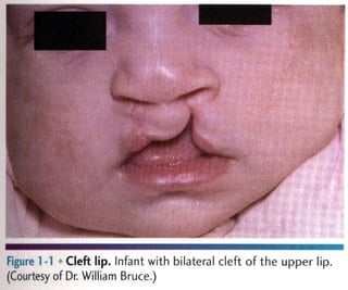
  • 1. Figure 1 >1 Cleft Infant with bilateral Cleft of the upper lip. (Courtesy of Dr, William Bruceā€˜)
  • 2. FlSlllc I 7 Paramedian lip pits. Bilateral piis (arrows) on lhe lower lip in a patient with van der Woude syndrome.
  • 3. m Double lip. Redundant fold of (issue on the upper lip in a patient with Ascher syndrome. (Courtesy of RC. Zeigā€˜er.)
  • 4. Figure Iā€™ll Fordyce granules. Lesions at the commissure
  • 5. Fxgmv l H,Ā» Microglossia. Abnormally small tongue associated with constricted mandibular arch.
  • 6. m.ā€˜ .mmmā€˜ Flā€˜hā€™ullā€˜ I I Microglossia. Associated constriction of the maxi Iary arch in he same patient shown in Figure 1716.
  • 7. Figure 1-19 ' Ankyloglossia. Abnormal attachment of the lingual frenum, limitlng tongue mobility.
  • 8. w Fugum I ~22 Fissured tongue. Moderate ļ¬ssuring of the dorsal tongue. (From Allen CM, Camisa C: Diseases of the mouth and lipsā€˜ In Sams WM, Lynch P, editors: Principles ofdermatology, New York, 1990, Churchill Livingstone.)
  • 9. Figure 1724 H tongue. Eiongated. black-ļ¬‚aming ļ¬hform papillae on the posterior dorsai tongue.
  • 10. figure lĀ»27 >Van'cosities. Multiple purple dilated veins on the ventval and lateral surfam of the tongue.
  • 11. Figure 1735 Exostoses. Multiple buccal exostoses of thc maxilr law and mandlbular alveolar ridges.
  • 12. Figure lĀ»38 Torus palatinus. Midline bony nodule of the palatal vault.
  • 13. Figure 1-39 Torus palatinus. Large, lobulated palatal mass.
  • 14. Figure 1-40 Torus palatinus. Asymmetric. lobulated bony mass.
  • 15. Figure 1741 Torus mandibularis. Bllulmdl Iobulatod bony protuberames of the mandibular lingual alveolav ridge.
  • 16. Figure 1742 Torus mandibularis. Masswe "kissmg" (on meet in the midline.
  • 17. Figure 1 -48 ~ Stafne defect. Anterior radiolucent lesion ofthe body of the mandible associated with the sublingual gland.
  • 18. Figurel-67 ā€˜ Dermoid cyst. Fluctuant midline swelling in the ļ¬‚oor ofthe mouth (From Budnick SD: Handbook afpediatrir oral pathology. Chicago, 1981, Year Book Medical.)
  • 19. Figure 1-69 Thyroglossal duct cyst. Swelling (arrow) of the anterior midline ofthe neckā€˜ (Courtesy of Drl Philip Sprinkle.)
  • 20. Figure L77 Hemihyperplasia. Enlargement of the right sbde of the face. (Courtesy of Dr George Blazis)
  • 21. Figure 1-78 Hemihyperplasia. Same patient as depicted in Figure 1777, with associated enlargement of the right half of the tongue. (Courtesy of Dr. George Blozis.)
  • 22. Figure 2-1 > Environmental enamel hypoplas . Bilaterally symmetric pattern of horizontal enamel hypoplasia of the anterior dentition. Maxillary central incisors have been restored previously (From Neville BW, Damm DD, White DK: Color atlas ofclinicul oral pathology. ed 2, Baltimore, 1999. Williams & Wi ins.)
  • 23. Figure 2-2 Environmental enamel hypoplasia. Same patient as depicted in Figure 271ā€˜ Note the lack of enamel damage on bicuspids, (From Neville BW, Damm DD, White DK: Color atlas of clinical oral pathology, ed 2, Baltimore, 1999, Williams & Wilkins.)
  • 24. Turnerā€™s hypoplasia. Extensive enamel hypoplasia of mandibular ļ¬rst bicuspid secondary to previous inļ¬‚ammatory process associated with overlying ļ¬rst deciduous molar. (From Halstead CL, Blozis CG, Drinnan AJ, et al: Physical evaluation ofthe dental patient, St Louis, 1982, Mosbyt)
  • 25. F ure 2-6 - Turner's hypopla 3. Extensive corona! hypopfasia of permanent maxillary left (entral incisor secondary to previous trauma to deciduous ce (ral incisor,
  • 26. Figure 2A8 Dental ļ¬‚uorosis. Dentition exhibiting lusterless, white, and opaque enamel.
  • 27. Figure 2-9 ' Dental ļ¬‚uoros White opaque alteration of the bicuspids and second molars in a patient who also exhibits discoloration of the teeth secondary to (etracycline use. Patient moved to area of endemic ļ¬‚uorosis at 3 years of age.
  • 28. Figure 2-10 ~ Attrition. Extensive loss of coronal tooth height WIthout pulp exposure in patient with anterior edge-to-edge occlusionv
  • 29. Figure 2-11 * Abrasion Horizontal cervical nokhes on the anterior mandibular dentition
  • 30. Figure 2-12 Abrasion. Extensive recession and loss of buccal radlculardentin. Note visible pulp Canals that have been ļ¬lled wllh lertiary dentin.
  • 31. Abrasion. Notching of the right central incisor caused by improper use of bobby pins. The patient aiso exhibits environmental enamel hypopiasia of the anterior dentition. (Courtesy of Dr. Robert]. Corlin.)
  • 32. Figure 2 14 Abra on. Notching ofthe ante or dentition on the right side caused by long-term use of tobacco pipe.
  • 33. Erosion Extensive loss of buccal and occlusalh 'rnz tooth structure. Note that the amalgam margins are above the surface of the dentin.
  • 34. mum Figure 2716 Erosion. Occlusal surface of the mandibular dentition exhibiting concave dentin depressions surrounded by eievated rims of enamel.
  • 35. Erosion. Extensive loss of enamel and dentin on the buccal surface of the maxillary bicuspids. The patient had sucked chronically on tamarinds (an acidic fruit).
  • 36. Figure 2-18 Erosion. Palatal surfaces of the maxillary dentition in which the exposed dentin exhibits a concave surface and a peripheral white line ofenamel. The patient suffered from bulimia
  • 37. Figure 2-19 Abfraction. Deep and rrow enamel cervncal defects on the facial surface of the mandibular dentition (From Neville BW. Damm DD. White DK: Coloratlus of clinical omā€˜l pathology. ed 2, Baltimore, 1999, Williams & Wilkinsl)
  • 38. m mĀ» , Internal resorption (pink tooth of Mummery). Pmk disrolomh'on of H19 maxillary (Pntml indsor,
  • 39. Exgm'e 1 21 Internal resorption. Same patient as depicted in Figure 2-21ā€˜ Note extensive resorption of both maxil|ary central incisors,
  • 40. Internal resorption. Balloonlike enlargement of the root canal,
  • 41. Flgure 2-24 0 External resorption. Extensive Irregular destrucā€” tion of both roots of1he mandibular second molar associated wixh chronic periodontitis. (Courtesy of Dr. Tommy Shimer)
  • 42. w Figure 2<25 ā€˜ External resorp Ion. ā€œMotheatenā€ radiolucent alteration of the maxillary left central incisor The tooth had been reimplanted after traumatic avulsion. (Courtesy of Drā€˜ Harry Meyers)
  • 43. Figure 227 ~ External resorption. Diffuse external resorption of radicular dentin of maxillary dentition. This prOCESS arose after initiation of orthodontics.
  • 44. Figure 2-40 Hypodont . MultIple developmentally mlssmgpel manent teeth and several retained deciduous teeth in a female adult
  • 45. Figure 2742 Hypodontia, Developmentally missing maxillary lateral incisors. Radiographs revealed no underlying teeth, and there was no history of trauma or extraction.
  • 46. Figure 2-43 * Hyperdon a (mesiodens). Erupted superr numerary, rudl'memary tooth of the anterior maxilla
  • 47. Figure 2-44 Hyperdon a (mesiodens). U teral :uper- numerary tooth of the anterior maxilla, which has altered the eruption path of the maxil|ary right permanent central incisor.
  • 48. supernumerary teeth of the anterior maxilla. Figure 2ā€”45 % Hyperdontia (meslodens). Bulateral Inverted
  • 49. Figure 2ā€”46 '-Ā» Hyperdontia. Right mandibular dentition exhibiting four erupted bicuspids.
  • 50. Figure 2ā€”47 v> Paramolar. Rudimentary tooth situated palatal to a maxiilary meter in a patient who also exhibits hypodontia.
  • 51. Figure 2-48 + Paramolar. Radiograph of the same patient depicted in Figure 2-4.7 Note the fully formed tooth overlying crown of the adjacent molar.
  • 52. Flgure 2749 Natal teeth. Mandihu|ar rentral I'n(i<ors (hat were erupted at birth.
  • 53. Figure Z~50 ' Diffuse microdontla. Dentition in which the teeth are Smaller than normal and widely spaced within the arch
  • 54. Figure 2ā€”51 Ā» Isolated microdon Ia (peg lateral). Small, cone, shaped right maxillary lateral incisor.
  • 55. Figure 2-52 ā€™v Bilateral gemination. Two double teeth, The moth count was normal when each anomalous tooth was counted axoneā€˜
  • 56. Figure 2-53 Gemina n. Mandibular bicuspid exhibiting biļ¬d crowni
  • 57. Figme 2 SC Fusion. Double tooth in the place of the mandibular right lateral incisor and cuspid.
  • 58. Figure 2ā€”57 "v Fusnon. Bilateral double teeth in the place of the mandibular lateral incisors and cuspids.
  • 59. Figure 2-59 Concrescence. Union by cementum of adjacent maxillary molars.
  • 60. # Figure 2761 Concrescence. Cross photograph of the same teeth depicted in Figure 260. Histopathologir examination revealed that union occurred in the area of cemental repair previously damaged by a periapical inļ¬‚ammatory lesion.
  • 61. Figure 2 3 Talon cusp. Acce ory cusp present on the palatal surface of H19 maxill y loft rpntral incisor: Note the (hreer pronged pattern, which rezemlļ¬e: an eagle talon.
  • 62. Figure 2-70 Ā« Coronal den vag atus, type II. Mandibular lateral incisor exhibiting lingual bulbous enlargement at the site of (clonal opening of enamel invagination.
  • 63. F gure 2-7] x Coronal dens vag natus, type II. Radiograph of the mandibular lateral incisor depicted in Figure 2-70. Note the radiopaque and enamel-lined invagination extending below the level of the cementoenamel junction.
  • 64. Figure 2ā€”72 Coronal dens invaginatus, type M. Cross photo- graph ofa sectioned tooth. Note the dilated invagination with apical accumulation of dystrophic enamel.
  • 65. Figure 2773 Coronal dens nva atus, type III. Parulis overr lying vital maxillary cuspid and lateral incisor. The Luspld contained a dens Invaginatus that perforated t mesidl >urlace ofits root.
  • 66. Figure 2-74 Coronal dens invaginatus, type III. Maxillary cuspid exhibiting an enamel invagination that parallels the pulp canal and perforates the lateral root surface (Courlesy of Dr. Brian Blocher.)
  • 67. Figure 2ā€”76 ' Enamel pearl. Mass of ectopic enamel located in the furcation area ofa molar tooth. (Courtesy of Drā€˜ Joseph Beard.)
  • 68. m ā€˜Ā«w Ā§Ā§ Fegure 2779 Taurodontism. Mandibular molar teeth exhibiting increased pulpal apicoocclusal height With apically positioned pulpal floor and bifurcation. (Courtesy of Dr. Michael Kahn,)
  • 69. Figure 2-81 O Hypercemento Mandibular ļ¬rst molar exhibiting thickening and blunting of the roots.
  • 70. Figure 2-83 Ilacerahon. Maxlllary molar exhlbltlng sharp angulation of the rootsā€˜ Note the interradicular bone.
  • 71. Figure 2-87 v Supernumerary root. A, Cross photograph exhibiting a maxillary molar with a small supernumerary root. B, MeSIal-to-distal radiographic view exhibiting the a(cessory root with central pulp canal. lfa buccalitoJingual mdiographic view had been taken (as would be necessary in patient (are), the additional root would not have been evident.
  • 72. Figure 2789 Hypoplastic amelogenesu Imperfecta, general- ized pitted pattern. Note the numerous pinpoint pits scattered across the surface of the teeth. The enamel between the pits is of normal thickness. hardness. and coloration, (From Stewart RE. Prescott CH: Oml facial genetics, St Louis, 1976, Mosby.)
  • 73. Figure 2 95 Hypomaluration amelogenesis imperfecta. Denlition thibiting mottled, opaque white enamel with scattered areas 0{ brown discoloration.
  • 74. Figure 2 ~96 Hypomaturation amelogenesis imperfecta, snowcapped pattern. Dentition exhibiting zone of white opaque enamei in the incisal and occlusal one fourth of the enamel surface. (Courtesy of DLā€˜ Heddie O. Sedano.)
  • 75. Fugure 2-97 ' Hypocalciļ¬ed amelogenesis imperfecta. DentiĀ» lion exhibiting diffuse yellow-brown discoloration, Note numerous teeth wiļ¬‚l loss of coronaI enamel except for the cervical portion.
  • 76. ļ¬gure 2-101 Dentinogenesis imperfecta. Dentition exhib diffuse brownish discoloration and slight translucence.
  • 77. Figure Z-IOZ ~ā€™ Dentinogenesis imperfecta. Dentition exhibiting grayish discoloration with Signiļ¬cant enamel loss and attrition.
  • 78. Flgulā€˜e 3-1 Chm Ic hyperplastlc pulpltls. Erythematous granulation tissue extruding from the pulp chamber of the mandibular first molar:
  • 79. Figure 3716 Periapical granuloma. WEN-deļ¬ned radiolucency associated with the apex of the maxillary ļ¬rst bicuspid, (Courizsy of Drā€˜ Frank Beylotā€˜tel)
  • 80. Figure 3-] er plcal granuloma. Large, well-defined radiolui cency associated with the apices of the mandibular ļ¬rst molarā€˜ (Courtesy of Drā€˜ Robert E. Loy.)
  • 81. * Figure 3-24 Ā« Periap al cyst. Wechircumscribed radiolucency intimater associated with the apex of the mandibular central incisor. Note the loss oflamina dura in the area of the lesion
  • 82. . . Nā€˜N,Ā§ Figure 3-25 ~ Periapical cyst. Radiolucency associated with the maxillary central incisor, which exhibits signiļ¬cant root resorption.
  • 83. Figure 3-36 ' Periapical abscess. Bilateral soft-tissue swelling of the anterior palate.
  • 84. ļ¬gure 3-42 - Parulls. Asymptomatic yellowush nodule of the anurior mandibular alveolar ridge. Adjacent teeth were normal clinically and also asymptomatic
  • 85. Figure 3-43 * Periapical abscess. Same patient as depicted in Figure 3742ā€˜ Periapical radiolucency associated with the nonvital mandibular lateral incisor.
  • 86. Figure 3 Ā»45 Ludwig's angina. Soft-tissue swelling of the rig?ā€œ submandibular region (Courtesy of Drā€˜ Brian Blocher.)
  • 87. Figure 3-46 Cellu 5 involving canine space. Erythematous and edematous enlargement of the left side of the face with involvement of the eyelids and conjunctiva. Patients with odonto- genic infections involving the canine space are at risk for cavernous sinus thrombosis. (Courtesy of Dr. Richard Ziegle )
  • 88. Figure 348 . Acute osteomyelitis with sequestruml Radior lucency of the right body of the mandible with central radiopaque mass of necrotic bone. (Courtesy of Dr. Michael Meyrowitz.)
  • 89. Figure 3-52 t lefuse sclerosmg osteomyelitis. Duffuse area of increased mdiodensity of the right body of the mandible in the tooth-bearing area. No other quadrants were involved (Courtesy of Dr. Louis M. Beta)
  • 90. Figure 3753 : Condensing osteitis. Increased areas of radioā€” density surrounding the apices of the nonvital mandibular ļ¬rst molar.
  • 91. Figure 4718 Nifedipine-related gingival hyperp|asia. Diffuse, fibrotic gingival hyperplasia after 1 month ofintensive oral hygiene. Significant erythema, edema, and increased enlargement were present before intervention,
  • 92. Figure 4-22 Phenytoin-related ginglval hyperplasia. Signiļ¬cant gingival hyperplasia almost totally covers the crowns of the posterior maxillary dentition (Courtesy of Dr. Ann Drummond and Dr. Timothy Johnsonā€˜)
  • 93. 'i-ā€˜guā€˜yo 1: 3 , s. Painful erythematous enlargement of the soll tissues overlying the crown of the partially elupted right mandlbular third molar.
  • 94. Flgulā€˜e 5"ā€œ Hutchinson's incisors of congenital syphilis.Den tition exhibiting crowns tapering toward the incisal edges. (me Halstead CL, Blozis CG, Drinnan A], Cier RE: Physicai evaiuntionq' the dental patient. St Louis, 1982, Mosby.)
  • 95. Figure 7-31 Mumps. Bilateral parotid enlargement. (From Neville BW. Damm DD, White DK: Color atlas ofclim'ral oml pathology, ed 2, Baltimore, 1999, Williams & Wilkinsā€˜)
  • 96. ļ¬guraļ¬ S Asp rin bur . Extensive area of white epithelial mosisulthe left buccal muwsa caused by aspirin placement in maltempl to alleviate dental pain,
  • 97. Figure 8719 Cotton roll burn. Zone ofwhite epithelial ne(rosis and erythema of the maxillary alveolar mucosa.
  • 98.
  • 99. F ure 8727 Osteoradionecrosis. Same patient as depicted in Figure 8725. Note fistula formation of the left submandibular area resulting from osteoradionecrosis of the mandibular body.
  • 100. K 3'! Amalgam tattoo. Area of mucosal discoloralioml the mandibular alveolar ridge immediately below the bridge politic
  • 101. Lg. . 3 Minor aphthous ulcerations. Two ulcerations of different Sizes located on the maxillary labial mucosi
  • 102. Figure 9-7 Major aphthous ulcerat on. A, Large ulceration of the left anterior buccal mucosa. B, Same lesion after 5 days of therapy with betamethasone syrup used in a SWIshā€”and-swallow method. The patient was free of pain by the second day of therapy. The ulcerahoh healed com- pletely during the foHowmg weekā€˜
  • 103. _ Allergic mucosal reaction to systemic drug administration. Large irregular erosion ofā€™tlā€™le right ventral surr fare of the tongue. The lesion arose secondary to use of oxapmzin, a nonsteroidal antHnļ¬‚ammatory drug.
  • 104. Figure 9-33 0 Contact stomatitis from cinnamon ļ¬‚avoring. Oblong area of sensitive erythema with overlying shaggy hyperkeratosis.
  • 105. . saw Ā«m %ļ¬Ā¢Ć©ā€™x wa , ā€œa 'r u; n 4: Oral mucosal contact readion to dental amalgam. Radiating pattern of hyperkeratotic striae on the pos- terior buccal mucosa that contacts a large distobuccal amalgam of the permanent mandibular second molar.
  • 106. Figure 9740 Angioedema. lefuse upper llp swelling that arose rapidly.