1. Update on the Tympanic Membrane Displacement Technique
Cherith Webb, Robert Marchbanks and Tony Birch
Neurological Physics, part of the Imaging Group, Department of Medical Physics and Bioengineering, University Hospital Southampton
NHS Foundation Trust . Contact: cherith.webb@uhs.nhs.uk
Current project funded by
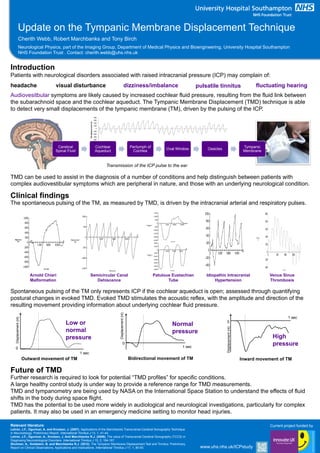
Cerebral
Spinal Fluid
Cochlear
Aqueduct
Perilymph of
Cochlea
Oval Window Ossicles
Tympanic
Membrane
Transmission of the ICP pulse to the ear
Introduction
Patients with neurological disorders associated with raised intracranial pressure (ICP) may complain of:
Audiovesitbular symptoms are likely caused by increased cochlear fluid pressure, resulting from the fluid link between
the subarachnoid space and the cochlear aqueduct. The Tympanic Membrane Displacement (TMD) technique is able
to detect very small displacements of the tympanic membrane (TM), driven by the pulsing of the ICP.
headache visual disturbance fluctuating hearingdizziness/imbalance pulsatile tinnitus
Future of TMD
Further research is required to look for potential “TMD profiles” for specific conditions.
A large healthy control study is under way to provide a reference range for TMD measurements.
TMD and tympanometry are being used by NASA on the International Space Station to understand the effects of fluid
shifts in the body during space flight.
TMD has the potential to be used more widely in audiological and neurological investigations, particularly for complex
patients. It may also be used in an emergency medicine setting to monitor head injuries.
TMD can be used to assist in the diagnosis of a number of conditions and help distinguish between patients with
complex audiovestibular symptoms which are peripheral in nature, and those with an underlying neurological condition.
Clinical findings
The spontaneous pulsing of the TM, as measured by TMD, is driven by the intracranial arterial and respiratory pulses.
-500
-400
-300
-200
-100
0
100
200
300
400
TMdisplacement(nl)
Patulous Eustachian
Tube
Spontaneous pulsing of the TM only represents ICP if the cochlear aqueduct is open; assessed through quantifying
postural changes in evoked TMD. Evoked TMD stimulates the acoustic reflex, with the amplitude and direction of the
resulting movement providing information about underlying cochlear fluid pressure.
0
Bidirectional movement of TM
Normal
pressure
1 sec
Displacement(nl)
0
Outward movement of TM
Low or
normal
pressure
1 sec
Displacement(nl)
1 sec
0
Inward movement of TM
High
pressure
Displacement(nl)
Idiopathic Intracranial
Hypertension
Semicircular Canal
Dehiscence
PATIENTID:3498548DISPLACEMENTDATATest
No:322-Nov-200203:15PM
Time(mS)
Displacement
(nl)
0
200
400
600
800
1000
-200
-400
0 1500 3000 4500
Venus Sinus
Thrombosis
Arnold Chiari
Malformation
Relevant literature:
Lehrer, J.F., Ogunlusi, A. and Knutsen, J. (2007). Applications of the Marchbanks Transcranial-Cerebral Sonography Technique
in Neurootology: Preliminary Report. International Tinnitus J 13, 1, 41-44.
Lehrer, J.F., Ogunlusi, A., Knutsen, J. And Marchbanks R.J. (2009). The value of Transcranial-Cerebral Sonography (TCCS) in
Diagnosing Neurootological Disorders. International Tinnitus J 15, 2, 164-167.
Shulman, A., Goldstein, B. and Marchbanks R.J. (2012). The Tympanic Membrane Displacement Test and Tinnitus: Preliminary
Report on Clinical Observations, Applications and Implications. International Tinnitus J 17, 1, 80-93. www.uhs.nhs.uk/ICPstudy