
Recommended
Recommended
More Related Content
Similar to Blood Disorders and Cardiovascular System
Similar to Blood Disorders and Cardiovascular System (15)
More from ChantellPantoja184
More from ChantellPantoja184 (20)
Recently uploaded
Recently uploaded (20)
Blood Disorders and Cardiovascular System
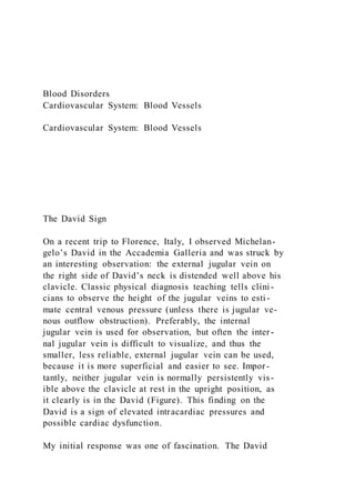
- 1. Blood Disorders Cardiovascular System: Blood Vessels Cardiovascular System: Blood Vessels The David Sign On a recent trip to Florence, Italy, I observed Michelan- gelo’s David in the Accademia Galleria and was struck by an interesting observation: the external jugular vein on the right side of David’s neck is distended well above his clavicle. Classic physical diagnosis teaching tells clini - cians to observe the height of the jugular veins to esti - mate central venous pressure (unless there is jugular ve- nous outflow obstruction). Preferably, the internal jugular vein is used for observation, but often the inter - nal jugular vein is difficult to visualize, and thus the smaller, less reliable, external jugular vein can be used, because it is more superficial and easier to see. Impor- tantly, neither jugular vein is normally persistently vis- ible above the clavicle at rest in the upright position, as it clearly is in the David (Figure). This finding on the David is a sign of elevated intracardiac pressures and possible cardiac dysfunction. My initial response was one of fascination. The David
- 2. is an awe-inspiring work with incredible attention to de- tail and accuracy in physical anatomy. With the knowledge of a cardiologist, it appeared illogical to me for the David to have jugular venous distention. I felt a chill go down my back as I realized that this observation and its implications were very possibly new and unrecognized. In addition, this message (so to speak) from Michelangelo needed an ex- planation. Immediately, my response was to document the finding with multiple photographs for study and review. I remember speaking with my girlfriend that day after we visited the Accademia Galleria and discussing that I thought I observed something very unique about the sculpture— something that appears to have been hiding in plain sight for more than 500 years. When I returned home, I reviewed textbooks and the medical literature and discussed this with a few medi - cal and physiology colleagues. We had many very inter- esting discussions trying to understand this paradox. I also knew that I needed to know more about Michel- angelo and his work. Michelangelo, like some of his artistic contempo- raries, had anatomical training,1,2 and in looking at the David, one can clearly see that the external jugular vein and the sternocleidomastoid muscle are anatomically correct. Michelangelo’s sculpture depicts David just be- fore his battle with Goliath. In reviewing other sculp- tures of Michelangelo, I found that he demonstrates the same finding of supraclavicular external jugular venous distention again in his sculpture of Moses at the tomb of Pope Julius the Second. Most would agree that the sit- ting Moses is thought to be in an excited state. In con- trast, the recently deceased Christ in Michelangelo’s Pietà (1499) does not demonstrate jugular venous dis- tention. Michelangelo did demonstrate the previously
- 3. recognized distention of the dependent veins in the David, the Moses, and his Pietà. Dependent venous dis- tention was recognized previously and clearly present in earlier works, such as the Colossus of Constantine (circa 325), which he would have studied. (I did also notice in photographs what could be the finding of jugular ve- nous distention in the ancient sculpture Laocoön and His Sons, but this sculpture was found in 1506, after the Da- vid was completed; Michelangelo is said to have seen this sculpture.) In my review of Michelangelo’s work, I could find no mention of anyone writing about his depiction of jugular venous distention. To put this finding in context: at the time the David was created, in 1504, William Harvey had yet to de- scribe the true mechanics of the circulatory system. This did not occur until 1628. In addition, while Harvey pos- tulated that small vessels connected the arterial and ve- nous vessels, capillaries were not identified until 1661 (by Marcello Malpighi).3 At this point in my investigation, I realized that Mi- chelangelo must have noticed temporary jugular ve- nous distention in healthy individuals who are excited. In sculpture, one can only show a single image in time, and he must have wanted to express this observation in his work. I am amazed at his ability to recognize this find- ing and express it in his artwork at a time when there was such limited information in cardiovascular physiology. In- terestingly, even today, this phenomenon is not dis- cussed in typical cardiology textbooks. Recognizing this message from Michelangelo also had special meaning to me. I am a physician who has spent more than 4 decades in the study and practice of medi -
- 4. cine. Now nearing the end of my career, I have turned to Figure. An Image of Michelangelo’s David, Showing Jugular Venous Distention Photo credit: Daniel M. Gelfman, MD; image taken September 28, 2018. FROM THE HEART Daniel M. Gelfman, MD Division of Clinical Affairs, Marian University College of Osteopathic Medicine, Indianapolis, Indiana. Corresponding Author: Daniel M. Gelfman, MD, Division of Clinical Affairs, Marian University College of Osteopathic Medicine, 3200 Cold Spring Rd, Indianapolis, IN 46222 ([email protected] marian.edu). Opinion 124 JAMA Cardiology February 2020 Volume 5, Number 2 (Reprinted) jamacardiology.com © 2019 American Medical Association. All rights reserved.©
- 5. 2019 American Medical Association. All rights reserved. Downloaded From: https://jamanetwork.com/ by Roger Olson on 03/04/2020 Congenital Defects of the Heart Math Unit Plan Grade: Week 1 Monday Tuesday Wednesday Thursday Friday Lesson Title State Math Standards Learning Objectives
- 6. Instructional Strategy Summary of Instruction Differentiation Materials, Resources, and Technology Formative Assessment
- 7. Summative Assessment (a short description of the summative assessment) Part 2: Rationale © 2018 Grand Canyon University. All Rights Reserved. Respiratory System Henry’s Law Dalton’s Law Atmospheric Pressure = 760 mmHg Atmospheric Pressure = PN2 + PO2 + PCO2 + PH2O 597(PN2) + 159(PO2) + .3(PCO2) + 3.7(PH2O) = 760 mmHg 760 x .786 = 597 760 x .209 = 159 760 x .0004 = .3 760 x .005 = 3.7 Atmospheric Pressure = 760 mmHg Atmospheric Pressure = PN2 + PO2 + PCO2 + PH2O
- 8. 597(PN2) + 159(PO2) + .3(PCO2) + 3.7(PH2O) = 760 mmHg 760 x .786 = 597 760 x .209 = 159 760 x .0004 = .3 760 x .005 = 3.7 Sea level pressure = 760 mm Hg Sea level PO2 = 159 mm Hg Everest summit pressure = 253 mm Hg Everest summit PO2 = 43.1 mm Hg Atmospheric Pressure = 760 mmHg Atmospheric Pressure = PN2 + PO2 + PCO2 + PH2O 597(PN2) + 159(PO2) + .3(PCO2) + 3.7(PH2O) = 760 mmHg 760 x .786 = 597 760 x .209 = 159 760 x .0004 = .3 760 x .005 = 3.7 Arterial blood at sea level - 97% saturated Venous blood leaving peripheral tissues at sea level - 75% saturated
- 9. Arterial blood at summit of Everest - 77% saturated The Respiratory System • Image from 1601 text, De Vocis Auditusque Organis Historia Anatomica, by Giulio Casserio • Professor of head and neck anatomy at University of Padua • William Harvey was one of his students • How many cardiorespiratory structures can you identify? CLASSICS IN THORACIC SURGERY Leonardo da Vinci and the Sinuses of Valsalva Francis Robicsek, MD The Carolinas Heart Institute, the Heineman Medical Research Laboratory, and the Carolinas Medical Center, Charlotte, North Carolina
- 10. Recent studies indicate that eddy currents generated by the sinuses of Valsalva play an important role in the physiologic closure of the aortic valve. This process is briefly discussed and evidence is presented that this fact was well known and elaborated upon by the renaissance artist Leonardo da Vinci. This fact is illustrated with his words and drawings. (Ann Thoruc Surg 1991;52:328-35) God geornetricizes Leonardo da Vinci he semilunar valves, in contrast to atrioventricular T valves, have no direct attachment to the myocar- dium; therefore, their function is generally thought to be entirely passive responding to fluctuation of the pressure between the left ventricle and the aorta. Whenever the pressure generated by ventricular systole exceeds that reigning in the thoracic aorta, the aortic valve opens; whenever the left ventricular pressure decreases to less than the pressure in the aorta, the valve shuts. Recent research, however, indicates that this process, which occurs about 115,000 times a day, is far more complex. As has been demonstrated by Clark [1] and others [ 2 4 ] , although the leaflets are the most dynamic parts of the aortic valve, the motion of other associated structures such as the vascular wall and the expansion of the entire valve complex itself [ P 6 ] also play an important role. Even more important in aortic valve function than these factors are some particular features of the blood flow induced by the presence of ellipsoidal sacculations of the aortic wall, named after the great Italian anatomist, An- tonio Valsalva .
- 11. If one may ask a cardiac surgeon what the aortic valve consists of, the answer will likely be: “Three leaflets and three commissures.” The same inquiry directed to a physiologist would probably evoke a different response: “Three leaflets, three commissures, and three sinuses.” This emphasis on the functional significance of the si- nuses of Valsalva is becoming more and more clear to those who study circulatory dynamics, but is still not appreciated by those who perform reconstructive opera- tion at the aortic root. The first scientific study in modern times on the role of these sinuses in the closure of the aortic valve was made by Henderson and Johnson [7], who demonstrated in hydrodynamic model studies that the closure of the aortic valve under pulsatile flow is not an abrupt event triggered by sudden drop in ventricular pressure alone but rather a Address reprint requests to Dr Robicsek, Heineman Medical Research Center, PO Box 35457, Charlotte, NC 28235. gradual process in the course of which during decelera- tion of flow the valve leaflets move gradually toward closure. These observations have received additional sup- port by van Steenhoven [&-lo], Peskin [ll, 121, Bellhouse [1>15], and their associates. In elegant experiments Bell - house constructed a rigid model of the aortic root with a flexible valve and perfused it with a pulsatile flow of water. The flow pattern was outlined by dye injection and recorded by serial cinematography. The principal obser - vations made in these studies were that after a rapid and full opening of the leaflets, some of the blood ejected from the left ventricle coils back at the sinus edge, then decel - erates, reverses its direction along the sinus wall, and forms vortices in the sinuses of Valsalva before it rejoins
- 12. the mainstream of forward flow. He further stipulated that due to these eddy currents and deceleration, the pressure exerted at the lateral aspects of the leaflets exceeds that on their central surfaces and causes the leaflets to approximate even before the systole is com- pleted [14]. Because of this process only minimal reversed flow is required for final valve closure, and regurgitation does not occur (Figs 1, 2). Nearly identical experiments were performed and sim- ilar conclusions were drawn by Leonardo da Vinci in 1513 (Fig 3). Leonardo was not only a superb painter and sculptor, he also was a Renaissance man in the true sense of the word: an exceptional architect, a talented mechanical and hydraulic engineer, as well as the founder of functional anatomy. His heritage includes about 200 richly annotated anatomical sketches, all but a few now housed in the private collection of Her Majesty the Queen of England [161. The lion’s share of Leonardo’s work on anatomy falls into two distinct periods. His earlier drawings were made about 1487 to 1493, mostly in Florence and in Milan. They show his preoccupation with the structure of the skull and the eye, which he called the ”window of the soul.” His later work began around 1506 and continued until his death in 1519 in France [17]. This period encompasses studies on other organs and is permeated with the recog- nition as to how mechanics relate to human physiology. His views as an architect-engineer opened a heretofore unseen functional perspective into the study of the hu- man body, which he regarded as a God-created structure ”which feels and moves” but also as an “edifice governed by the laws of mechanics” [18]. After comprehending
- 13. function, Leonardo proceeded with attempts to enhance the same; he designed life belts and webbed gloves to enable man to swim better, and fabricated wings and parachutes to make him float in the air. 0 1991 by The Society of Thoracic Surgeons 0003- 4975/91/$3.50 Angioplasty Blood Vessel Disorders Coronary Artery Bypass Grafting (CABG) CABG Movie Congenital Defects of the Heart Rene Laennec, 1781 - 1826 Inventor of the Stethoscope 80 In 1816, I was consulted by a young woman laboring under general symptoms of diseased heart, and in whose case percussion and the application of the hand were of little avail on account of the great degree of fatness. The other method just mentioned [direct auscultation] being rendered inadmissible by the age and sex of the patient, I happened to recollect a simple and well-known fact in acoustics, . . . the great distinctness with which we hear the scratch of a pin at one end of a piece of wood on applying our ear to the other. Immediately, on this suggestion, I rolled a quire of paper
- 14. into a kind of cylinder and applied one end of it to the region of the heart and the other to my ear, and was not a little surprised and pleased to find that I could thereby perceive the action of the heart in a manner much more clear and distinct than I had ever been able to do by the immediate application of my ear. Rene Laennec, August 1819 In the preface to, De l'Auscultation Médiate 81 82 83 First stethoscope in the United Kingdom Brought from Paris in 1822 by Thomas Hodgkin Hodgkin’s Disease Displayed at Gordon Museum of Pathology, King’s College London Normal Resting Values 6000 ml/min or 6.0 l/min = 75/beats/min X 80 ml/beat Moderate Exercise Values 13.44 l/min = 120/beats/min X 112 ml/beat Heavy exercise
- 15. CO can be 18-30 l/min Elite athletes CO can increase ≈ 700% to 40 l/min!!!! 98 92 Blood Objectives • List the components of the cardiovascular • Describe the important components and major functions of blood. • List the characteristics and functions of red blood cells. • Describe the structure of hemoglobin and indicate its functions. • Discuss red blood cell production and maturation. • Explain blood typing and the basis for ABO and Rh incompatibilities. • Categorize the various white blood cells on the basis of structure and function. • Describe the structure, function, and production of platelets. • Describe the reaction sequences responsible for blood clotting. The cardiovascular system • A mechanism for rapid transport of nutrients, waste products, gases, and cells Blood • Fluid connective tissue • Functions include
- 16. • Transporting dissolved gases, nutrients, hormones, and metabolic wastes • Regulating pH and ion composition of interstitial fluids • Restricting fluid loss at injury sites • Defending the body against toxins and pathogens • Regulating body temperature by absorbing and redistributing heat • Physical Characteristics of Blood • Thicker & heavier than H2O • Temp. = 100.4°F (38°C) • Volume = 5-6 liters for males, 4-5 liters for females • Blood = 8% of total body weight The composition of blood • Plasma (45%) and formed elements (55%) comprise whole blood • Red blood cells (RBC) • White blood cells (WBC) • Platelets • Can fractionate whole blood for analytical or clinical purposes Hemopoiesis • Process of blood cell formation • Hemocytoblasts are circulating stem cells that divide to form all types of blood cells 93 Plasma • 92 percent of plasma is water
- 17. • Higher concentration of dissolved oxygen and proteins than interstitial fluid Plasma proteins • More than 90 percent are synthesized in the liver • Albumins • 60 percent of plasma proteins • Responsible for viscosity and osmotic pressure of blood Additional Plasma Proteins • Globulins • ~35 percent of plasma proteins • Include immunoglobins which attack foreign proteins and pathogens • Include transport globulins which bind ions, hormones, and other compounds • Fibrinogen • Converted to fibrin during clotting • Removal of fibrinogen leaves serum Abundance of RBCs • Erythrocytes account for 99.9 percent of the formed elements • Hematocrit measures the percentage of whole blood occupied by formed elements • Commonly referred to as the volume of packed red cells Structure of RBCs • Biconcave disc, providing a large surface to volume ration • Shape allows RBCs to stack, bend, and flex • RBCs lack organelles • Typically degenerate in about 120 days
- 18. Hemoglobin • Molecules of hemoglobin account for 95 percent of the proteins in RBCs • Hemoglobin is a globular protein, formed from two pairs of polypeptide subunits • Each subunit contains a molecule of heme which reversibly binds an O2 molecule • Damaged or dead RBCs are recycled by phagocytes RBC life span and circulation • Replaced at a rate of ≈ 3,000,000 new blood cells entering the circulation per second • Components of hemoglobin individually recycled • Heme stripped of iron and converted to biliverdin, then bilirubin • Iron is recycled 94 RBC production • Erythropoeisis = the formation of new red blood cells • Occurs in red bone marrow • Process speeds up with in the presence of EPO (erythropoietin) • RBCs pass through reticulocyte and erythroblast stages Blood types • Determined by the presence or absence of surface antigens (agglutinogens) • Antigens A, B, and Rh (D)
- 19. • Antibodies are in plasma (agglutinins) that do not react with antigens • Cross-reactions only occur when antigens meet antibodies in incompatible transfusions • Incompatible blood transfusions cause donated RBC's to be attacked causing agglutination (clump) • These cells lodge, swell & rupture: hemolysis • ***Hint: Donors RBC must be compatible with recipient’s plasma antibodies*** Blood Type Compatible Donor Types Incompatible Types A A, O B, AB B B, O A, AB AB A, B, AB, O --- O O A, B, AB Rh System First worked out on the rhesus monkey - Also based on antigens on RBC surface Those with Rh antigens are Rh+ - Those without Rh antigens are Rh- Normally plasma does not contain anti-Rh antibodies Antibodies develop only upon greater exposure to Rh antigens Donation reactions would occur only upon multiple incompatible transfusions The most common problem is during pregnancy
- 20. Rh+ fetus & Rh- mother can cause problems in later pregnancies Mom will be exposed to Rh+ blood during delivery and will then make anti-Rh antibodies A second pregnancy will cause mom's anti-Rh antibodies to cross placenta If fetus is Rh- there is no problem - If fetus is Rh+ hemolysis will occur in fetal blood This is erythroblastosis fetalis (hemolytic disease of the newborn [HDN]) Kids born with this receive gradual transfusions before or after birth (with Rh-) Injections to mom after first pregnancy can prevent problems w/ second 95 Leukocytes • Have nuclei and other organelles • Defend the body against pathogens • Remove toxins, wastes, and abnormal or damaged cells • Are capable of amoeboid movement (margination) and positive chemotaxis • Some are capable of phagocytosis Types of WBC • Granular leukocytes • Neutrophils – 50 to 70 percent total WBC population
- 21. • Eosinophils – phagocytes attracted to foreign cells that have reacted with antibodies • Basophils – migrate to damaged tissue and release histamine and heparin Types of WBC • Agranular leukocytes • Monocytes – become macrophage • Lymphocytes – includes T cells, B cells, and NK cells Differential count • Indicates a number of disorders by determining which WBC is responding to disease WBC Production • Granulocytes and monocytes are produced by bone marrow stem cells • Divide to create progenitor cells • Stem cells may originate in bone marrow and migrate to peripheral tissues • Lymphocytes originate in bone but develop in lymphoid tissue (thymus, spleen . .) Platelets • Flattened discs • Circulate for 9-12 days before being removed by phagocytes Platelet functions • Transporting chemicals important to clotting • Forming temporary patch in walls of damaged blood vessels • Contracting after a clot has formed Platelet production (thrombocytopoiesis)
- 22. • Megakaryocytes release platelets into circulating blood 96 Hemostasis • Prevents the loss of blood through vessel walls • Three phases: • Vascular phase • Platelet phase • Coagulation phase Hemostasis • Vascular phase • Local blood vessel constriction (vascular spasm) • Platelet phase • Platelets are activated, aggregate at the site, adhere to the damaged surfaces • Coagulation phase • Factors released by platelets and endothelial cells interact with clotting factors to form a clot • Basically: Fibrinogen in the presence of thrombin converts to Fibrin • Fibrin is the basic framework of a blood clot 115
- 23. The Respiratory System Objectives • Describe the primary functions of the respiratory system. • Identify the organs of the respiratory system and describe their functions. • Define and compare the processes of external and internal respiration. • Summarize the physical principles governing the movement of air into the lungs and the diffusion of gases into the blood. • Explain the important structural features of the respiratory membrane. • Describe how O2 and CO2 are picked up, transported, and released in the blood. Functions of the respiratory system • Gas exchange between air and circulating blood • Moving air across the exchange surface of the lungs • Protection of respiratory surfaces • Production of sound • Provision for olfactory sensations Organization of the respiratory system • Upper respiratory system • Nose, nasal cavity, paranasal sinuses, pharynx • Lower respiratory system • Larynx, trachea, bronchi, bronchioles, alveoli The Respiratory tract • Passageways carrying air to and from the alveoli • Upper respiratory passages filter and humidify incoming air
- 24. • Lower passageways include delicate passages and alveolar exchange surfaces Respiratory mucosa • Respiratory epithelium and underlying connective tissue supported by lamina propria • Lines conducting portion of respiratory tract • Protected from contamination by respiratory defense system The nose and nasal cavity consists of • External nares • Nasal cavity • Vestibule • Superior, middle, and inferior meatuses • Hard and soft palates • Internal nares • Nasal mucosa 116 The Pharynx • Shared by the digestive and respiratory systems • Divided into three sections: • Nasopharynx – superior portion • Oropharynx – continuous with the oral cavity • Laryngopharynx – between the hyoid bone and the esophagus The Larynx • Air passes through the glottis on the way to the lungs • Cartilages of the larynx • Three large cartilages • Thyroid, cricoid, and epiglottis
- 25. • Paired cartilages • Arytenoids, corniculate, and cuneiform • Folds of the larynx • Inelastic vestibular folds • Delicate vocal folds Sound production • Air passing through the glottis vibrates the vocal folds producing sound waves • Pitch depends on conditions of vocal folds • Diameter • Length • Tension The laryngeal musculature • Muscles of the neck and pharynx position and stabilize the larynx • When swallowing, these muscles • Elevate the larynx • Bend the epiglottis over the glottis The Trachea • Extends from the sixth cervical vertebra to the fifth thoracic vertebra • A tough, flexible tube running from the larynx to the bronchi • Held open by C-shaped tracheal cartilages in submucosa • Mucosa is similar to the nasopharynx The primary bronchi • Trachea branches in the mediastinum into right and left bronchi
- 26. • Bronchi enter the lungs at the hilus 117 The Lungs Lobes and surfaces of the lungs • Lobes of the lung are separated by fissures • Right lung has three lobes • Left lung has two lobes • Concavity on medial surface = cardiac notch The bronchial tree • System of tubes formed from the primary bronchi and their branches • Primary bronchi branch into secondary or lobar bronchi • Secondary bronchus goes to each lobe of the lungs • Secondary bronchi branch into tertiary bronchi • Tertiary bronchi supply air to a single bronchopulmonary segment The bronchioles • Ultimately branch into terminal bronchioles • Delivers air to a single pulmonary lobule • Terminal bronchiole becomes respiratory bronchioles Alveolar ducts and alveoli • Respiratory bronchioles end in ducts and sacs Respiratory membrane
- 27. • Simple squamous epithelium • Endothelial cell lining an adjacent capillary • Fused basal laminae Cells of the respiratory membrane include • Septal cells • Produce surfactant which keep alveoli open • Alveolar macrophage • Patrol epithelium and engulf foreign particles The pleural cavities and pleural membranes • Each lung covered by one pleura • Pleura—serious membranes lining the pleural cavity • Parietal—attaches to the walls of the pleural cavity • Visceral—adheres to the surface of the lungs • Pleural fluid—fills and lubricates the space between the pleura 118 Respiratory physiology • External respiration • Exchange of gases between interstitial fluid and the external environment • The steps of external respiration include • Pulmonary ventilation – moving air between external environment and lungs • Gas diffusion – moving gases across respiratory membrane and into blood
- 28. • Transport of O2 and CO2 – in blood from lungs into the systems • Internal respiration • Exchange of gases between interstitial fluid and cells Pulmonary ventilation • Movement of air depends upon • Boyle’s law • Pressure and volume inverse relationship • Volume depends on movement of diaphragm and ribs • Pressure and airflow to the lungs • Compliance – an indication of the expandability of the lungs Respiratory volumes - Spirometry • Tidal Volume (VT) • Amount of air inhaled or exhaled with each breath • Inspiratory Reserve Volume (IRV) • Inhaled air above VT • Expiratory Reserve Volume (ERV) • Exhaled air below VT • Vital Capacity (VC) • VT + IRV and ERV • Residual Volume (RV) • Air left in lungs after maximum exhalation • Total Lung Capacity (TLC) • VC + RV
- 29. Gas Diffusion • Dalton’s law and partial pressure • Individual gases in a mixture exert pressure proportional to their abundance Atmospheric pressure (760 mm Hg) = PO2 + PCO2 + PN2 + PH2O Atmospheric PO2 = 21% .21 x 760 mm Hg = 160 mm Hg Atmospheric PCO2 = .04% .0004 x 760 mm Hg = .3 mm Hg Atmospheric PN2 = 78.5% .785 x 760 mm Hg = 597 mm Hg 119 Transportation of O2 and CO2 Oxygen transport • 3% dissolved in plasma • 97% carried in hemoglobin (heme portion) as oxyhemoglobin (HbO2) • The amount of oxygen hemoglobin can carried is dependent upon • PO2 • pH – higher values result in less saturation • temperature – higher temps cause Hb to release O2 Carbon dioxide transport • 7 % dissolved in plasma • 23 percent bound to hemoglobin
- 30. • carbaminohemoglobin • 70 % carried as carbonic acid, which dissociates into a hydrogen ion and bicarbonate ion • Carbon dioxide plus water reversibly reacts to form a hydrogen ion and bicarbonate ion • CO2 + H2O ⇔H+ + HCO3- • Most of the H+ is taken up by Hb, which therefore serves as a buffer • The + HCO3- move out of RBC and into plasma in exchange for a Cl-, this is the chloride shift The efficiency of the respiratory system decreases with age as: • Elastic tissue deteriorates causing lower lung compliance and vital capacity • Chest movements are restricted by arthritic changes • Some degree of emphysema normally occurs 97 The Heart Objectives • Describe the organization of the cardiovascular system. • Describe the location and general features of the heart, including the pericardium.
- 31. • Discuss the differences between nodal cells and conducting cells, and describe the components and functions of the conducting system of the heart. • Identify the electrical events associated with a normal electrocardiogram. • Explain the events of the cardiac cycle including atrial and ventricular systole and diastole, and relate the heart sounds to specific events in the cycle. • Define cardiac output, heart rate, and stroke volume and describe the factors that influence these variables. • Explain how adjustments in stroke volume and cardiac output are coordinated at different levels of activity. The cardiovascular system is divided into two circuits • Pulmonary circuit • blood to and from the lungs • System circuit • blood to and from the rest of the body • Vessels carry the blood through the circuits • Arteries carry blood away from the heart • Veins carry blood to the heart • Capillaries permit exchange Anatomy of the Heart The pericardia
- 32. • Visceral pericardium or epicardium • Parietal pericardium • Pericardial fluid between the above two • Superficial fibrous pericardium is anchored to mediastinum Superficial anatomy of the heart • The heart consists of four chambers • Two atria and two ventricles • Major blood vessels of the heart include • Inferior and superior vena cavae • Aorta and pulmonary trunk The heart wall • Components of the heart wall include 98 • Epicardium – visceral pericardium • Myocardium – cardiac muscle • Endocardium – simple squamous epithelium Internal anatomy and organization • Atria • Thin-walled chambers that receive blood from the veins (VC & PV) • Pump blood into ventricles • Ventricles • Thick-walled chambers separated from the atria by AV valves • Pump blood into arteries (PA & Aorta)
- 33. • Chordae tendineae • Tendinous fibers attached to the AV valves and papillary muscles Blood flow through the heart • Right atrium • Tricuspid valve • Right ventricle • Pulmonary valve – pulmonary trunk – pulmonary arteries • Pulmonary circuit – pulmonary veins • Left atrium • Bicuspid valve(mitral valve) • Left ventricle • Aortic valve • Aorta and systemic circuit – vena cava – right atrium Heart chambers and valves • Structural Differences in heart chambers • The left side of the heart is more muscular than the ri ght side • Functions of valves • AV valves prevent backflow of blood from the ventricles to the atria • Semilunar valves prevent backflow into the ventricles from the pulmonary trunk and aorta Blood supply to the heart • Coronary arteries include the right and left coronary arteries,
- 34. marginal arteries, anterior interventricular artery (anterior descending), and the circumflex artery • Coronary veins include the great cardiac vein, anterior and posterior cardiac veins, the middle cardiac vein, and the small cardiac vein, and the coronary sinus 99 Cardiac physiology • Two classes of cardiac muscle cells • Specialized muscle cells of the conducting system (control heartbeat) • Contractile cells (produces powerful contractions that propels blood) The conducting system • The conducting system includes • Sinoatrial (SA) node • Atrioventricular (AV) node • Conducting cells • Atrial conducting cells are found in internodal pathways • Ventricular conducting cells consist of the AV bundle, bundle branches, and Purkinje fibers Impulse conduction through the heart
- 35. • SA node begins the action potential • Stimulus spreads to the AV node • Impulse is delayed at AV node • Impulse then travels through ventricular conducting cells (AV bundle, bundle branches, and Purkinje fibers) • Then distributed by Purkinje fibers The electrocardiogram (ECG) • A recording of the electrical events occurring during the cardiac cycle • The P wave accompanies the depolarization of the ventricles • The QRS complex appears as the ventricles depolarize (masks atrial repolarization) • The T wave indicates ventricular repolarization Contractile cells • Resting membrane potential of approximately –90mV • Action potential • Rapid depolarization • A plateau phase unique to cardiac muscle • Repolarization • Refractory period follows the action potential Calcium ion and cardiac contraction • Cardiac action potentials cause an increase in Ca2+ around myofibrils • Ca2+ enters the cell membranes during the plateau phase • Additional Ca2+ is released from reserves in the sarcoplasmic reticulum
- 36. 100 The cardiac cycle • The period between the start of one heartbeat and the beginning of the next • During a cardiac cycle • Each heart chamber goes through systole and diastole • Correct pressure relationships are dependent on careful timing of contractions Pressure and volume changes: atrial systole • Rising atrial pressure pushes blood into the ventricle • Atrial systole • The end-diastolic volume (EDV) of blood is in the ventricles Pressure and volume changes: ventricular systole • Isovolumetric contraction of the ventricles: ventricles are contracting but there is no blood exiting chamber • Ventricular pressure increases forcing blood through the semilunar valves Pressure and volume changes: ventricular diastole • The period of isovolumetric relaxation when all heart valves are closed • Atrial pressure forces the AV valves open Heart sounds • Auscultation – listening to heart sound via stethoscope • Four heart sounds • S1 – “lubb” caused by the closing of the AV valves
- 37. • S2 – “dupp” caused by the closing of the semilunar valves • S3 – a faint sound associated with blood flowing into the ventricles • S4 – another faint sound associated with atrial contraction Cardiodynamics - stroke volume and cardiac output • Cardiac output – the amount of blood pumped by each ventricle in one minute • Cardiac output equals heart rate times stroke volume CO cardiac output (ml/min) = Hr heart rate (beats/min) X SV stroke volume (ml/beat) Normal Resting Values 6000 ml/min or 6 l/min = 75/beats/min X 80 ml/beat Moderate Exercise Values 13.44 l/min = 120/beats/min X 112 ml/beeat Heavy exercise can increase CO to 18-30 l/min
- 38. Elite athletes can increase CO ≈ 700% to 40 l/min!!!! 101 SV ⇑ from ⇑ in End-Diastolic Volume (120ml resting) venous pressure ⇑ ventricular volume This stretches the LV myocardium = ⇑ force of contraction Starling's Law *Why does RHR ⇓ for athletes? The proportion of blood ejected from the LV compared to total volume is termed ejection fraction (65%) Summary: regulation of heart rate and stroke volume • Sympathetic stimulation increases heart rate • Parasympathetic stimulation decreases heart rate • Circulating hormones, specifically E, NE, and T3, accelerate heart rate • Increased venous return increases heart rate • EDV is determined by available filling time and rate of venous return • ESV is determined by preload, degree of contractility, and afterload Heart Disease mostly from poor coronary circulation Ischemia decreased O2 supply Angina Pectoris chest pain from ischemia w/o death to myocardium. Caused by: stress (Ψ or phys.), atherosclerosis, ↑BP, nicotine...
- 39. Myocardial Infarction (MI) Heart attack - #1 cause of death in USA Necrosis of myocardium. Congenital Heart Disease #1 cause of full term newborn fatality Atrial Septal Defect (ASD) Foramen ovale remains open after birth Ventricular Septal Defect (VSD) Hole in interventricular septum Patent Ductus Arteriosus (PDA) Patent fetal Ductus Arteriosus Tetrolagy of Fallot VSD, aorta arising from RV or VSD, pulmonic RV hypertrophy Largest contributor to "blue baby" Transposition of the Great Vessels Aorta arises from the RV Pulmonary artery arises from the LV Other Heart Diseases Rheumatic heart disease, congestive heart disease, arrhythmic heart disease
- 40. 102 Blood Vessels and Circulation Objectives • Distinguish among the types of blood vessels. • Describe fluid and dissolved material transport of the cardiovascular system. • Describe the factors that influence blood pressure and blood pressure regulation. • Discuss the movement of fluids between capillaries and interstitial spaces. • Describe how blood flow and pressure in tissues is regulated. • Identify the principle blood vessels of each circuit and the areas they serve. • Describe fetal circulation and the changes at birth and during aging. Structure of vessel walls • Walls of arteries and veins contain three distinct layers • Tunic intima • Tunica media • Tunica externa (adventitia) Differences between arteries and veins • Compared to veins, arteries • Have thicker walls • Have more smooth muscle and elastic fibers • Are more resilient Arteries • Undergo changes in diameter
- 41. • Vasoconstriction – decreases the size of the lumen • Vasodilation – increases the size of the lumen • Classified as either elastic (conducting) or muscular (distribution) • Small arteries (internal diameter of 30 um or less) are called arterioles Capillaries • An endothelial tube inside a basal lamina • These vessels • Form networks • Surround muscle fibers • Radiate through connective tissue • Weave throughout active tissues • Capillaries have two basic structures • Continuous • Fenestrated • Flattened fenestrated capillaries = sinusoids 103 Capillary beds • An interconnected network of vessels consisting of • Collateral arteries feeding an arteriole • Metarterioles • Arteriovenous anastomoses • Capillaries • Venules
- 42. Veins • Collect blood from all tissues and organs and return it to the heart • Are classified according to size • Venules • Medium-sized veins • Large veins Venous valves • Venules and medium-sized veins contain valves • Prevent backflow of blood Distribution of blood • Total blood volume is unevenly distributed • Venoconstriction maintains blood volume • Veins are capacitance vessels • Capacitance = relationship between blood volume and pressure Circulatory pressure • Circulatory pressure is divided into three components • Blood pressure (BP) • Capillary hydrostatic pressure (CHP) • Venous pressure Resistance (R) • Resistance of the cardiovascular system opposes the movement of blood • For blood to flow, the pressure gradient must overcome total peripheral resistance
- 43. • Peripheral resistance (PR) is the resistance of the arterial system Overview of cardiovascular pressures • Factors involved in cardiovascular pressures include • Vessel diameter • Cross-sectional area of vessels • Blood pressure • Blood viscosity 104 Arterial blood pressure • Arterial blood pressure • Maintains blood flow through capillary beds • Rises during ventricular systole and falls during ventricular diastole • Pulse is a rhythmic pressure oscillation that accompanies each heartbeat • Pulse pressure = difference between systolic and diastolic pressures • Example of Pulse Pressure for BP of 120/90: PP = 120 – 90; PP = 30 • Mean arterial pressure (MAP) = diastolic pressure + (pulse pressure ÷ 3) Example of MAP for BP of 120/90 MAP = 90 + (30 ÷ 3) MAP = 90 ÷ 10 MAP = 100 mm Hg
- 44. Capillary exchange • Flow of water and solutes from capillaries to interstitial space Processes that move fluids across capillary walls • Diffusion • Filtration • Hydrostatic pressure (CHP) • Reabsorption Venous pressure and venous return • Assisted by two processes • Muscular compression • The respiratory pump Cardiovascular regulation • Autoregulation • Neural mechanisms • Endocrine mechanisms Autoregulation of blood flow within tissues • Local vasodilators accelerate blood flow in response to: • Decreased tissue O2 levels or increased CO2 levels • Generation of lactic acid • Release of nitric acid • Rising K+ or H+ concentrations in interstitial fluid • Local inflammation • Elevated temperature 105 Neural mechanisms • Adjust CO and PR to maintain vital organ blood flow
- 45. • Medullary centers of regulatory activity include • Cardiac centers • Vasomotor centers control • Vasoconstriction via adrenergic release of NE • Vasodilation via direct or indirect release of NO Reflex control of cardiovascular function • Baroreceptors reflexes monitor stretch • Atrial baroreceptors monitor blood pressure • Chemoreceptor reflexes monitor CO2, O2, or pH levels Hormones and cardiovascular regulation • Antidiuretic hormone – released in response to decreased blood volume • Angiotensin II – released in response to a fall in blood pressure • Erythropoietin – released if BP falls or O2 levels are abnormally low • Natriuretic peptides – released in response to excessive right atrial stretch Exercise and the cardiovascular system • Light exercise results in • Extensive vasodilation • Increased venous return • A rise in cardiac output • Heavy exercise results in • Increased blood flow to skeletal muscles • Restriction of blood flow to nonessential organs Cardiovascular response to hemorrhaging: short term
- 46. • Carotid and aortic reflexes increase CO and peripheral vasoconstriction • Sympathetic nervous system elevates blood pressure • E and NE increase cardiac output and ADH enhances vasoconstriction Cardiovascular response to hemorrhaging: long term • Decline in capillary blood pressure recalls fluids from interstitial spaces • Aldosterone and ADH promote fluid retention • Increased thirst promotes water absorption across the digestive tract • Erythropoietin ultimately increases blood volume and improves O2 delivery 106 Identify the Below Landmarks on the Heart Chambers Right atrium Right ventricle Left atrium Left ventricle Valves Right AV (tricuspid) valve Left AV (bicuspid or mitral) valve Chordae tendineae Papillary muscles Pulmonary semilunar valve Aortic semilunar valve Fibrous skeleton
- 47. Vessels Superior vena cava Inferior vena cava Pulmonary trunk Pulmonary arteries Pulmonary veins Right coronary artery Left coronary artery Anterior interventricular (anterior descending) coronary artery Great cardiac vein Coronary sinus, and it’s opening to right atrium Other Structures Foramen ovale Fossa ovalis Ductus arteriosus Ligamentum arteriosum Parietal pericardium Visceral pericardium (epicardium) Myocardium Endocardium Interatrial septum Interventricular septum Sinoatrial (SA) node Internodal pathways Atrioventricular (AV) node AV bundle Bundle branches Purkinje fibers Identify the Below Systemic Arteries
- 48. Ascending aorta Thoracic aorta Brachiocephalic trunk Subclavian Axillary Brachial Deep brachial Radial Ulnar Common Carotid Internal Carotid External Carotid Facial Superficial Temporal Occipital Vertebral Basilar Cerebral arterial circle (circle of Willis) Anterior communicating Anterior cerebral Posterior communicating Posterior cerebral Celiac trunk Common hepatic Superior mesenteric Renal Inferior mesenteric Common iliac Internal iliac External iliac Femoral Popliteal Anterior tibial
- 49. Posterior tibial 107 Identify the Below Systemic Veins Superior vena cava Brachiocephalic trunk Subclavian Axillary Cephalic Brachial Basilic Radial Ulnar Median cubital Internal jugular External jugular Temporal Facial Vertebral Inferior vena cava Hepatic Renal Gonadal Common iliac Internal iliac External iliac Femoral Poplilteal Posterior tibial Great saphenous Hepatic portal circulation
- 50. Superior mesenteric Inferior mesenteric Splenic Hepatic portal vein 108 Special circulation • The brain • Four arteries which anastomose insuring constant blood flow • Cerebral arterial circle (Circle of Willis) • The heart • Coronary arteries arising from the ascending aorta • The lungs • Pulmonary circuit, regulated by local responses to O2 levels • Opposite other tissues (declines in O2 cause vasodilation) • Hepatic portal system • Contains substance absorbed by the stomach and intestines • Delivers these compounds to the liver for • Storage • Metabolic conversion • Detoxification and Excretion • Fetal Circulation • Fetal blood flow to the placenta is supplied via paired umbilical arteries • A single umbilical vein drains from the placenta to the ductus venosus
- 51. • Collects blood from umbilical vein and liver • Empties into the inferior vena cava • No need for pulmonary function in the fetus • Two shunts bypass the pulmonary circuit • Foramen ovale • Ductus arteriosus • Cardiovascular changes at birth • Lungs and pulmonary vessels expand • Ductus arteriosus constricts and becomes ligamentum arteriosum • A valvular flap closes the foramen ovale forming fossa ovalis • Ductus venosus closes and forms ligamentum arteriosum Age-related changes in blood may include • Decreased hematocrit • Constriction or blockage of peripheral veins by a thrombus • Pooling of blood in the veins of the legs Vessels are less elastic, prone to Ca2+ deposits and thrombi formation