2. HISTORY
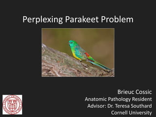
• Female, 50 g, 2 year old, opal red-rumped parakeet
(Psephotus haematonotus)
• Part of a larger flock with 7 recently-acquired birds
• All 7 birds developed severe respiratory distress
• All 7 + 6 parakeets died
• No improvement with antibiotics
18. Discussion
• Cause of death
– Respiratory distress, occlusion of the upper
respiratory tract
– Anorexia, dehydration
19. Parakeets die-off
- Similar lesions reported in birds quarantined in
south Florida
- Our birds may have:
- Contracted the virus in Florida
- Contracted the virus in upstate New-York
The stress of the travel and the introduction in a new
flock may have facilitated the infection
20. Avipoxviruses
• Described in 278 bird species from 70 families
and 20 orders (32 psittacines):
– Poultry industry
– Pet birds industry
– Endangered species
– Ecosystems
• Worldwide distribution
21. Avipoxviruses
• Double stranded DNA virus
• Two major forms: cutaneous and diphtheric
• Production of epidermal growth factor-like
• Transmission is usually direct
– Indirect is less common (fomites, mosquitos, mites)
22. Ancillary tests results
• PCR negative:
- Time in formalin / fixation
- Time in decalcification
solution
- Sequence of the primers
Psittacinepox and fowlpox are in different clades
Jarmin et al., (2006)
23. Giemsa
Tripathy et al., 1973. Immunoperoxidase technique for detection of Fowlpox
Antigen. Avian diseases, 17(2):274-278.
25. Bollinger bodies
Dr. Otto Bollinger
German pathologist
(1843-1909). Wikipedia.
Otto Bollinger, (1873), was the first to demonstrate a relationship between the
lesions and the inclusions bodies, several years before the discovery of the first
virus by Dmitry Ivanosky, in 1892
26. Acknowledgments
• Dr. Teresa Southard
• Dr. Nicholas Wolfer for submitting this case
• Drs. Elizabeth Buckles and Jarra Jagne for their
help
• Necropsy and histology staff
27. References
• Beaufrere H., Bhaskaran M., Jankowski G, et al. 2009.
What's your diagnosis? Journal of Avian Medicine and
Surgery 23(4):325-328
• Hernandez M., Sanchez C., Margarita E.G., et al., 2001.
Avian pox infection in Spanish Imperial eagles (Aquila
adalberti). Avian Pathology, 30:1, 91-97
• Jarmin S., Manvell R., Gough R.E., et al. 2006. Avipoxvirus
phylogenetics: identification of a PCR length polymorphism
that discriminates between the two major clades. J, of
General Virol. 87(8), 2191-2201.
• Van Riper C. and Forrester D., 2007. Avian Pox. In: Infectious
Disease of Wild birds. Thomas N.J., Hunter D.B., Atkinson
C.T. Eds: Wiley-Blackwell, 131-176.
Dorsal aspect of the oral cavity
Parakeet was mildly underconditioned.
Left naris was occluded wit dried opaque yellow-white material
Choana and caudal oropharynx contained approximately 0.25 mL of mucoid, translucent green material.
Serial cross sections of the head after a couple of days in decalcification solution.
Bilaterally, affecting approximately 70% of the mucosa of the infraorbital sinuses,
are locally extensive areas of moderate to severe, epithelial hyperplasia, with marked thickening of the stratum spinosum, up to 8 times the normal thickness, and extensive lamellar, orthokeratotic hyperkeratosis
Numerous keratinocytes are markedly swollen with variably sized clear cytoplasmic vacuoles (ballooning degeneration) and others contain prominent, 10-30 μm in diameter, round to frequently ring-shaped, granular, eosinophilic, intracytoplasmic, inclusion bodies (Bollinger bodies). Randomly scattered individual keratinocytes are shrunken with hypereosinophilic cytoplasm and fragmented nuclei (necrosis).
Multifocally, within the nasal mucosa and the dermis is an infiltrate composed predominantly of plasma cells and lymphocytes with fewer heterophils and macrophages
Affecting approximately 30% of the overlying feathered skin
Multifocally, the overlying epidermis is eroded or ulcerated and covered with a crust composed of eosinophilic proteinaceous fluid, and cellular and karyorrhectic debris admixed with keratin.
We can apprectiate on this cross section that approximately 80 to 90 % of the nostrils are occluded by hyperplastic epithelium and debris
Giemsa: It is specific for the phosphate groups of DNA and attaches itself to regions of DNA where there are high amounts of adenine-thymine bonding. Used for protozoan blood parasites (Malaria, Trypanosoma).
Here, used to stained the intracytoplasmic inclusion bodies
Multifocally, in the stratum corneum, few intracytoplasmic inclusions stain blue. Other inclusion bodies are pale eosinophilic.
Microscopically, the histological findings are consistent with a mixed presentation, both diphtheric (wet pox) and cutaneous (dry pox), of an Avipoxvirus infection. The findings are also consistent with the gross observation of mucoid material in the nasopharynx and dried material in the left naris.
Unfortunately, we do not know the length or sequence of the primers used for the PCR.
The lesions observed grossly and histologically explain the clinical signs of respiratory distress eventually leading to the death of the animal. This etiology is also consistent with the history of a flock outbreak
The previous report of a die-off of red-rumped parakeets associated with poxvirus infection describes similar lesions with birds quarantined in south Florida (Beaufrere et al., 2009). In our case, the new birds also came from Florida, We hypothesize that the imported birds acquired the viral infection in Florida , where the insect vectors are more active during winter than in upstate New York. The stress of the travel may have compromised the birds’ immune systems leading to the onset of clinical signs in the imported birds and introduction of the virus to a naïve population. Alternatively, a latent poxvirus infection in the New York parakeet population may have been activated secondary to the stress of intermixing with the imported birds The establishment of the new dominance schemes within the flock may also have facilitated skin wounds, allowing the invasion of the epidermis by the virus.
Two major forms of the disease are commonly described, a cutaneous or dry form, and diphtheric or wet form, with frequent combination both, as in this case
The cutaneous form is characterized by nodular, discrete, proliferative lesions on feather-free areas of the body (comb, eyelids, around the beak, wattle, legs and wings). Mortality rate are usually low with this form. The diphtheric form is characterized by fibrinonecrotic and proliferative lesions on the mucosae of the upper respiratory and digestive tracts, eventually causing respiratory and digestive disturbances. Therefore, this form and the combined form, are associated with a much higher mortality rate
There could be several reasons to explain these results: the fixation and decalcification processes may have fragmented the viral DNA and depending on the size of the primers used for the PCR, impaired the polymerization reaction. The primers used were also fowlpox-specific, while the virus in this case is most likely a psittacinepox virus and potentially a ‘parakeetpox virus”. Virus isolation which can be performed by inoculating the chorioallantoic membrane of avian embryos (Gonzalez et al., 2008) was not attempted in this case.
Phylogenetic tree of the major avipoxvirus clades based on partial 4b core protein coding sequence analysis.
clade (from Ancient Greek: κλάδος, klados, "branch") is a group of organisms that consists of a common ancestor and all its lineal descendants, and represents a single "branch" on the "tree of life”. (Wiki)
Based on 2 core proteins 4b and p35.
(Meleagris gallopavo). Infection, Genetics and Evolution 35:221-229.
Giemsa stain preparation, blue-stained inclusions. 420X.
Tripathy et al., 1973. Immunoperoxidase technique for detection of Fowlpox Antigen. Avian diseases, 17(2):274-278.
Degradation of DNA or only viral proteins
Electron microscopy examination of the viral particles reveals characteristic brick-shaped nucleocapsids, with a convoluted outer membrane, lateral bodies and a biconcave (dumbbell-shaped) central core (Hernandez et al., 2001).
Electron micrograph of a keratinocyte showing an intracytoplasmatic membrane-bounded inclusion composed of numerous
viral particles, some of which appear free in the cytoplasm (F). ´ 7000. Bar = 2 mm.
Electron micrograph of viral inclusion at higher magnification, showing viral particles. The convoluted outer membrane
(m), lateral bodies (l) and dumbbell-shaped central core (c) are characteristic of a poxvirus. ´ 50 000. Bar = 0.5mm.
Hernandez et al., 2001. Avian poxvirus infection in Spanish Imperial eagles (Aquila adalberti). Avian Pathology, 30(1):91-97.
Four to six days post-infection, poxviruses produce variably-sized, pathognomonic, intracytoplasmic, eosinophilic, viral inclusions (Bollinger bodies), that contain smaller elementary bodies (Borrel bodies; OIE, 2008)