Radionuclide imaging of parathyroid glands
•
0 likes•36 views

how to use the gamma camera in diagnosing a parathyroid lesion ?! and which tracer should you use ??

Recommended
More Related Content
What's hot
What's hot (9)
Similar to Radionuclide imaging of parathyroid glands
Similar to Radionuclide imaging of parathyroid glands (20)
Recently uploaded
Recently uploaded (20)
Radionuclide imaging of parathyroid glands
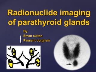
- 1. { Radionuclide imaging of parathyroid glands By Eman sultan Passant dorgham
- 2. Stimulates the gland to secrete PTH Increase ca mobility from bones (osteoclastic stimulation ) Kidney - Resorb ca - Increase Activation of vit. D Why parathyroid glands ??
- 3. What is parathyroid gland ? Embryology & Anatomy Develop at 6 weeks and migrate caudally at 8 weeks The paired superior parathyroid glands develop with the thyroid gland from the 4th branchial pouch .. Then migrate to coricothyroid cartilage level .. Lateral &posterior to the upper pole thyroid The paired inferior parathyroid glands develop with the thymus from the 3rd branchial pouch the n migrate to the level of aortic arch or fail to migrate
- 4. What is parathyroid gland ? anatomy Parathyroid glands usually eembeded between the posterior border of the thyroid gland and its fibrous capsule Each one measures 6 x 4 x 2 mm and weigh 25-40mg The number of the glands vary from 2 – 6
- 5. Blood supply : Branches from the inferior & superior thyroid arterys Venous drainage : to the superior , middle , inferior thyroid veins What is parathyroid gland ? anatomy
- 6. Ectopic locations of the parathyroid gland • Para-eosophageal • Intra-thymic • Thyro-thymic ligament • Intrathyroid • Carotid sheath • Undescended ( high cervical )/ retropharyngeal • Mediastinal
- 7. • Adenoma • Hyperplasia • Carcinoma primary • Hyperplasia (chronic renal failure, malabsorption) Secondary • Autonomous secondary (renal transplant pt ) Tertiary Hyperparathyroidism – pathophysiology
- 9. Dual phase planar imaging Thyroid ¶thyroid glands imaged at 15 min and 2 hours after tracer injection Tracers : 1. TC99m MIBI 2. TC99m TETR MIBI clears from thyroid with half life 30 min but retained by the abnormal parathyroid glands … unlike TETR which clear much slowly from thyroid
- 10. According to the “ differential washout” targeted to the back ground activity , the abnormal parathyroid tissue become more evident on delayed images
- 11. Dual isotope imaging sometimes used with dual phase imaging especially with history of thyroid disease or surgery This type compares pattern of TC99m MIBI or TETR localization to thyroid specific radiopharmaceutical to evaluate concordance (related to thyroid tissue ) versus discordance (indicative of parathyroid tissue ) called subtraction method . Dual isotope imaging
- 12. Parathyroid scintigraphy 90% sensitive for localizing adenomas but 60% sensitive for hyperplasia .
- 13. SPECT (Single-photon emission computed tomography) • To enlarge the gland • See the adenoma not seen on planar images • To confirm diagnosis & Define the position precisely • Thyroid adenoma or carcinoma can coexist and can retain tracer resulting in false positive study
- 14. Scintigraphy- reporting • patient indication for the study • Procedure - radiopharmaceuticals - timing of acquisition of images - planar or SPECT - time of detection of the lesion - definite location of the lesion • Study limitation (paatient movements) • Interpretation & computer subtraction