

Recommended
More Related Content
What's hot
What's hot (20)
Similar to Facial palsy
Similar to Facial palsy (20)
More from Balamurugan r
Recently uploaded
Recently uploaded (20)
Facial palsy
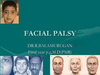
- 1. FACIAL PALSY DR.R.BALAMURUGAN Final year p.g,M.D(PMR)
- 3. FACIAL PARALYSIS • Commonly Unilateral Nuclear- from destruction of the nucleus • Central or cerebral orSupranuclear • Peripheral- from a lesion of the nerve • Supranuclear lesions usuallya part of hemiplegia, only the lower part of the face is paralysed. The upper part (frontalis and part of orbicularis oculi)escapes due to bilateral representation in the cerebral cortex.Infranuclear lesions- entire face is paralysed, as seen in bell’s palsy
- 4. Clinical features Weakness or paralysis of the upper and lower facial muscles of the affected side Drooping of ipsilateral eyelids Inability to close the eye completely Dry eye due to inability to close eyes completely Excessive tearing of the eye (epiphora) Drooping of the corner of the mouth Ipsilateral impaired/loss of taste sensation Difficulty with eating due to ipsilateral muscle weakness causing food to be trapped on the affected side of the mouth Dribbling of saliva Altered sensation on the affected side of the face Pain in or behind the ear Increased sensitivity to sound (hyperacusis) on affected side if stapedius muscle is involved
- 5. Clinical Testing of Facial Nerve Functions • 1. Observe patient Face • during rest & movement for : • • Asymmetry • • Hemi facial spasm • • Facial tics • • blinking
- 7. ETIOLOGIC CLASSIFICATON OF FACIALPALSY • Various classification have been suggested in this respect. • Based on: • Course of the nerve • Various etiologic causes • Degree of dysfunction observed
- 8. INTRACRANIAL (CENTRAL) CAUSES • Vascular abnormalities • CNS degenerative diseases • Tumours of the intracranial cavity • Trauma to the brain • Congenital abnormalities and agenesis
- 9. INTRATEMPORAL CAUSES • Bacterial and Viral infection • Cholesteatoma • Trauma- blunt temporal bone trauma, • longitudinal and horizontal fractures of the • temporal bone and gunshot wounds. • Tumours invading the middle ear, mastoid and • facial nerve • Iatrogenic causes
- 10. • NEUROLOGIC • Myasthenia Gravis • Multiple Sclerosis • Guillain Barre syndrome • NEOPLASTIC • Facial nerve tumours Glomus tumours • Meningiomas, acoustic neuroma • Parotid tumours Temporal bone/external • auditory meatus tumours
- 11. • INFECTIONS • Otitis media, mastoiditis • Bacterial causes • Viral causes
- 12. Recurrent facial palsy • Bell’s palsy, • Melkersson’s syndrome, • diabetes, • sarcoidosis • tumuors
- 13. BELL’S PALSY • First described more than a century ago by Sir Charles Bell Bell palsy is certainly the most common cause of facial paralysis worldwide
- 14. Viruses that have been linked to bells palsy • Cold sores and genital herpes (herpes simplex) Chickenpox and shingles (herpes zoster) • Mononucleosis (Epstein-Barr) • Cytomegalovirus infections • Respiratory illnesses (adenovirus) • German measles (rubella) • Mumps (mumps virus) • Flu (influenza B), Hand-foot-and-mouth disease (coxsackievirus
- 15. EXTRACRANIAL CAUSES • Malignant tumours of the parotid gland • Trauma • Iatrogenic causes • Primary tumours of the facial nerve • Malignant tumours of the ascending ramus of the mandible, pterygoid region and skin
- 16. Traumatic facial nerve palsy • Second most common cause of FN paralysis • - Represents 15% of all cases of FN paralysis • - Most common cause of traumatic facial nerve injury is temporal bone fracture
- 17. Bilateral simultaneous facial palsy • Moebius syndrome • GB Syndrome • Sarcoidosis • Myotonic dystrophy • Skull trauma • Infectious mononucleosis • CMV • Acute porphyria, Botulism • Lyme disease,Bell’s herpes simplex
- 18. International classification of Diseases • G51 Facial nerve disorders G51.0 Bell's palsy • G51.1 Geniculate ganglionitis • G51.2 Melkersson's syndrome • G51.3 Clonic hemifacial spasm – G51.31 …… right – G51.32 …… left – G51.33 …… bilateral – G51.39 …… unspecified
- 19. • G51.4 Facial myokymia • G51.8 Other disorders of facial nerve • G51.9 Disorder of facial nerve, unspecified
- 20. RAINER SCHMELZEISEN CLASSIFICATION • CONGENITAL • Moebius Syndrome • Myotonic dystrophy • Melkersson Rosenthal syndrome • Congenital Cholesteatoma • Birth injuries • Osteopetrosis
- 21. HOUSE-BRACKMAN(1985) CLASSIFICATION • Grade I-normal function without weakness. • Grade II-mild dysfunction with sligth facial asymmetry with a minor degree of synkinesis. • Grade III-moderate dysfunctions-obvious, but not disfiguring, asymmetry with contracture and/or • hemifacial spasm, but residual forehead motion and incomplete eye closure.
- 22. • • Grade IV-moderately severe dysfunction- obvious, disfiguring asymmetry with lack of forehead motion and incomplete eye closure. • • Grade V-severe dysfunction-asymmetry at rest and only slight facial movement. • • Grade VI-total paralysis-complete absence of tone ormotion.
- 23. Complications • Irreversible damage to your facial nerve • Misdirected regrowth of nerve fibers, resulting in involuntary contraction of certain muscles whenyou're trying to move others (synkinesis) — forexample, when you smile, the eye on the affected sidemay close • Partial or complete blindness of the eye that won't close, due to excessive dryness and scratching of the cornea, the clear protective covering of the eye
- 24. synkinesis • Most distressing consequences of facial paralysis. • Synkinesis refers to the abnormal involuntary facial movement that occurs with voluntary movement of a different facial muscle group. • •Abnormal regeneration of facial nerve fibers to the facial muscle groups
- 25. ASSESSMENT AND PLANNING • Cause of facial paralysis • Functional deficit/extent of paralysis • Time course/duration of paralysis • Likelihood of recovery • Other cranial nerve deficits • Patient’s life expectancy • Patient’s needs/expectations
- 26. Clinical Testing of Facial Nerve Functions • Blink test: Delay in blinking on one side • 3. Testing facial movement • i. Temporal branch: • To wrinkle forehead, To elevate eye brow • ii. Zygomatic branch: to screw up the eye • iii. Buccal branch: to wrinkle the nose • iv. Mandibular branch: to show the teeth, toblow out the cheeks • v. Cervical branch: by grimacing
- 27. EVALUATIONS OF NERVE FUNCTION • HISTORY is of vital importance to establish the onset characteristics,duration and degree of recovery. • • Previous trauma, surgery or infection may help in arriving at a diagnosis • • Examination of the face at rest and movement. • • Radiolologic evaluations • • Nerve excitability tests.
- 28. Pathophysiology of nerve injury • Neuropraxia : Blocks flow of axoplasm from stoma to distal axon. • Axonotemesis : Wallerian degeneration with intact endoneural tubules. Neurotemesis : Wallerian degeneration with loss of endoneural tubules . • Transection : Complete division of the nerve .
- 29. Minimal excitability test Compare the minimal current necessary to elicit minimal muscle contraction when applied tobranch of facial nerve on normal side to paralyze • Difference of 3.5mA Or greater between 2 side→ degeneration→surgical decompression
- 30. ELECTRICAL TESTING OF FACIAL NERVE • MAXIMUM STIMULATION TEST • Pulsed electric current is delivered through a cutaneous electrode • Short pulse will stimulate an intact nerve & elicit a muscular twitch. • In paralysed facial nerve, this indicates that lesion is neuropraxia & distal neurons have not undergone degeneration Hence differentiates between neuropraxia & axonotmesis:prognostic value.
- 31. • No value for 1st 72 h • Equal response • Reduced response • Absent response • Frequent testing • shows progressive ↑ • threshold →continuing • degeneration
- 32. NERVE EXCITABILITY TEST • Current required for stimulation on normal side is compared with paralysed side. • Disadv: even few intact fibres can elicit a response when rest in • undergoing degeneration. • Muscle twitch response is subjective • Uncomfortable procedure • Requires patient co-operation
- 33. ELECTRONEUROGRAPHY • Measures compound action potential in facial muscles in response to facial nerve stimulation. • Compare compound action potential of facial n. after Supramaximal stimulation of bothsides • The degree of degeneration is directly proportional to the amplitude loss of measured summation potential • Not useful during 1st72 h,90% or more degeneration indicate decompression within3 weeks
- 34. ELECTROMYOGRAPHY • The recording of spontaneous and voluntary muscle potentials by needles introduced into the muscle is called electromyography(EMG). • Records motor unit potentials of the orbicularis oculi & orbicularis oris muscle during rest &voluntary contraction
- 35. • EMG can be used to determine:- • 1.If a nerve in question is in fact in continuity(volitional activity recorded) • 2.Evidence of degenration ( fibrillation after 10- 14 days) • 3.If there are early sign of reinnervation (polyphasic innervation potentials after 4-6 weeks)
- 36. • Fibrillation potentials typically arises 2-3 weeks following injury • With regeneration of nerve after injury, polyphasic • reinnervation potential replaces fibrillation potential • Reinnervation potentials may precede clinical signs of recovery by 6-12 weeks
- 37. Limitation of electophysiological testing • Electric impulse can stimulate only normal/ neuropraxic fibres and can’t distinguish b/w axonotemesis or neurotemesis • Provides no useful information in cases of incomplete facial paralysis • It fails to provide information on the immediate post paralysis period( first 72 hours)
- 39. • Types of physical therapy interventions for facial palsy • Facial exercises, such as • – Strengthening and Stretching, • – Endurance, • – Therapeutic and facial mimic exercises ("mime • therapy")
- 40. • • Electrotherapy, • • Biofeedback, • • Transcutaneous electrical nerve stimulation (TENS) • • Thermal methods or massage, alone or in • combination with any other therapy
- 41. Exercise therapy • Simple movement retraining • • Expression training- mime • • Functional training • • PNF • • Massage-Efflureage,circular&fine thumb kneading improve circulation and prevent contracture • Active exercise in front of mirror prevent contracture
- 43. Electrotherapy • May have an adverse effect on recovery • • Avoid in acute stage • • Poor evidence to show it may be helpful in • chronic facial paralysis.
- 44. Feedback • Mirror feedback • EMG feedback • Lack of proprioceptors
- 45. Prognosis • The Copenhagen Facial Nerve Study found that around 71% of patients recover normal function without treatment. Around 13% are left with slight weakness and around 4% with severe weakness resulting in major facial dysfunction. Contracture of the facial muscles on the affected side was found in 17% and associated movements were found in 16%.6
- 46. STRAPPING
- 48. BIONIC FACE • An implantable neuroprosthetic device may one day provide a new approach to restoring more natural facial movement in patients with one- sided facial paralysis (hemifacial palsy) • Initial experiments in animals show promising results with a "bionic face" approach to facial reanimation -- using electrical signals from the uninjured side of the face to trigger muscle movement on the paralyzed side.
- 49. House-Brackmann scale of facial nerve weakness • 1-Normal • 2-Mild Weakness • 3-Weak but eye closes • 4-weak with incomplete eye closure • 5-Flicker of movement • 6-No movement
- 50. THANK YOU