
Recommended
More Related Content
Similar to Class 2 Carbohydrates 273837!$$($("(+$($.pdf
Similar to Class 2 Carbohydrates 273837!$$($("(+$($.pdf (20)
Recently uploaded
Recently uploaded (20)
Class 2 Carbohydrates 273837!$$($("(+$($.pdf
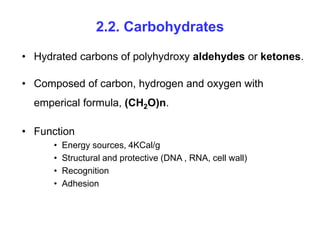
- 1. 2.2. Carbohydrates • Hydrated carbons of polyhydroxy aldehydes or ketones. • Composed of carbon, hydrogen and oxygen with emperical formula, (CH2O)n. • Function • Energy sources, 4KCal/g • Structural and protective (DNA , RNA, cell wall) • Recognition • Adhesion
- 2. • The 2-D extended structure is called Fischer projection and the 3-D ring one is called Haworth formula. • The ring structure of aldose and ketose are called hemiacetal and hemiketal, respectively. • Three classes • Monosaccharide • Oligosaccharide • Polysaccharide
- 3. Monosaccharides • Single polyhyrdoxy aldehyde or ketone unit. • Glucose is the most abundant monosaccharide. • Those with more than 4 carbon have cyclic structure. • Important fuel molecules & building blocks for nucleic acids. • The smallest monosaccharides, for which n = 3, are dihydroxyacetone and D & L glyceraldehyde. – Thus they are trioses.
- 4. Functional groups Ketone = R-CO-R Aldehyde = R-CO-H
- 6. The D and L isomers are enantiomers or mirror images of each other
- 8. • Monosaccharides with 4, 5, 6, and 7 C-atoms are called tetroses, pentoses, hexoses, and heptoses, respectively. • Because these molecules have multiple asymmetric carbons, exist as diastereoisomers (optically active isomers that are not mirror images) as well as enantiomers. • Note that D-glucose and D-mannose differ in configuration only at C-2, such sugars are called epimers. • Thus, D-glucose and D-mannose are epimeric at C-2; D- glucose and D-galactose are epimeric at C-4.
- 10. • Unmodified glucose reacts with oxidizing agents such as Cu2+ (Fehling's solution) because the open-chain form has a free aldehyde group that is readily oxidized. • Sugars that react are called reducing sugars; those that do not are called non-reducing sugars. • Non-reducing sugars do not have a free aldehyde group and so cannot react with Cu2+. • Monosaccharides are reducing sugars.
- 12. Ring formation • The predominant forms of ribose, glucose, fructose, and many other sugars in solution exist as rings. • In the process of cyclization in general, an aldehyde can react with an alcohol to form a hemiacetal. • For an aldohexose such as glucose, the C-1 aldehyde in the open-chain form of glucose reacts with the C-5 hydroxyl group to form an intramolecular hemiacetal.
- 13. • The resulting cyclic hemiacetal, a six-membered ring, is called pyranose because of its similarity to pyran. • Analogous to Pyran: O pyran O H OH H OH H OH H OH CH2OH H 1 2 3 4 5 6
- 14. • The aldehyde C atom now becomes asymmetric during ring formation. • This new asymmetric carbon atom formed during cyclization is called the Anomeric carbon. • The cyclic structure containing the -OH group on the right of the anomeric carbon is known as α-D-glucopyranose; on the left side is called β-D-glucopyranose.
- 15. • Haworth projections: – Can be written from the Fischer projection. – C1 drawn on the right (anomeric C). – The cyclic structure of a D-isomer has the last CH2OH group located above the ring (C6). – –OH groups on the left are drawn up (C3). – –OH groups on the right are drawn down (C2, C4).
- 16. • Haworth projections: • C atoms are not shown. • The designation α means that the hydroxyl group attached to C-1 is below the plane of the ring; β means that it is above the plane of the ring. • The C-1 carbon atom is called the anomeric carbon atom, and the α and β forms are called anomers.
- 19. • Similarly, a ketone can react with an alcohol to form a hemiketal. • The C-2 keto group in the open-chain form of a ketohexose, such as fructose, can form an intramolecular hemiketal by reacting with either the C-6 -OH group to form a six-membered cyclic hemiketal or the C-5 -OH group to form a five-membered cyclic hemiketal, respectively. • The five-membered ring is called a furanose because of its similarity to furan.
- 20. Ring formation …
- 22. Ring formation …
- 23. Ring formation: Pentose sugars
- 25. Monosaccharides join with amines and alcohols through glycosidic bonds • The anomeric C-atom reacts with the –OH group of methanol to form two products, methyl α & β -D-glucopyranoside. • The new bond formed between the anomeric carbon atom of glucose and the -OH oxygen atom of methanol is called a glycosidic bond specifically, an O-glycosidic bond. • The anomeric carbon atom of a sugar can also be linked to the nitrogen atom of an amine to form an N-glycosidic bond.
- 27. Modified monosaccharides Usually expressed on cell surfaces
- 29. Complex carbohydrates formed from monosaccharides • Because sugars contain many hydroxyl groups, glycosidic bonds can join one monosaccharide to another. • Oligosaccharides are built by the linkage of two or more monosaccharides by O-glycosidic bonds. • The wide array of these linkages in concert with variety of monosaccharides and their many isomeric forms makes complex carbohydrates information-rich molecules.
- 30. • Complex forms include: – Disaccharides – Oligosaccharides – Polysaccharides Disaccharides • Consist of two sugars joined by an α-glycosidic bond. • Most common ones • Sucrose: • Lactose: • Maltose
- 31. • Sucrose: formed by glycosodic linkage between α-D- glucopyranose and β-D-fructofuranose. – The enzyme sucrase breaks the disaccharides. • Lactose: formed by β-1-4 glycosidic linkage between galactose and glucose. – In humans lactase and in bacteria β-galactosidase enzyme breaks the linkage. • Maltose: two D-glucose residues are joined by a glycosidic linkage b/n the α-anomeric form of C-1 on one sugar and the -OH oxygen atom on C-4 of the adjacent sugar. – Such a linkage is called an α -1,4-glycosidic bond. – Hydrolysed by maltase.
- 33. Disaccharides …
- 34. Polysaccharides • Large polymeric oligosaccharides, formed by the linkage of multiple monosaccharides are called polysaccharides. • Polysaccharides play vital roles in energy storage and in maintaining the structural integrity of an organism. • If all of the monosaccharides are the same, these polymers are called homopolymers.
- 35. Glycogen • The most common homopolymer in animal cells is glycogen. • This storage form of glucose is a very large, branched polymer of glucose residues. • Most of the glucose units in glycogen are linked by α -1,4- glycosidic bonds. • The branches are formed by α -1,6-glycosidic bonds, present about once in 10 units.
- 36. Starch • The nutritional reservoir in plants is starch, of which there are two forms: • Amylose, the unbranched type of starch, consists of glucose residues in α -1,4 linkage. • Amylopectin, the branched form, has about 1 α -1,6 linkage per 30 α -1,4 linkages, in similar fashion to glycogen except for its lower degree of branching. • More than half the carbohydrate ingested by human beings is starch. • Both amylopectin and amylose are rapidly hydrolyzed by α -amylase, an enzyme secreted by the salivary glands and the pancreas.
- 37. Strach and glycogen: energy storage polysaccharides
- 38. Amylopectine
- 39. Cellulose • Cellulose is major polysaccharide of glucose found in plants, serves a structural rather than a nutritional role. • Cellulose is one of the most abundant organic compounds in the biosphere. • It is an unbranched polymer of glucose residues joined by β- 1,4 linkages.
- 40. Cellulose… • The α configuration allows cellulose to form very long, straight chains. • Fibrils are formed by parallel chains that interact with one another through hydrogen bonds. • The α-1,4 linkages in glycogen and starch produce a different molecular architecture from that of cellulose. • A hollow helix is formed instead of a straight chain in glycogen and starch.
- 41. • These differing consequences of the α and β linkages are biologically important. • The straight chain formed by β linkages is optimal for the construction of fibers having a high tensile strength. • In contrast, the open helix formed by α linkages is well suited to forming an accessible store of sugar. • Mammals lack cellulases and therefore, cannot digest wood and vegetable fibers. Cellulose…
- 42. Cellulose: a structural polysaccharide Favour straight chain Favour bent structures
- 43. Chitin • Same as cellulose, except –OH on C2 replaced with acetamide. – Amino sugar – Homopolymer of N-acetyl-D-glucosamine • Very strong. • Structural component of exoskeleton of arthropods
- 44. Glycoproteins • Covalent attachment to proteins: glycosylation. • Linkage through • N atom of R chain of Asparagine (Asn) (N-linked) to from GlcNac :N acetyl glucose amine or • O atom of R chain of Serine or Threonine residues (O- linkage) to form GalNac: Nacetyl galactoamine. • Asn accept an oligosaccharide if • Asn-X-Ser or Asn-X-Thr (X = any residue). • Thus potential glycosylation sites can be detected within aa sequences in the polypeptide chains.
- 46. Glycoproteins … • All N-linked glycopeptides have in common • A pentasaccharide core consisting of – 3 mannose – 2 N-acetylglucoseamine residues • Additional sugars attached to this core – Form great variety of glycoproteins
- 49. • Carbohydrates are linked to some soluble proteins as well as membrane proteins. • In particular, many of the proteins secreted from cells are glycosylated. • Most proteins present in the serum component of blood are glycoproteins (Eg: elastase). • Furthermore, N-acetylglucosamine residues are O-linked to some intracellular proteins. • The role of these carbohydrates, which are dynamically added and removed, is under active investigation.
- 50. Elastase
- 51. • Protein glycosylation takes place inside the lumen of the endoplasmic reticulum (ER) and the Golgi complex, organelles that play central roles in protein trafficking. • Elastase, which is secreted by the pancreas as a zymogen, is synthesized by ribosomes attached to the cytoplasmic face of the ER membrane. • The peptide chain is inserted into the lumen of the ER as it grows, guided by a signal sequence of 29 amino acids at the amino terminus.
- 52. • This signal sequence, is then cleaved from the protein in the transport process into the ER. • After the protein has entered the ER, the glycosylation process begins. • The N-linked glycosylation begins in the ER and continues in the Golgi complex, whereas the O-linked glycosylation takes place exclusively in the Golgi.
- 54. • The Golgi complex is the sorting center in the targeting of proteins to lysosomes, secretory vesicles, and the plasma membrane. • The cis face of the Golgi complex receives vesicles from the ER, and the trans face sends a different set of vesicles to target sites. • Vesicles also transfer proteins from one compartment of the Golgi complex to another.
- 56. Mannose 6-phosphate target lysosomal enzymes to their destination … • Active enzymes are synthesized from Golgi • But exported rather than directed to lysosomes • Enzymes mislocated in I-cell disease • These enzymes normally contain • Mannose 6-phophate residue • In I-cell the mannose is unmodified • Mannose 6-phophate is a marker which direct many hydrolytic enzymes from Golgi to lysosomes.
- 57. • A glycoprotein destined for delivery to lysosomes acquires a phosphate marker in the cis Golgi compartment in a two- step process. – First, a phosphotransferase adds a phospho-N-acetylglucosamine unit to the 6-OH group of a mannose residue. – Then a phosphodiesterase removes the added sugar (Glu Nac) to generate a mannose 6-phosphate residue in the core oligosaccharide. • I-cell patients are deficient in the phosphotransferase catalyzing the first step in the addition of the phosphoryl group.