
Recommended
Recommended
More Related Content
What's hot
What's hot (20)
Similar to Middle ear anatomy and physiology
Similar to Middle ear anatomy and physiology (20)
Recently uploaded
Recently uploaded (20)
Middle ear anatomy and physiology
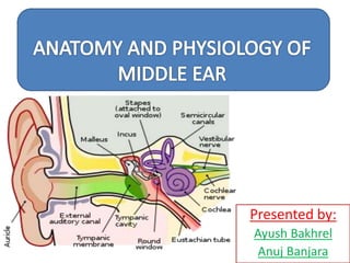
- 1. Presented by: Ayush Bakhrel Anuj Banjara
- 2. What is Middle Ear? Middle Ear is an air-filled, mucous membrane lined space in the temporal bone between the tympanic membrane laterally and the lateral wall of the internal ear medially .
- 3. Embryology First pharyngeal pouch Pharyngotympanic tube Middle ear cavity Mastoid antrum First pharyngeal arch Malleus Incus Tensor tympani muscle Ligament of malleus
- 4. Embryology Second pharyngeal arch Stapes Stapedius muscle First pharyngeal membrane Tympanic membrane
- 6. Division of Middle Ear
- 9. • Epitympanum(Attic)- Lying above pars tensa but medial to the Shrapnell’s membrane and bony lateral attic wall) • Mesotympanum- Lyinng opposite to the pars tensa • Hypotympanum – lying below the level of Pars tensa Division of Middle Ear
- 10. Tympanic Cavity
- 11. Boundaries of Middle Ear Middle Ear consists of 1. Roof 2. Floor 3. Anterior wall 4. Posterior Wall 5. Medial Wall 6. Lateral Wall
- 14. Roof ( Tegmental wall) The tegmental wall (roof) of the middle ear consists of a thin layer of bone, which separates the middle ear from the middle cranial fossa. This layer of bone is the tegmen tympani on the anterior surface of the petrous part of the temporal bone.
- 16. ROOF(TEGMENTAL WALL) = TERRACE= TEGMEN TYMPANI
- 17. • Floor (Jugular wall) Jugular wall (floor) of the middle ear consists of a thin layer of bone that separates it from the internal jugular vein. • Near the medial border of the floor is a small aperture,through which the tympanic branch from the glossopharyngeal nerve [IX] enters the middle ear.
- 19. ROOF(TEGMENTAL WALL) = TERRACE= TEGMEN TYMPANI FLOOR ( JUGULAR WALL) Internal Jugular Vein
- 20. Membranous wall (Lateral wall) The membranous (lateral) wall of the middle ear consists almost entirely of the tympanic membrane, but because the tympanic membrane does not extend superiorly into the epitympanic recess , the upper part of the membranous wall of the middle ear is the bony lateral wall of the epitympanic recess.
- 21. ROOF(TEGMENTAL WALL) = TERRACE= TEGMEN TYMPANI FLOOR ( JUGULAR WALL) Internal Jugular Vein Lateral Wall = Tympanic membrane
- 23. Mastoid wall(Posterior) The mastoid (posterior) wall of the middle ear is only partially complete. The lower part of this wall consists of a bony partition between the tympanic cavity and mastoid air cells . Superiorly, the epitympanic recess is continuous with the aditus to the mastoid antrum . Associated with the mastoid wall are : • the pyramidal eminence, a small elevation through which the tendon of the stapedius muscle enters the middle ear • the opening through which the chorda tympani nerve, a branch of the facial nerve [VII] , enters the middle ear.
- 25. ROOF(TEGMENTAL WALL) = TERRACE= TEGMEN TYMPANI FLOOR ( JUGULAR WALL) Internal Jugular Vein Lateral Wall = Tympanic membrane Mastoid antrum Pyramidal eminence(Staped ius muscle enters middle ear) Chorda tympani
- 26. Anterior wall The anterior wall of the middle ear is only partially complete. The lower part consists of a thin layer of bone that separates the tympanic cavity from the internal carotid artery. Superiorly, the wall is deficient because of the presence of: • a large opening for the entrance of the pharyngotympanic tube into the middle ear • a smaller opening for the canal containing the tensor tympani muscle. The foramen for the exit of the chorda tympani nerve from the middle ear is also associated with this wall
- 28. ROOF(TEGMENTAL WALL) = TERRACE= TEGMEN TYMPANI FLOOR ( JUGULAR WALL) Internal Jugular Vein Lateral Wall = Tympanic membrane Mastoid antrum Pyramidal eminence(Staped ius muscle enters middle ear) Chorda tympani Pharyngotympanic tube Tensor tympani Internal Carotid Artery Chorda tympani
- 29. Labyrinthine wall(Medial wall) The labyrinthine (medial) wall of the middle ear is also the lateral wall of the internal ear •A prominent structure on this wall is a rounded bulge (the promontory) produced by the basal coil of the cochlea, which is an internal ear structure involved with hearing.
- 30. Labyrinthine wall Associated with the mucous membrane covering the promontory is a plexus of nerves (the tympanic plexus) . Additionally, a branch of the tympanic plexus (the lesser petrosal nerve) leaves the promontory and the middle ear, travels across the anterior surface of the petrous part of the temporal bone, and leaves the middle cranial fossa through the foramen ovale .
- 31. ROOF(TEGMENTAL WALL) = TERRACE= TEGMEN TYMPANI FLOOR ( JUGULAR WALL) Internal Jugular Vein Lateral Wall = Tympanic membrane Mastoid antrum Pyramidal eminence(Staped ius muscle enters middle ear) Chorda tympani Pharyngotympanic tube Tensor tympani Internal Carotid Artery Promontory Chorda tympani
- 32. Oval window and Round Window
- 33. Oval Window The oval window is posterosuperior to the promontory. It is the point of attachment for the base of the stapes (footplate), and ends the chain of bones that transfer vibrations initiated by the tympanic membrane to the cochlea of the internal ear.
- 34. ROOF(TEGMENTAL WALL) = TERRACE= TEGMEN TYMPANI FLOOR ( JUGULAR WALL) Internal Jugular Vein Lateral Wall = Tympanic membrane Mastoid antrum Pyramidal eminence(Staped ius muscle enters middle ear) Chorda tympani Pharyngotympanic tube Tensor tympani Internal Carotid Artery Promontory Oval window (Posterosuperiorly) Chorda tympani
- 35. The round window is posteroinferior to the promontory. Posterior and superior to the oval window is the prominence of the facial canal which is a ridge of bone produced by the facial nerve as it passes through the temporal bone. Above and posterior to the prominence of the facial canal is a prominence of the lateral semicircular canal produced by the lateral semicircular canal.
- 37. ROOF(TEGMENTAL WALL) = TERRACE= TEGMEN TYMPANI FLOOR ( JUGULAR WALL) Internal Jugular Vein Lateral Wall = Tympanic membrane Mastoid antrum Pyramidal eminence(Staped ius muscle enters middle ear) Chorda tympani Pharyngotympanic tube Tensor tympani Internal Carotid Artery Promontory Oval window (Posterosuperiorly) Round window Prominence of facial canal Prominence of lateral semicircular canal Chorda tympani
- 38. Mastoid Area Posterior to the epitympanic recess of the middle ear is the auditus to the mastoid antrum, which is the opening to the mastoid antrum . The mastoid antrum is a cavity continuous with collections of air-filled spaces (the mastoid cells) , throughout the mastoid part of the temporal bone, including the mastoid process. The mastoid antrum is separated from the middle cranial fossa above by only the thin tegmen tympani. The mucous membrane lining the mastoid air cells is continuous with the mucous membrane throughout the middle ear. Therefore infections in the middle ear can easily spread into the mastoid area.
- 40. Pharyngotympanic tube The pharyngotympanic tube connects the middle ear with the nasopharynx and equalizes pressure on both sides of the tympanic membrane. Its opening in the middle ear is on the anterior wall. It consists of: • a bony part (the one-third nearest the middle ear) • a cartilaginous part (the remaining two-thirds ) .
- 42. Auditory Ossicles Consists of 3 main ear ossicles: 1. Malleus (hammer) 2. Incus(anvil) 3. Stapes(stirrup)
- 43. Malleus(Hammer, largest) Largest of the auditory ossicles. It is attached to the tympanic membrane. Its part are: •Head •Neck •Anterior and lateral process •Handle The anterior process is attached to the anterior wall of the middle ear by a ligament. • The lateral process is attached to the anterior and posterior malleolar folds of the tympanic membrane.
- 45. Incus It consist of : • Body of the incus • Long and short limbs • The enlarged body of the incus articulates with the head of the malleus. • The long limb extend downward and articulated with the stapes. • The short limb extends posteriorly and attached by a ligament to upper posterior wall of middle ear.
- 46. Stapes(Smallest) It is the most medial bone -it is attached to the oval window. - It consists of: - Head - Anterior and Posterior limb - Base • The head : directed laterally and articulates with the long process of the incus. • The two limbs separate from each other and attach to the oval base. • The base of the stapes fits into the oval window .
- 47. Muscle Origin Insertion Innervation Function Tensor tympani Cartilaginous part of pharyngotympanic tube, greater wing of sphenoid, its own bony canal Upper part of handle of malleus Branch from mandibular nerve [V3] Contraction pulls handle of malleus medially, tensing tympanic mem brane Stapedius Attached to inside of pyramidal eminence Neck of stapes Branch of facial nerve [VII] Contraction pulls stapes posteriorly, preventing excessive oscillation
- 48. • Major vessels – Anterior tympanic branch of maxillary artery – tympanic membrane – Stylomastoid branch of posterior auricular artery – middle ear and mastoid air cells • Minor vessels – Petrosal and superior tympanic branch of middle meningeal – Branch of artery of pterygoid canal – Tympanic branch of internal carotid Arterial Supply
- 49. Venous Supply • Pterygoid venous plexus • Superior petrosal sinus Lymphatic Drainage Retropharyngeal nodes—upper jugular chain
- 50. • Function – Couples sound energy to cochlea – Physical protection for cochlea – Impedance matching • Hydraulic action of tympanic membrane • Lever action of ossicles • Eustachian tube-equalizes pressure and keep tympanic membrane in neutral positon – Attenuation of sound. Physiology
- 52. • CSOM(Chronic Suppurative Otitis Media) – Perforated tympanic membrane with persistent drainage from the middle ear (lasting >3months ) – Conductive hearing loss • Cholesteatoma – Abnormal, noncancerous skin growth that can develop in the middle section of your ear, behind the eardrum – Acquire ear infections repeatedly early in life Clinical Aspect
- 53. • Gray’s anatomy for student • Guyton and Hall Text Book of Physiology(13th edition) • Ear, Nose and Throat – PL Dhingra(6th edition)