
Recommended
More Related Content
What's hot
What's hot (20)
Similar to Normas of skull
Similar to Normas of skull (20)
More from Ayshah Hashimi
Recently uploaded
Recently uploaded (20)
Normas of skull
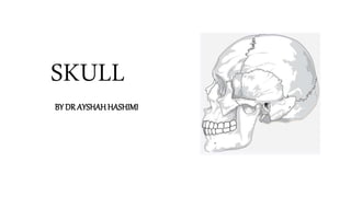
- 1. SKULL BY DR AYSHAH HASHIMI
- 2. • The skull is a bony structure that forms the head. It supports the structures of the face and provides a protective cavity for the brain • The skull is made up of a 22 fused flat bones • Contains many foramina, fossae, processes, and several cavities or sinuses • Bones in skull develops from intramembranous ossification • The joint in the skull are mostly sutures and few are 1° cartilaginous • Only temporomandibular joint is synovial in nature that permits us to speak, eat, drink and laugh • Skull lodges brain, cochlear and vestibular apparatus, retina, olfactory mucosa and taste buds
- 3. Skull is divided into two parts • Cranium/Braincase/Calvaria/skullcap • 8 bones forms the cranium 2 Parietal 2 Temporal Frontal Occipital Sphenoid Ethmoid Bone Frontal Bone Parietal Bone Temporal Bone Sphenoid Occipital Bone Ethmoid
- 4. • Facial skeleton composed of 14 bones 2 Maxilla 2 Zygomatic 2 Nasal 2 Lacrimal 2 Palatine 2 Inferior Nasal Concha Mandible Vomer
- 5. Methods of studying skull • Superior view or Norma Verticalis • Posterior view or Norma occipitalis • Anterior view or Norma Frontalis • Lateral view or Norma Lateralis • Inferior view or Norma Basalis
- 6. Exterior of Skull Norma Verticalis • Anteriorly frontal bone • Posteriorly occipital bone • On each side 2 parietal bone Sutures • Coronal suture (b/w frontal and parietal bone) • Sagittal suture (b/w 2 parietal bone) • Lambdoid suture (b/w parietal and occipital bone) • Metopic suture (inter-frontal suture)
- 7. • Vertex- highest point on sagittal suture • Vault- dome shaped roof of the skull • Obelion- midpoint on sagittal suture between the 2 parietal foramen • Bregma is a point attachment of coronal and sagittal suture. Also called a Anterior fontanelle. It is the site of membranous gap that closes at 18 months of age • Lambda is a point between the sagittal and lambdoid suture. Also called a Posterior fontanelle. It is the membranous gap that closes at 2-3 months of age • Parietal tuber/eminence is the area of maximum convexity of the parietal bone • Frontal eminence is the area of maximum convexity of the frontal bone • Parietal foramen is 2.5-4 cm in front of lambda Bregma Lambda Obelion Parietal Tuber
- 8. Norma occipitalis • Superiorly parietal bone • Inferiorly occipital bone • On each side mastoid part of temporal bone Sutures • Lambdoid suture • Sagittal suture • Occipitomastoid suture • Parietomastoid suture
- 9. • External occipital protuberance is a median protuberance. It marks the junction of the head and the neck • Inion is the most prominent point on external occipital protuberance • Superior nuchal line is bony ridges passing laterally from the protuberance • Highest nuchal line- 1 cm above from superior nuchal line • Inferior nuchal line • External occipital crest- straight vertical prominent line from ext. occipital protuberance to inferior nuchal line • Mastoid foramen • Lambda parietal foramen, obelion
- 10. Attachment • Trapezius arises from external occipital protuberance and medial 1/3 of superior nuchal line • SCM (sternocleidomastoid) is inserted laterally on superior nuchal line and splenius capitis below • Occipitalis arises from highest nuchal line Trapezius Occipitalis Splenius capitis Sternocleidomastoid
- 12. Norma Lateralis Bones- • Frontal • Parietal • Occipital • Temporal • Sphenoidal • Zygomatic • Mandible • Maxilla • Nasal • Lacrimal Sutures • Coronal suture • Lambdoid suture • Fronto-zygomatic suture • Fronto-sphenoid suture • Spheno-parietal suture • Parieto-temporal suture • Spheno-temporal suture • Occipito-mastoid suture
- 13. Features • Temporal lines begins at zygomatic process of frontal bone, arcs backward and backwards and cross the frontal bone and parietal bone • Supramastoid crest is continuation of temporal lines posteriorly where it turns downward and forward • Zygomatic arch is articulation of zygomatic process with zygomatic bone • External acoustic meatus Temporal lines Supramastoid crest Zygomatic Arch External acoustic meatus
- 14. • Suprameatal triangle is a small depression posterosuperior to the meatus • Asterion (Mastoid fontanelle) is a point where lambdoid, occipitomastoid and Parietomastoid suture meets • Temporal fossa is a depression that lie between temporal line and zygomatic process • Pterion is H- shaped suture where four bones adjoin each other • Mastoid process • Styloid process Suprameatal Triangle Asterion Pterion Mastoid Process Styloid Process
- 16. Attachment • Temporalis muscle arises from temporal fossa • Masseter arises from medial and lower border of zygomatic arch • Sternocleidomastoid, splenius capitis and longissimus capitis are inserted into mastoid process Temporalis Masseter
- 17. Norma Frontalis Bones • Frontal bone • Mandible • Rt & Lt Maxilla • Rt & Lt Nasal bone • Rt & Lt Zygomatic bone Parts • Frontal region • Orbit • Bony aperture of the nose • Lower part of the face Sutures • Internasal suture • Frontonasal suture • Nasomaxillary suture • Lacrimo-maxillary suture • Fronto-maxillary suture • Intermaxillary suture • Zygomaticomaxillary suture • Zygotico-frontal suture
- 18. Frontal Region • Superciliary arch are rounded, curved elevation above the orbits • Supraorbital notch • Glabella is a median elevation connecting the two arches • Nasion is a median point at which internasal suture meets with frontonasal suture • Frontal eminence Bony aperture of the nose • Pear shaped, narrow above and wide below • Most fractured bone of the face • Above- lower border of nasal bone • Below- nasal notch of maxilla on each side Lower part of the face • Maxilla • Mandible • Zygomatic bone Superciliary arch Supraorbital Notch Nasion Glabella
- 19. Orbit Bones that forms orbit- • Maxilla • Zygomatic bone • Frontal bone • Sphenoid • Ethmoid • Lacrimal bon Boundaries • Superiorly- Frontal Bone • Medially- Frontal process of Maxilla • Laterally- Zygomatic Bone Sphenoid Ethmoid Frontal Maxilla
- 20. Openings Structure Supraorbital foramen Supraorbital vessels and nerve Infraorbital Groove Infraorbital vessels and nerve Nasolacrimal Canal Nasolacrimal duct Superior Orbital fissure 3rd, 4th, 5th, 6th cranial nerve, Superior ophthalmic vein Inferior Orbital fissure Maxillary nerve, inferior ophthalmic vein Anterior ethmoidal foramen Anterior ethmoidal vessels and nerve Posterior ethmoidal foramen Posterior ethmoidal vessels and nerve Optic Canal Optic and ophthalmic Nerve Supraorbital foramen Infraorbital groove SOF IOF AEF PEF OPTIC CANAL
- 21. Facial muscles • These are striated skeletal muscles supplied by facial nerve. Also called as mimetic muscles • The muscles of the face controls facial expression and are responsible for mastication, laughing, talking etc. • These muscles are categorised into several groups 1. Muscles of mouth 2. Muscles of nose 3. Muscles of orbit
- 22. Norma Basalis Bones of inferior view of skull • Maxilla bone • Zygomatic bone • Temporal bone • Occipital bone • Palatine bone • Vomer • Sphenoid Studied under three parts: • Anterior • Middle • Posterior Maxilla Palatine Vomer Sphenoid Sphenoid Occipital Temporal Temporal
- 23. Anterior part of Norma Basalis • Formed by hard palate, bounded by alveolar arches • Hard palate is formed by maxilla anteriorly and palatine bone posteriorly • There are three canals or foramen in the hard palate- incisive fossa, greater and lesser palatine foramen • Hard palate has cruciform suture- intermaxillary, interpalatine and palatomaxillary suture • Posterior border of hard palate is called as posterior nasal spine • Palatine crest are curved ridges
- 24. Middle part of Norma Basalis • Lie between the posterior nasal spine and an imaginary transverse line passing through the anterior border of foramen magnum • Divided into two parts median and lateral parts • Median part comprises of vomer bone anteriorly and Clivus posteriorly • Lateral part is formed of wings of sphenoid bone, pterygoid processes and petrous part of temporal bonetemporal bone
- 25. Median part of middle area of Norma Basalis Vomer Bone • Vomer is a part of nasal septum and divides the nasal cavity into two equal half • It articulates with four bones Maxilla (anteroinferior) Palatine (posteroinferior) Sphenoid (posterosuperior) Ethmoid (superior) • Posteriorly Vomer separates the post nasal aperture into two • Inferiorly it articulate with bony plate while superiorly it articulates with the sphenoid bone • Posterior border is free • Clivus is bony area formed by articulation of body of sphenoid bone posteriorly with the basilar part of the occipital bone
- 26. Lateral part of middle area of Norma Basalis • Greater wings and Pterygoid process of Sphenoid bone forms the lateral part • Medial plate has depression called as Scaphoid fossa and a projection called as pterygoid Hamulus • Lateral plate has depression called as Pterygoid fossa • Sulcus tubae is an opening between greater wing of sphenoid and petrous part of temporal bone through which auditory tube passes • Petrous part of temporal bone lie between greater wings of sphenoid and occipital bone
- 27. Posterior part of Norma Basalis • Lie posterior to foramen magnum • Divided into two parts median and lateral parts • Median part comprises of Foramen magnum Occipital Condyles External occipital crest External occipital protuberance • Lateral part is formed by Squamous part of occipital bone Nuchal lines (Superior & Inferior) Styloid process Mastoid process Jugular foramen
- 28. Interior of skull • The cranium is lined internally by endocranium and externally by pericranium • Both are continuous through the foramina and sutures • Cranial bones consists of Outer compact layer (thick and tough) Middle spongy layer Inner compact layer (thin and brittle) • Skull derive their blood supply mostly from the meningeal arteries and drain by 4 diploic veins • Divided under sub-headings Internal surface of Cranial Vault Internal surface of the Base of the skull
- 29. Internal surface of Cranial Vault • Thin & brittle • Presents markings produced by meningeal vessels, venous sinuses, arachnoid granulations. • Frontal crest lies anteriorly in the median plane. It projects backwards • Sagittal sulcus runs backwards and becomes progressively wider to lodge the superior sagittal sinus • Granular Foveolae are deep irregular large pits situated along the length of the superior sagittal sinus • The vascular markings • Parietal foramen
- 30. Internal surface of the Base of the skull • Natural subdivision Anterior cranial fossa Middle cranial fossa Posterior cranial fossa • Duramater is firmly adherent to the floor of the fossae and is continuous with the pericranium through the foramina and fissures
- 31. Anterior Cranial Fossa • Boundaries Anteriorly- Frontal bone Posteriorly- sphenoid bone (lesser wings, anterior clinoid process and sulcus Chiasmaticus) • Floor Anteriorly- Cribriform plate of the ethmoid bone Posteriorly- body of sphenoid On each side- Frontal bone and lesser wings of Sphenoid • Other features: Foramen cecum Crista galli • Clinical Anatomy: Fracture causes bleeding and discharge of CSF through nose and black eye.
- 32. Middle Cranial Fossa • Boundaries Anteriorly- Sphenoid bone (lesser wings, anterior clinoid process and sulcus Chiasmaticus) Posteriorly- Superior border of petrous part of temporal bone, Sella turcica Laterally- Greater wing of sphenoid, anteroinferior angle of parietal bone, squamous part of temporal bone • Clinical Anatomy Fracture causes bleeding and discharge of CSF through ear and damage to 7th & 8th cranial nerve Most commonly fractured
- 33. Posterior Cranial Fossa • Boundaries Anteriorly- Sella turcica of Sphenoid bone Posteriorly- Occipital bone Laterally- Mastoid part of temporal bone • Floor Anteriorly- Clivus Middle- foramen magnum Posteriorly- Occipital bone • Clinical Anatomy Fracture causes bruising over mastoid region extending down to sternocleidomastoid muscle
- 34. Structures Passing Through Various Foramen An opening that allows the passage of structures from one region to another Opening Structures passing Cribriform plate Olfactory nerve (1st CN) Optic canal Optic nerve (2nd), ophthalmic artery Sup. orbital fissure 3rd, 4th, 5th, 6th CN, Sup. ophthalmic vein Foramen Rotundum Maxillary nerve Foramen Ovale Mandibular nerve Int. Acoustic Meatus 7th & 8th CN Jugular Foramen 9th, 10th, 11th CN, Sigmoid & Inf. Petrosal sinus Foramen spinosum Middle meningeal vessels Hypoglossal Canal 12th CN Foramen Magnum Medulla, meninges, vertebral arteries and
Editor's Notes
- Bregma is anterior fontanelle and it closes at 18 months Lambda is posterior fontanelle and closes at 2-3 months