Lasers and its applications in conservative dentistry
•
5 likes•440 views

detail description about laser, its properties and its application in conservative dentistry and most importantly in endodontic field.

Recommended
Recommended
More Related Content
What's hot
What's hot (20)
Similar to Lasers and its applications in conservative dentistry
Similar to Lasers and its applications in conservative dentistry (20)
Recently uploaded
Recently uploaded (20)
Lasers and its applications in conservative dentistry
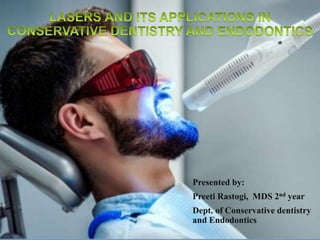
- 1. Presented by: Preeti Rastogi, MDS 2nd year Dept. of Conservative dentistry and Endodontics
- 2. Introduction Laser physics Component of laser device Laser delivery system Classification of lasers Mechanism of laser tissue interaction Wavelength of laser used in dentistry Laser used in conservative dentistry Laser used in Endodontics Laser hazards Laser hazard control measures Advantage and disadvantage of laser Conclusion References LASERS AND ITS APPLICATION IN CONSERVATIVE AND ENDODONTIC DENTISTRY 2
- 3. Most patients still associate the sound and vibration of the drill with pain. However, with the new advancements in this field, several options have become available to progressive dentists to allay fears and offer patients state of the art treatment. One such advancement is the advent of the laser technology presenting new vistas for dentists in fields of dentistry. • Laser is a device that transforms light of various frequencies into a chromatic radiation in the visible, infrared and ultraviolet regions with all the waves in a phase capable of mobilizing immense heat and power when focused at a close range LASER is acronym for ‘Light Amplification by Stimulated Emission of Radiation.’ The rapid development in the field of laser technology, modern lasers with a wide range of characteristics are now available and being used in the field of dentistry, most importantly in the field of conservative dentistry. LASERS AND ITS APPLICATION IN CONSERVATIVE AND ENDODONTIC DENTISTRY 3
- 4. In 1917, Albert Einstein laid the foundation for the invention of the laser by theorizing that photoelectric amplification could emit a single frequency, or stimulated emission. A predecessor of the laser, called the MASER ,for "Microwave Amplification by Stimulated Emission of Radiation",was developed in 1954 at Columbia University by Charles Townes and Jim Gordon and in Russia by Nicolay Basov. The term LASER was first introduced to the public in 1959, in an article by a Columbia University graduate student, Gordon Gould. In early 1960s, the first working laser was invented by Theodore Maiman at the Hughes Research Laboratories in Malibu,built the first functioning laser who inserted a ruby rod into a photographic flashlamp by using a mixture of helium and neon. The second laser to be developed was the neodymium laser by Snitzer in 1961. LASERS AND ITS APPLICATION IN CONSERVATIVE AND ENDODONTIC DENTISTRY 4 Albert Einstein [DCNA -2004]
- 5. LASERS AND ITS APPLICATION IN CONSERVATIVE AND ENDODONTIC DENTISTRY 5 Design and built of the first working ruby laser Dr. Theodore H. Maiman July 11, 1927 - May 5, 2007
- 6. In 1971, The first use of lasers in endodontics was reported by Weichman and Johnson, as they utilize high power infrared CO2 laser to seal the apical foramen in vitro In 1962, the argon laser was developed, whereas, the ruby laser became the first medical laser to coagulate retinal lesions, when it was used in 1963. In 1964, Ralph Stern and Reidar Sognnaes used the ruby laser to vaporise enamel and dentine. The first laser that had truly both hard and soft tissue application was the CO2 laser, invented by Patel in 1964. The Nd:YAG laser was also developed in 1964 by Geusic, which is thought to have a better interaction with dental hard tissues. The first report of laser exposure to a vital human tooth was given in 1965 by Leon Goldman . In 1970’s, researchers began to find the clinical oral soft tissue uses of medical CO2 and neodymium doped: yttrium aluminum garnet (Nd:YAG) lasers. In 1969 Leon Goldman used the laser clinically on enamel and dentine. LASERS AND ITS APPLICATION IN CONSERVATIVE AND ENDODONTIC DENTISTRY 6
- 7. LASER PHYSICS BASIC LASER SCIENCE
- 8. LIGHT The basic unit of this radiant energy is called a photon; wave of photons travels at the speed of light and it can be defined by two basic properties. The first is amplitude, which is defined as the total height of the wave oscillation from the top of the peak to the zero line on a vertical axis. This is an indication of the amount of intensity in the wave: larger the amplitude, the greater the amount of useful work that can be performed. The second property is the wavelength, which is the horizontal distance between any two corresponding points on the wave. Dental lasers have wavelengths in the order of much smaller units by using the terminology of either nanometres (10-9 m) or microns (10-6 m). As the waves travel, they oscillate several times per second and this is termed as ‘frequency’. Frequency is inversely proportional to the wavelength: The shorter the wavelength, the higher the frequency and viceversa. Light is a form of electromagnetic energy that exists as a particle, travel in waves at a constant velocity. LASERS AND ITS APPLICATION IN CONSERVATIVE AND ENDODONTIC DENTISTRY 8
- 9. LASERS AND ITS APPLICATION IN CONSERVATIVE AND ENDODONTIC DENTISTRY 9
- 10. LASERS AND ITS APPLICATION IN CONSERVATIVE AND ENDODONTIC DENTISTRY 10 Comparison between ordinary visible light and laser light Ordinary light usually appearing white, is the sum of the many colors of the visible spectrum – violet, blue, green, yellow, orange and red. Multiple wavelength = white light (Polychromatic) Non-directional Non-focused Unorganised , incoherent Low Intensity 0.1 W/cm2 Laser energy is one specific color, a property called “ monochromaticity” in dental applications that color may be visible or invisible. Highly focused and directional- collimated beam Organised , Coherent Energy High Intensity 108-1016 W/cm2 REGULAR LIGHT IS POLYCHROMATIC LASER LIGHT IS MONOCHROMATIC
- 11. The German physicist Max Planck introduced quantum theory in 1900 which was further conceptualized as relating to atomic architecture by Neils Bohr, a physicist from Denmark. When a quantum, the smallest unit of energy, is absorbed by the electrons of an atom and a molecule, a brief excitation occurs. Since natural orders prefers substances to be in a resting state, the quantum is soon released, a process called spontaneous emission. The emitted energy packet was previously described as a photon. In 1916 Albert Einstein theorized that an additional photon travelling in the field of the excited atom that has the same excitation energy level would result in a release of two quanta, or coherent wave of two photons, a phenomenon he termed stimulated emission. LASERS AND ITS APPLICATION IN CONSERVATIVE AND ENDODONTIC DENTISTRY 11
- 12. If this process were to continue, more atoms would be energized, more identical photons would be emitted and further propagation of this stimulatory wave would result. At some point, a population inversion occurs, meaning that a majority of the atoms of the active medium are in the elevated rather than the resting state. A pumping mechanism offering a constant supply of energy is necessary to maintain this excitation. The photons are reflected back and forth within the active medium to further enhance stimulated emission and successive passes through the active medium increase the power of and ultimately collimate the photon beam. This the process of amplification. LASERS AND ITS APPLICATION IN CONSERVATIVE AND ENDODONTIC DENTISTRY 12
- 13. The laser energy produced is radiated in a specific form of electromagnetic energy. The entire array of wave energy is described by the electromagnetic spectrum, with a range from gamma rays, whose wavelength are typically less than10-10m, to radiowaves, whose wavelength can be thousand of meters in size. Very short wavelengths below approximately 350nm are termed ionizing and can deeply penetrate biologic tissue, produce charged atoms and molecules and have a mutagenic effect on cellular DNA. Wavelength greater than 350nm cause excitation and heating of the tissue with which they interact. The accepted dividing line between ionizing and non- ionizing wavelengths is at the junction of ultraviolet and visible violet light on the spectrum. All available dental laser devices are classified as non- ionizing because their emission wavelength exceed 350nm. LASERS AND ITS APPLICATION IN CONSERVATIVE AND ENDODONTIC DENTISTRY 13
- 14. • Monochromatic • Coherence • Collimation • Monochromatic means that the light produced by a particular laser will be of a characteristic wavelength. If the light produced is in the visible spectrum (400 nm to 750 nm), it will be seen as a beam of intense color. It is important to have this property to attain high spectral power density of the laser. • Coherence means that the light is all perfectly in phase as they leave the laser. That means that unlike a normal light source, their individual contributions are summated and reinforce each other. In an ordinary light source, much of the energy is lost as out of phase, waves cancel each other. LASERS AND ITS APPLICATION IN CONSERVATIVE AND ENDODONTIC DENTISTRY 14 Common principles on which all lasers work is generation of monochromatic, Coherent and collimated beam
- 15. • Collimation means that the laser light beam is perfectly parallel when leaving the laser aperture. This property is important for good transmission through delivery systems. • The main differentiating characteristic of lasers is wave length which depends on the laser medium and excitation diode, i.e. continuous wave or pulsed mode. LASERS AND ITS APPLICATION IN CONSERVATIVE AND ENDODONTIC DENTISTRY 15 Collimated and uncollimated beam
- 16. All laser devices have the following main constituents: a) Active medium:- A laser medium can be a solid, liquid or gas. Lasing medium decides the wavelength of emitted light from the laser and the laser is named after the lasing medium Eg: CO2 laser: If CO2 is used as active medium/lasing medium. b) External power source:- It excites or pumps the atom in the laser medium to their higher energy levels. This causes the population inversion. A population inversion happens when there are more atoms in the excited state pumped by the electrical charge rather than a non-excited state. Atoms in the excited state spontaneously emit photons of light, which bounce back and forth between the two mirrors in the laser tube. As they bounce within the laser tube, they strike other atoms, stimulating more spontaneous emission. Photons of energy of the same wavelength and frequency escape through the transmitive mirror as the laser beam. c) Optical cavity:- To contain and amplify the photon chain reaction that results from stimulated emission in a population of excited atoms, it is necessary to place this reaction within an optical cavity. An optical cavity contains two parallel mirrors placed on either side of the lasing medium. In this configuration, photons bounce off the mirrors and re-enter the medium to stimulate the release of more photons. If some form of energy is provided to continuously pump atoms up to the excited state, the population inversion can be maintained and high intensity light circulating back and forth between two mirrors can be generated. LASERS AND ITS APPLICATION IN CONSERVATIVE AND ENDODONTIC DENTISTRY 16
- 17. d) Cooling system:- Heat production is a by-product of laser light propagation. It increases with the power output of the laser and hence, with heavy-duty tissue cutting lasers, the cooling system represents the bulkiest component. Co-axial coolant systems may be air or water-assisted. e) Mirror:- The mirror collimates the light, photons hits perpendicular to the mirrors and re-enters the active medium, while those off-axis leave the lasing process. If one mirror is totally reflective and other mirror is partially transmissive, the light that escapes through partially transmissive becomes the laser beam. The lasing medium is located within resonating chamber, which has a cylindrical structure with a fully reflecting mirror on one side and partially transmissive mirror at other site. These mirrors are precisely mounted so that they are exactly parallel to one another. This arrangement allows for the reflection of photons of light back and forth across the chamber, eventually resulting in the production of an intense photo resonance within the medium. . LASERS AND ITS APPLICATION IN CONSERVATIVE AND ENDODONTIC DENTISTRY 17
- 18. In clinical practice, a laser must be able to effectively deliver laser energy to the target site. The existing range of laser delivery systems involves: • Articulated arms:- Laser light can be delivered by articulated arms, which are very simple but elegant devices. • Mirrors are placed at 45 degree angle to the tube carrying the laser light. The tubes can rotate above the normal axis of the mirrors. LASERS AND ITS APPLICATION IN CONSERVATIVE AND ENDODONTIC DENTISTRY 18 Articulated arm delivery system
- 19. • Hollow waveguides:- The laser energy is reflected along this tube and exits through a handpiece at the surgical end with the beam striking the tissue in a noncontact mode. • An accessory tip of sapphire or hollow metal can be connected to the end of the waveguide for contact with the surgical site. LASERS AND ITS APPLICATION IN CONSERVATIVE AND ENDODONTIC DENTISTRY 19 Waveguide delivery system
- 20. • Fiber optics:- The fiber-optic system can be used in both contact or noncontact modes. Mostly it is used in contact mode, directly touching the surgical site. All conventional dental instruments: hand or rotary, physically touch the tissue being treated. LASERS AND ITS APPLICATION IN CONSERVATIVE AND ENDODONTIC DENTISTRY 20 Fiberoptic cables
- 21. a. 'Constant on' Emission: It is the continuous wave in which the beam is emitted at only one power level for as long as the operator depresses the foot switch. b. 'Pushed on and off' Emission: This is a gated pulse mode having periodic alterations of the Laser energy, much like a blinking light. This mode is achieved by the opening and closing of a mechanical shutter in front of the beam pathway of a continuous wave emission. c. Free running pulse mode: In this mode a very large Laser energy is emitted for an extremely short span, in microseconds followed by a relatively long time in which the Laser is off. LASERS AND ITS APPLICATION IN CONSERVATIVE AND ENDODONTIC DENTISTRY 21 The dental Laser device can emit the light energy in three modalities as a function of time:
- 22. • This is the interaction of photons with the atoms or molecules of the target. Lasers have five different interactions with the target tissue. 1) Photobiological interactions 2) Photochemical interactions 3) Photothermal interactions 4) Photomechanical interactions 5) Photoelectrical interactions LASERS AND ITS APPLICATION IN CONSERVATIVE AND ENDODONTIC DENTISTRY 22
- 23. LASERS AND ITS APPLICATION IN CONSERVATIVE AND ENDODONTIC DENTISTRY 23 1. Reflection: During reflection, the incident laser beam redirects itself without affecting the target tissues. 2. Absorption: The second interaction is the absorption of laser energy by the intended target tissue. This effect is desirable and the amount of energy that is absorbed by the tissue depends on the tissue characteristics, such as pigmentation and water content; and on the laser wavelength and emission mode. Argon has a high affinity for melanin and haemoglobin in soft tissue. 3. Transmission: The third interaction is the transmission of laser energy directly through the tissue without affecting it. 4. Scattering: The fourth interaction is scattering. Scattering of the reflected light weakens the intended energy and possibly produces no useful biologic effect. PHOTOBIOLOGICAL INTERACTIONS FOUR BASIC TYPES OF LASER INTERACTION OCCURS WHEN LIGHT HITS THE TARGET TISSUE
- 24. Photochemical interaction:- It include biostimulation, which described the stimulatory effects of laser light on biochemical and molecular processes that normally occur in tissues such as healing and repair. Photothermal interaction:- It manifest clinically as photoablation or the removal of tissue by vaporization and superheating of tissue fluids; coagulation and hemostasis and photopyroysis or the burning away of tissues. LASERS AND ITS APPLICATION IN CONSERVATIVE AND ENDODONTIC DENTISTRY 24
- 25. Photomechanical interaction:- It include photo-disruption or photo-disassociation, which is the breaking apart of structures by laser light. Photo-acoustic interactions, which involve the removal of tissue with shock-wave generation. Photoelectrical interaction: It include photo-plasmolysis which describes how tissue is removed by the formation of electrically charged ions and particles that exist in a semi-gaseous, high-energy state. LASERS AND ITS APPLICATION IN CONSERVATIVE AND ENDODONTIC DENTISTRY 25
- 26. 1. Lasers can be classified according to its Hardness, Spectrum of light and Material used. 2. Lasers are also classified as: a) Soft tissue lasers b) Hard tissue lasers 3. Classification of laser based on its spectrum: 4. Classification of laser according to the material used: LASERS AND ITS APPLICATION IN CONSERVATIVE AND ENDODONTIC DENTISTRY 26
- 27. a) Soft tissue lasers Soft tissue lasers generally utilize diodes. Clinical applications includes: healing of localized osteitis, healing of aphthous ulcers, reduction of pain and treatment of gingivitis. Soft tissue lasers in clinical use are: 1. Helium-Neon (He-N) at 632.8 nm (red, visible). 2. Gallium-Arsenide (Ga-As) at 830 nm (infra-red, invisible). b) Hard tissue lasers Hard tissue lasers (surgical) can cut both soft and hard tissues. Newer variety can transmit their energy via a flexible fiber optic cable. Commonly used hard lasers are: 1. Argon lasers (Ar) at 488 to 514 nm 2. Carbondioxide lasers (CO2) at 10.6 micro-meter 3. Neodymium-doped Yttrium Aluminium Garnet (Nd:YAG) at 1.064 micrometer. 4. Neodymiumm Yttrium-Aluminum-Perovskite (Nd:YAP) at 1,340 nm. LASERS AND ITS APPLICATION IN CONSERVATIVE AND ENDODONTIC DENTISTRY 27
- 28. 1) UV Light a. Spectrum is 100 nm – 400 nm b. Use: Not used in dentistry. 2) Visible Light a. Spectrum is 400 nm – 750 nm b. Use: Most commonly used in dentistry (Argon and Diagnodent) 3) Infrared light a) Spectrum is 750 nm – 10000nm b) Use: Most dental lasers are in this spectrum. LASERS AND ITS APPLICATION IN CONSERVATIVE AND ENDODONTIC DENTISTRY 28
- 29. A. Gas lasers: • Argon • Carbon-dioxide B. Liquid: • Dyes C. Solid: • Nd:YAG • Erbium: Yttrium Aluminum Garnet (Er: YAG) • Diode D. Semiconductor: • Hybrid silicon laser E. Excimers: • Argon-fluoride • Krypton-fluoride • Xenon-fluoridesers LASERS AND ITS APPLICATION IN CONSERVATIVE AND ENDODONTIC DENTISTRY 29
- 30. • This laser has an active medium of ionized argon gas, energized by a high current electrical discharge, and the laser light is delivered fiber-optically in continuous wave and gated pulsed modes. • There are two emission argon laser wavelengths used in dentistry: 488 nm (blue) and 514 nm (blue green). • Both wavelengths are poorly absorbed in the enamel and dentin which is advantageous during cutting and sculpting gingival tissues as there is minimal interaction with the dental hard tissue without causing any damage to the tooth surface. • Both wavelength is used as an aid for caries detection When the argon laser light illuminates the tooth, the carious area appears as a dark orange-red color and is easily discernible from the surrounding healthy structures. • Argon lasers are indicated in periodontics, as they possess bactericidal properties against Prevotella intermedia and Porphyromonas gingivalis and also used to treat vascular malformations such as a hemangioma. • Potential complications of this laser treatment include granuloma formation, bleeding or non- resolution of the lesion. LASERS AND ITS APPLICATION IN CONSERVATIVE AND ENDODONTIC DENTISTRY 30
- 31. • The diode laser is manufactured from solid semiconductor crystals made from a combination of aluminum (with wavelength of 800 nm) or indium (900 nm), gallium and arsenic. • These wavelengths penetrate deep into the mucosa and highly attenuated by the pigmented tissue, although hemostasis is slow as compared with the argon laser. • These lasers are excellent soft tissue surgical lasers, so surgery can be performed safely as these wavelengths are poorly absorbed by the dental hard tissue. • This laser is indicated for gingivoplasty, sulcular debridement and deeper coagulation process on gingival and mucosa. • The chief advantage of the diode lasers is one of a smaller size, portable instrument. These lasers can also stimulate fibroblastic proliferation at low energy levels. LASERS AND ITS APPLICATION IN CONSERVATIVE AND ENDODONTIC DENTISTRY 31
- 32. • Nd:YAG has a solid active medium, which is a garnet crystal combined with rare earth elements yttrium and aluminum, doped with neodymium ions. • The available dental wavelength of 1064 nm is indicated for various soft-tissue procedures such as cutting and coagulating of gingival and sulcular debridement. • This laser provides good hemostasis provides a clear operating field during soft-tissue procedures. • The laser is also indicated for the removal of incipient caries, although the working efficiency is less in comparison with the Er: YAG, or Er, chromium (Cr):YSGG lasers. • When used in a noncontact, defocused mode, this wavelength can penetrate several millimeters which can be used for procedures such as treatment of aphthous ulcers or pulpal analgesia. • However, due to a decrease in pulpal function, sometimes damage to the dental pulp can results LASERS AND ITS APPLICATION IN CONSERVATIVE AND ENDODONTIC DENTISTRY 32
- 33. LASERS AND ITS APPLICATION IN CONSERVATIVE AND ENDODONTIC DENTISTRY 33
- 34. Erbium, Cr:YSGG (2780 nm) • Erbium, Cr: YSGG (2780 nm) which has an active medium of a solid crystal of yttrium scandium gallium garnet doped with erbium and chromium. Erbium:YAG (2940 nm) • Erbium: YAG (2940 nm) which has an active medium of a solid crystal of yttrium aluminum garnet doped with erbium. • Both lasers aid in caries removal. The laser produces clean, sharp margins during cavity preparation. • Since, depth of penetration of laser wavelength is less, so pulpal damage is minimal. During caries removal, since the laser has an anesthetic effect, the analgesia is not routinely indicated in the majority of patients. • The laser also assists in removal of endotoxins from root surfaces so providing an anti-microbial effect. • These lasers are comfortable to the patients as vibration produces from the laser are less severe in comparison to the conventional high-speed drill. Thus, they are less likely to provoke intraoperative discomfort or pain LASERS AND ITS APPLICATION IN CONSERVATIVE AND ENDODONTIC DENTISTRY 34
- 35. The Er-Cr:YSGG Laser • This laser is widely indicated in restorative and etching procedures. • During cavity preparation, the laser provides rough surfaces for bonding without causing any significant cracking in the dental hard tissue. • The advantage of this laser for restorative dentistry is that a carious lesion in close proximity to the gingiva can be treated and the soft tissue recontoured with the same instrumentation. • Furthermore, tissue retraction for uncovering implants is safe with this wavelength, because there is minimal heat transferred during the procedure. • However, the rough surface produced during etching procedures will have a wide range of strengths of enamel bonds which is unreliable. • Therefore, the procedure still requires acid etching to obtain equivalent bond strength LASERS AND ITS APPLICATION IN CONSERVATIVE AND ENDODONTIC DENTISTRY 35 Er: YAG Laser unit (screen)
- 36. • The CO2 laser is water or air cooled gas discharge, containing a gaseous mixture with CO2 molecules, that helps in producing a beam of infrared light. • The light energy, whose wavelength is 10,600 nm, well absorbed by water and is delivered through a hollow tube-like waveguide in continuous or gated pulsed mode. • The laser wavelength can easily assists in cutting and coagulation of soft tissue, thus providing a clear operating field. • The laser is indicated for the treatment of mucosal lesion, since it has a limited penetration depth. • Post-operative pain usually is minimal to none as it reduced pain by inducing local neural anesthesia as a function of neuron sealing and decreased pain mediator release. • CO2 laser also has some disadvantages. However, there is delayed wound healing for a few days, as a result of delayed re-epithelization and a different pattern of wound contraction. • Furthermore, the loss of tactile sensation could pose a disadvantage for the surgeon, but the tissue ablation can be precise with careful technique. LASERS AND ITS APPLICATION IN CONSERVATIVE AND ENDODONTIC DENTISTRY 36 CO2 Laser unit
- 37. The following limitation of CO2 and Nd: YAG Laser led to the development of Excimer laser. • Vaporization and carbonization of dental tissue. • Thermal side effect, which may damage pulp and periodontal tissue (Temperature may rise up to 65°C). • Caries removal leads to structural changes such as cracks, zones of necrosis, etc. Excimers work by the process of ablative photo decomposition, which implies bond breaking of molecules with minimal thermal side effects (Temperature increase is approximately 12°C). Residual energy is converted into kinetic energy by expansion of residual gaseous phase. Excimers are emitted in UV spectral range. LASERS AND ITS APPLICATION IN CONSERVATIVE AND ENDODONTIC DENTISTRY 37 The available Excimers are: ArF KrF XeCl XeF 193nm 248nm 308nm 351 nm
- 38. • Argon-fluorine Excimer laser has the following advantages: • Thermal effects are minimal (The temperature rise of pulp was 5°C). • Prepare cavities in dental hard tissue without side effects. • It has bacteriological effect on prepared surface. • The possibility of tissue identification during the treatment process in selective removal of affected tissues. • The zone of necrosis is small so there is no residual debris. • The experiments have observed no carcinogenic effect. LASERS AND ITS APPLICATION IN CONSERVATIVE AND ENDODONTIC DENTISTRY 38
- 39. Preventive effects (CO2, Nd:YAG, Excimers) Caries treatment (CO2, Nd:YAG, Excimers, Er:YAG) Composite Resin Light Curing (Argon, Dye) Tooth surface Conditioni ng (Excimers, CO2, Nd:YAG, Er:YAG) LASERS AND ITS APPLICATION IN CONSERVATIVE AND ENDODONTIC DENTISTRY 39
- 40. Laser fluorescence:- Laser fluorescence (LF) can be used as an integrated with standard methods for detection of occlusal caries. The portable diode laser- based system explain the emitted fluorescence on the occlusal surface agree with the extent of demineralization in the tooth. LASERS AND ITS APPLICATION IN CONSERVATIVE AND ENDODONTIC DENTISTRY 40
- 41. The Diagnodent is used for caries and calculus detection by emitting a non ionising laser beam at a wavelength of 655nm (at a 900 angle) towards a specific darkened groove on the occlusal surface of a patient’s tooth where bacterial decay is suspected, or along the long axis of a root surface to detect the presence of a bacteria-laden calculus. This diagnostic technology, in which the photons of this laser wavelength are absorbed into any existing bacteria in these areas of the patient’s tooth, is called Laser induced fluorescence. The instrument’s digital display indicates the number of bacteria in this area of the tooth and it may correspond to the extent of decay of the tooth. LASERS AND ITS APPLICATION IN CONSERVATIVE AND ENDODONTIC DENTISTRY 41 DIGNODENT
- 42. • Commonly used Lasers for this purpose are CO2, Nd:YAG and Excimers. • CO2 laser is thought to be ideally suitable since its long wavelength can easily be absorbed by enamel. Only the shallow depth is affected thereby minimizing possible harmful effects on the pulp. • The most effective wavelength for inhibiting artificial caries was found to be 9.3 μm to 10.6 μm. • Nd:YAG Laser is used as pit and fissure sealant. It removes inorganic and organic debris in pit and fissure without injuring the surrounding healthy enamel. The laser effect would weld hydroxyl apatite crystals blocking the pits/fissures. • Following two features are important for Lasers to help in caries prevention: • Minimum energy density is required to avoid damaging the soft tissues especially dental pulp. • The ability to direct the Laser beam within the restricted space by means of a flexible beam guide. • Earlier authors opined that Nd:YAG Laser may increase the acid resistance of tooth enamel. This effect is caused by melting of a thin glaze like surface layer of enamel. LASERS AND ITS APPLICATION IN CONSERVATIVE AND ENDODONTIC DENTISTRY 42
- 43. LASERS AND ITS APPLICATION IN CONSERVATIVE AND ENDODONTIC DENTISTRY 43 Nelson, et al (1978) was of the view that caries preventive effect of Laser was due to reduction of enamel permeability and also reduction of enamel solubility. Oho and Morio (1990) observed that the lased enamel was found to have a high positive birefringence, which might be due to presence of micropores. Laser act in the following ways: •The enamel surface gets sealed and becomes less permeable to diffusion by ions into and out of enamel during demineralization. •The composition of enamel is altered (reduced carbonate content of apatite crystals), which reduces its solubility and permeability. •The application of Laser is most effective when the emitted Laser light is matched to the absorption of target tissue. •The degree of difference between these two wavelengths determine the amount of Laser energy, i.e. reflected or absorbed into the target tissue; for example, when applied to enamel and dentin, treatment of tooth is different. •Enamel and dentin have different peak absorption values of 9.6 μm and 2.9 μm respectively. • As a result, no single Laser has proven fully versatile when applied to oral tissues. Unfortunately, commercial available Lasers operate at one predetermined wavelength that cannot be changed to accommodate the absorption values of treated teeth.
- 44. • The "Laser Drill" has been successful in replacing the conventional bur for cavity preparation. • CO2 Laser has been successfully used to remove carious dentin without damage to pulp. • Nd:YAG Laser is being used to remove caries. A small amount of Laser initiatior was applied to enamel to facilitate absorption of laser energy. • The patient feels slightly warm sensation. • When dentition is vaporized by Laser, its surface becomes darkened by carbonization. This can be easily removed by applying a steady stream of water while simultaneously lasing the area. • Due to alteration of surface structure, the lased tooth becomes resistant to decay. In case of root caries, the placement of restorative material may not be necessary after Laser application. LASERS AND ITS APPLICATION IN CONSERVATIVE AND ENDODONTIC DENTISTRY 44
- 45. • The Er:YAG laser has shown more promising results. • It is absorbed by water and hydroxyapatite, which partially accounts for its efficiency in cutting enamel and dentin. • When cutting the vital tooth, it can be lased for about 10 seconds and after that cutting or cavity preparation can be carried out without discomfort. • Cozean, et al (1997) reported that Er:YAG laser was equivalent to high speed drill in its ability to make cavity preparations in enamel and dentin. The procedure can be carried out without anaesthesia. LASERS AND ITS APPLICATION IN CONSERVATIVE AND ENDODONTIC DENTISTRY 45
- 46. • The first mechanism implies the direct effect of laser irradiation on the electric activity of nerve fibers within the dental pulp, whereas the second involves modification of the tubular structure of the dentin by melting and fusing of the hard tissue or smear layer and subsequent sealing of the dentinal tubules. • The lasers used for the treatment of dentinal hypersensitivity are divided into two groups: • low output power lasers (helium neon and gallium/ diode) and • middle output power lasers (Nd:YAG and CO2). • Low output power laser therapy is used to support wound healing, shows anti-inflammatory effect, has the ability to stimulate nerve cells. • Low output laser uses an output power of only 6 mW, which do not affect the morphology of the enamel or dentin surface but allows a small fraction of the energy to reach the pulp tissue. • He-Ne laser affects the peripheral A-delta or C-fiber nociceptor. • Laser energy of Nd:YAG are indicating thermally mediated effects and pulpal analgesia. CO2 lasers mainly seal the dentinal tubules as well as reduce the permeability. LASERS AND ITS APPLICATION IN CONSERVATIVE AND ENDODONTIC DENTISTRY 46
- 47. LASERS AND ITS APPLICATION IN CONSERVATIVE AND ENDODONTIC DENTISTRY 47 v. Treatment of tooth erosion • Dental erosion is caused by a series of extrinsic and intrinsic factors. Extrinsic factors largely include the consumption of acidic foods. • Carbon dioxide lasers have been mostly used in the prevention of erosion, due to its efficient interaction with hydroxyapatite crystals. vi. For removal of restorative materials • The Er:YAG laser is capable of removing all dental cements and composite resin restorations. • Lasers should not be used to ablate amalgam restorations, because of the potential release of mercury vapour. • The Er:YAG laser is incapable of removing gold crowns, cast restorations and ceramic materials because of the low absorption of these materials and the reflection of the laser light. • These limitations highlight the need for adequate operator training in the use of lasers.
- 48. • Laser etching has been evaluated as an alternative to the acid etching of enamel and dentine. • Er:YAG , CO2 and Nd :YAG Laser are being used for etching of enamel. • The Er:YAG laser produces micro-explosions during hard tissue ablation that result in microscopic and macroscopic irregularities. • These micro-irregularities make the enamel surface microretentive and offer a mechanism of adhesion without acid etching to composite resin. • However, it has been shown that adhesion to the dental hard tissues after Er:YAG laser etching is inferior to that, which is obtained with conventional acid etching. • CO2 laser is preferred because of its favorable absorption characteristics in dental hard tissue. In addition, CO2 laser can be placed at localized area when used with appropriate delivery system. This results in etching to a specific area of enamel. • A black dye is placed on enamel surface to increase absorption of Laser beam. • Total clinical time is reduced (seven seconds for conditioning and fifteen seconds for acid etching) as there is no need of washing or drying. LASERS AND ITS APPLICATION IN CONSERVATIVE AND ENDODONTIC DENTISTRY 48
- 49. Argon laser [488nm of wave length] is a promising source, as the wavelength of the light which is emitted by this laser is optimal for the initiation of polymerisation of the composite resin. • Argon laser wavelength activates camphorquinone, a photo-initiator that causes polymerisation of the resin composites. • The argon laser radiation is also able to alter the surface chemistry of both the enamel and root surface dentin, which reduces the probability of the recurrent caries. • Advantages • Shorter curing time • Better physical properties • Increased depth of cure • Better polymerization • Reduced polymerization shrinkage n composite resins. Studies also showed the positive effects of the argon laser compared to halogen light: 1) Increased hardness, 2) Increased diametral tensile strength, transverse flexural strength, and compressive strength of the polymerised composite resin, 3) Increased depth of curing 4) reduction in the residual monomer of composite resin. LASERS AND ITS APPLICATION IN CONSERVATIVE AND ENDODONTIC DENTISTRY 49
- 50. • The light source increases gradually during the curing process to create the best bond available in dentistry today. • Most dental curing lights deliver light in that is between 400 and 500 nm • Appointment length is also reduced because it is 500% more powerful than standard equipment. • Less than 1% of dental offices nationwide have this instrument, making it one of the newest tools in dentistry. • It is established that dentinal bonding is substantially increased (up to 300%) if the dentin is pretreated with a pulsed CO2 laser prior to bonding. • Improved dentin bonding with Argon or Nd :YAG Lasers has also been reported. • The enamel surface to receive composite can first be Laser etched to provide enamel the desired roughness. • After restoration, the restoration-tooth interface is Laser treated to reduce the marginal gap, subsequently the recurrent caries. LASERS AND ITS APPLICATION IN CONSERVATIVE AND ENDODONTIC DENTISTRY 50
- 51. Two types of Lasers are usually used: • The Argon Laser that emits a visible blue light and • CO2 Laser that emits invisible infrared light. • The Laser energy rapidly decompose H2O2 to O2 and H2O. Argon laser is best for removal of initial dark stains than CO2 laser. LASERS AND ITS APPLICATION IN CONSERVATIVE AND ENDODONTIC DENTISTRY 51
- 52. LASERS AND ITS APPLICATION IN CONSERVATIVE AND ENDODONTIC DENTISTRY 52 •The objective of laser bleaching is to achieve the ultimate power bleaching process using the most efficient energy source while avoiding any adverse effects. •Using the 488-nm argon laser as an energy source to excite the hydrogen peroxide molecule offers more advantages than other heating instruments. •The argon laser rapidly excites the already unstable and reactive hydrogen peroxide molecule; the energy then is absorbed into all intermolecular and reaches eigenstate vibrations. •Lasers can enhance bleaching by photo-oxidation of colored molecules in the teeth or by interaction with the components of the bleaching gel through photochemical reactions. The result is a visually whitened tooth surface.
- 53. • This technology eliminates the need for conventional intra-oral impression materials. • Instead, laser scanners take an optical impression of a prepared tooth and the opposing dentition, and they take a bite registration to produce an interactive three dimensional image. • This three-dimensional laser-based imaging technology enables the dentist to take an optical impression and to create a computer file with this data. • A virtual model is created, based on the transmitted data and a precise master model is made. • The physical model is sent to the laboratory where a final restoration is made. LASERS AND ITS APPLICATION IN CONSERVATIVE AND ENDODONTIC DENTISTRY 53
- 54. The use of lasers in endodontics has been studied since the early 1970s, and lasers have been more widely used since the 1990s 1. Lasers as a Diagnostic Tool for Endodontics 2. Analgesic Effects of Laser 3. Laser & Dentin Hypersensitivity 4. Treatment of vital pulpal tissue-Pulpotomy and Direct pulp capping and pulp amputation 5. Root canal disinfection and irrigation- • Access cavity preparation and root canal orifice enlargement. • Root canal wall preparation. • Sweeping of Root canal and irrigation. • Sterilization or disinfection of infected canals. • Obturation with gutta percha or resin. • Removal of temporary cavity sealing materials, root canal sealing materials, and fractured instruments in root canals 6. Vertical root fracture diagnosis and treatment 7. Laser Assisted Obturation and removal of gutta percha obturation material. 8. Endodontic surgery- • Flap preparation – incision of soft tissue to prepare a flap and expose the bone. • Cutting bone to prepare a window access to the apex (apices) of the roots • Apicoectomy – amputation of the root end • Root end preparation for retro fill amalgam or composite • Removal of pathological tissues (i.e., cysts, neoplasm or abscess) and hyperplastic tissues (i.e., granulation tissue) from around the apex. LASERS AND ITS APPLICATION IN CONSERVATIVE AND ENDODONTIC DENTISTRY 54
- 55. a. Laser Doppler Flowmetry (LDF): LDF was developed to assess blood flow in microvascular systems. This technique uses helium-neon and diode lasers at a low power of 1 or 2 mW. Electrical vitality testing works on stimulating nerve ending but LDF detects blood circulation in pulp potentially a much more reliable and less uncomfortable for the patient. • The laser beam is directed through the crown of the tooth to the blood vessels within the pulp. Moving red blood cells causes the frequency of the laser beam to be Doppler shifted and some of the light to be backscattered out of the tooth . • The reflected light is detected by a photocell on the tooth surface and its output is proportional to the number and velocity of the blood cells. • The main advantage of this technique, in comparison with electric pulp testing or other vitality tests, is that it does not rely on the occurrence of a painful sensation to determine the vitality of a tooth. • . LASERS AND ITS APPLICATION IN CONSERVATIVE AND ENDODONTIC DENTISTRY 55 Laser doppler line scanning procedure
- 56. • Laser Doppler flowmetry has some limitations. • It may be difficult to obtain laser reflection from certain teeth. • Generally, the anterior teeth, in which the enamel and dentin are thin, do not present a problem. • Molars, with their thicker enamel and dentin and the variability in the position of the pulp within the tooth, may cause variations in pulpal blood flow. • Furthermore, differences in sensor output and inadequate calibration by the manufacturer may dictate the use of multiple probes for accurate assessment. • Laser Doppler flowmetry assures objective measurement of pulpal vitality. • When equipment costs decrease and clinical application improves, this technology could be used for patients who have difficulties in communicating or for young children whose responses may not be reliable. LASERS AND ITS APPLICATION IN CONSERVATIVE AND ENDODONTIC DENTISTRY 56
- 57. Thermal testing: • In this pulsed Nd: YAG laser is applied instead of hot burnisher or hot gutta-percha. • Pain produced by laser is mild and tolerable when compared to conventional pulp tester. • Differential diagnosis of normal pulp and acute pulpitis: • On stimulation by Nd: YAG laser at 2 W and 20 pulses per second, at distance of 1 cm from the tooth, pain occurs within 20-30 seconds but also disappears soon after laser is removed. • But in case of acute pulpitis pain lingers on even after removal of laser. LASERS AND ITS APPLICATION IN CONSERVATIVE AND ENDODONTIC DENTISTRY 57
- 58. • The role of LASER in direct and indirect pulp capping has proved to be useful in endodontics. • Melcer et al in 1987 first described laser treatment of exposed pulp tissues using the CO2 laser in dogs to achieve hemostasis. • The first laser pulpotomy was performed using CO2 lasers in dogs in 1985. • Melcer et al showed that the CO2 laser produced new mineralized dentin formation without cellular modification of pulpal tissue when tooth cavities were irradiated in beagles and primates. • Moritz et al used a CO2 laser in patients in whom direct pulp capping treatment was indicated. An energy level of 1 W at 0.1-second exposure time with 1-second pulse intervals was applied until the exposed pulps were completely sealed. They were then dressed with calcium hydroxide . In the control group, the pulps were capped with calcium hydroxide only. Symptoms and vitality were examined after 1 week and monthly for 1 year: 89% of the experimental group had no symptoms and responded normally to vitality tests versus only 68% of the control group. LASERS AND ITS APPLICATION IN CONSERVATIVE AND ENDODONTIC DENTISTRY 58
- 59. • In cases of deep and hypersensitive cavities, indirect pulp capping should be considered. A reduction in the permeability of the dentin, achieved by sealing the dentinal tubules, is of paramount importance. Nd:YAG and 9.6 µm CO2 lasers can be used for this purpose. • The 9.6 µm CO2 laser energy is well absorbed by the hydroxyapatite of enamel and dentin, causing tissue ablation, melting, and resolidification. • The effect of Nd:YAG laser energy on intrapulpal temperature was investigated by White et al. • They found that the use of a pulsed Nd:YAG laser with an energy level of below 1 W, a 10-Hz repetition rate, and an overall 10-second exposure time did not significantly elevate the intrapulpal temperature. • According to their results, these parameters may be considered safety parameters because the remaining dentinal thickness in cavity preparations cannot be measured in vivo. • It is therefore recommended that clinicians choose laser parameters lower than these safety limits. LASERS AND ITS APPLICATION IN CONSERVATIVE AND ENDODONTIC DENTISTRY 59
- 60. • Endodontic instrumentation produces organic and mineral debris on the walls of the root canal. • Although this smear layer can be beneficial in that it provides obstruction of the tubules and decreased dentinal permeability. It may also harbor bacteria and bacterial byproducts. • For these reasons, use of laser for the removal of the smear layer and its replacement with the uncontaminated chemical sealant or sealing by melting the dentinal surface has become a goal. • The removal of smear layer and debris by lasers is possible however, it is hard to clean all root canal walls because the laser is emitted straight ahead, making it impossible to irradiate lateral wall. LASERS AND ITS APPLICATION IN CONSERVATIVE AND ENDODONTIC DENTISTRY 60
- 61. • The long-term success of an endodontic therapy often fails due to remaining bacteria in the root canal or dentin tubules, which cannot be sufficiently eliminated through the classical root canal preparation technique or through rinsing solutions. • Laser light which penetrates up to > 1000 μm into the dentin has scope for complete canal sterilization. • The laser is an effective tool for killing microorganisms because of the energy and wavelength characteristics. • Sterilization: Commonly used laser are Nd: YAG, argon CO2, Er: YAG and semiconductor diode. PAD (Photoactivated Disinfection of root canals). • Laser irradiation has the ability to kill the bacteria, remove debris, and smear layer from the root canal walls following biomechanical instrumentation. • Infected root canals are an indication for this laser treatment, but its application in extremely curved and narrow infected root canals appears difficult. • The application of excimer lasers in dentistry for the treatment of dental root canals is reported in studies. High-energy UV radiation emitted by a XeCl excimer laser (308 nm) and delivered through suitable optical fibers can be used to remove residual organic tissue from the canals. LASERS AND ITS APPLICATION IN CONSERVATIVE AND ENDODONTIC DENTISTRY 61
- 62. • LAI with an erbium laser has been introduced as a method for activating the irritant. • The effect is based on cavitation; in water, activation of the laser at sub-ablative settings may result in the formation of large elliptical vapor bubbles, which expand and implode. • These vapor bubbles may cause a volumetric expansion of 1600 times the original volume, which increases pressure and drives fluid out of the canal. • When the bubble implodes after 100–200 μs, an under pressure develops and sucks fluid back into the canal, inducing secondary cavitation effects. • Therefore, the laser works as a fluid pump. LASERS AND ITS APPLICATION IN CONSERVATIVE AND ENDODONTIC DENTISTRY 62
- 63. • As microorganisms play a crucial role in the development of pulpal and periapical disease, the prognosis of endodontic therapy is intimately related to the presence of bacteria within the root canal system. • Microorganisms may persist in the apical region of the root canal system despite chemomechanical preparation. • The usefulness of Nd: YAG, diode, potassium titanyl phosphate [KTP], and Er: YAG for photothermal disinfection of the root canal has been demonstrated in numerous studies. • An alternative approach to microbial killing in the root canal system by laser light involves the use of low-power lasers to drive a photochemical reaction that produces reactive oxygen species, a technique termed PAD. LASERS AND ITS APPLICATION IN CONSERVATIVE AND ENDODONTIC DENTISTRY 63
- 64. • Using exogenous photosensitisers such as tolonium chloride, killing of all types of bacteria can be achieved. • In vitro studies of PAD have demonstrated its ability to kill photosensitized oral bacteria (such as E. faecalis), and more recently, microbial killing in vivo in the root canal system has been demonstrated. • While PAD can be undertaken as part of the routine disinfection of the root canal system, it also has potential use for eradicating persistent endodontic infections for which conventional methods have been unsuccessful. LASERS AND ITS APPLICATION IN CONSERVATIVE AND ENDODONTIC DENTISTRY 64
- 65. • Root canal shaping represents an important step in the endodontic procedure as it aids in removal of organic tissues and facilitates irrigation and canal obturation • Ar, CO2 and Nd:YAG laser have been used to soften gutta-percha. • The 308 nm excirmer laser is the only system that offers precise ablation of tissue, fiber delivery and bactericidal effects. • Good transmission through water and enamel surface conditioning in one system. • It is useful to use lasers as adjuncts to conventional treatment, but it is not possible to use lasers alone for treatment. LASERS AND ITS APPLICATION IN CONSERVATIVE AND ENDODONTIC DENTISTRY 65 Laser beam is directed towards root canal for endodontic therapy
- 66. Lasers are using in repairing incomplete vertical fractures by causing fusion of the fracture. • If laser is used for surgery, a bloodless surgical field should be easier to achieve. If the cut surface is irradiated, it gets sterilized and sealed. • Clinically the use of Er: YAG laser resulted in improved healing and diminished postoperative discomfort. LASERS AND ITS APPLICATION IN CONSERVATIVE AND ENDODONTIC DENTISTRY 66
- 67. This laser replaces traditional surgery for many gum and soft tissue dental applications and is gentler than traditional surgical procedures. This laser used for : • Improve treatment results for gum disease • Contour gums for smile enhancement • Surgically correct oral abnormalities. • Surgically assist in arresting herpes lesions and canker sores • Assist in biopsies LASERS AND ITS APPLICATION IN CONSERVATIVE AND ENDODONTIC DENTISTRY 67
- 68. • Patient may sometimes experience pain the day after endodontic treatment. This is particularly common after the treatment of chronic complaints. • This can be managed by Low Level Laser therapy (LLLT). LLLT includes light- emitting diodes and other light sources. • It is effective for reducing pain and inflammation after endodontic treatment and can be used as a diagnostic tool for pulp hyperemia. • Laser irradiation increases circulation, and thus, a patient will feel a sharp pain when the laser is applied to a tooth with a hyperemic pulp. • LLLT seems to be an effective and non-pharmacological approach for the reduction of post-endodontic treatment pain. LASERS AND ITS APPLICATION IN CONSERVATIVE AND ENDODONTIC DENTISTRY 68
- 69. • LLLs refer to the use of red-beam or near-infrared lasers with a wavelength between 600 and 1000 nm power from 5 to 500 mW. • These lasers of its effect is unknown, it is theorized that, due to the low absorption by human skin, the laser light can penetrate deeply into the tissues where it has a photobiostimulation effect. • Light in infrared spectrum at specific wavelength penetrates the tissue and is absorbed where the light energy is converted into biochemical energy, restoring normal cell function. • The therapy performed with such lasers is often called LLLT, and the lasers are called “therapeutic lasers.” • The appropriate dose appears to be between 0.3 and 19 J/cm2. LASERS AND ITS APPLICATION IN CONSERVATIVE AND ENDODONTIC DENTISTRY 69 Dental LLLT unit Intraoral application of LLLT
- 70. • Analgesic effect of the laser • In vivo studies of the analgesic effect of LLLT on nerves supplying the oral cavity have shown that LLLT decreases the firing frequency of the nociceptors, with a threshold effect seen in terms of the irradiance required to exert maximal suppression. • There have been claims that successful analgesia following oral surgery can be achieved with all major LLLT wavelengths from 632 nmto 904 nm. • Local CO2 laser irradiation will reduce the pain associated with orthodontic force application, without interfering with tooth movement. LASERS AND ITS APPLICATION IN CONSERVATIVE AND ENDODONTIC DENTISTRY 70
- 71. Nerve repair and regeneration • Low level laser therapy has been seen to reduce the production of inflammatory mediators of the arachadonic acid family from injured nerves, and to promote neuronal maturation and regeneration following injury. • The LLLT protocols used, typically involve daily irradiation for prolonged periods, for example, 10 days at 4.5 J per day. • The direct application of this technique to dentistry has yielded positive results in promoting the regeneration of inferior dental nerve (IDN) tissue, damaged during surgical procedures. Post surgical pain • A single episode of LLLT (irradiance 0.9-2.7 J) is 100% effective for apical periodontitis following root canal treatment and post-extraction pain. • There are conflicting results with regard to pain reduction post extraction by LLLT verses placebo controls. LASERS AND ITS APPLICATION IN CONSERVATIVE AND ENDODONTIC DENTISTRY 71
- 72. • Laser can be used in variety of other fields such as: • Nd:YAG Laser is widely used for welding • CO2, Nd:YAG, Argon Laser can be used to sterilize dental instruments and to kill bacteria on culture media, glass slides, etc. • Exposure of dentin to Laser leads to activation of dentinogenesis. • Laser can produce 3-D images of objects. These images are called holograms. The study casts, etc. can be saved as holograms. • The three dimensional co-ordinates of a crown can be relayed to a computer, which controls a milling machine designed to produce the final restoration. • The cavity treated with laser offers better adaptation of glass-ionomer cements. • Laser produces effective analgesia and has replaced the need for local anaesthesia. • Laser is effective in soft tissue management during cavity and crown preparations. • Effective in detecting vertical root fractures. LASERS AND ITS APPLICATION IN CONSERVATIVE AND ENDODONTIC DENTISTRY 72
- 73. It is painless, bloodless that results in clean surgical field and fine incision with precision. The risk of infection is reduced as a more sterilized environment is created, as laser kills microorganisms - Sterilization of operating field No post-operative discomfort, minimal pain and swelling, generally doesn't require medication. Superior and faster healing, offers better patient compliance Reduced operator chair time Minimal invasive cavity preparation Bactericidal effect Haemostatic effect Increased depth of penetration; makes it possible to cure thicker increments of composite resin. LASERS AND ITS APPLICATION IN CONSERVATIVE AND ENDODONTIC DENTISTRY 73 [DCNA -2004 & INGLE]
- 74. High working speed. As fast as the high-speed turbine Outstanding precision Soft, quiet, vibration-free operation No risk of cross-infection Fewer cracks than with turbine Multiple quadrant dentistry No need for etching Pulsing minimizes charring and thermal necrosis LASERS AND ITS APPLICATION IN CONSERVATIVE AND ENDODONTIC DENTISTRY 74
- 75. Relatively high cost. Requires specialized training for the clinician. Modification of clinical technique is required. Harmful to eyes and skin of both clinician and patients if exposed adversely. No single wavelength of laser will optimally treat all dental diseases Lasers don't completely eliminate the need for anesthesia LASERS AND ITS APPLICATION IN CONSERVATIVE AND ENDODONTIC DENTISTRY 75
- 76. It requires additional training and education for various clinical applications and types of lasers. High cost required to purchase equipment, implement technology and invest in required education. More than one laser may be needed since different wavelengths are required for various procedures. LASERS AND ITS APPLICATION IN CONSERVATIVE AND ENDODONTIC DENTISTRY 76
- 77. A risk is something which is potential to cause injury. Their are a number of risks related to the use of lasers in clinical environment, the most used the laser light itself. The Centre for Devices and Radiological Health (CDRH) of Food and Drug Administration (FDA) of USA sets forth the standards governing the manufacturing of laser equipment in the Code of Federal Regulations(CFR). 1. Ocular Hazards: Eye abrasion occurs either by direct emission from laser source or by the iatrogenic reflection of laser light from mirror surfaces used during dental treatment. Dental instruments have ability to create reflections that may result in tissue injury to both the clinician and patient. 2. Tissue Damage: Thermal interaction of laser radiant energy with tissue proteins can result in injury to the skin and other non target tissues (oral tissue). Temperature elevations of 21ºC above the normal body temperature (37ºC) can create cell destruction by denaturation of cellular enzymes and structural proteins, which interrupt basic metabolic processes. 3. Respiratory Hazards: It includes the potential inhalation of air borne biohazard: materials are delivered as a result of surgical application of lasers. These secondary hazards belong to a group of ‘potential laser hazards’ (also called as ‘non beam hazards’). LASERS AND ITS APPLICATION IN CONSERVATIVE AND ENDODONTIC DENTISTRY 77
- 78. 4. Combustion Hazards: Lasers can cause combustion in the presence of flammable materials. The dental surgical setting can be easily erupted, if exposed to the laser beam which is used by flammable solids, liquids and gases. 5. Electrical Hazards: Laser systems include high potential, high power electrical supplies. The most serious accidents with lasers have been electrocution. There are various associated hazards that may be potentially harmful . Electrical hazards are grouped as: 1. Shock hazards 2. Fire hazards or explosion hazards. LASERS AND ITS APPLICATION IN CONSERVATIVE AND ENDODONTIC DENTISTRY 78
- 79. LASERS AND ITS APPLICATION IN CONSERVATIVE AND ENDODONTIC DENTISTRY 79 Class Description I Low powered lasers that are safe to oral II Low powered visible lasers that are hazardous when viewed for larger than 0.25sec III Low powered visible lasers that are hazardous when viewed for larger than 1000sec IIIa Medium powered lasers that are normally not hazardous if craves for less than 0.25sec without magnifying optics IIIb Medium powered lasers (0.5w) that can be hazardous viewed directly IV High powered lasers (>0.5w) that produce ocular, skin, and fire hazards. [DCNA -2004]
- 80. 1. According to OSHA guidelines and ANSI standards, for the safe use of lasers in dentistry, Control measures are: A. Engineering Controls: Engineering controls are planned and built into the laser equipment to give safety. Engineering controls involves enclosures, interlocks and beam stops. Very constructive at removing hazards. B. Personal protective equipment: All personal within the dental treatment room must wear adequate eye protection, including the patient. Eyewear is an integral part of the protection plan for both the patient and clinical staff. The safety glasses must meet specifications with the most important criteria being optical density. This eyewear has to meet a standard that allows the wearer to be able to gaze directly at the laser beam. C. Procedural controls: Some dental procedures require general anesthesia. If general anaesthesia is used during dental procedure, in place of the standard PVC intubation tube, a red rubber or silastic tube should be used. A wax spatula or periosteal elevator should be used to shield the tissue near the teeth. Always check the foot switch before each procedure to make sure it does not get stuck in position while operating. Many laser accidents can be easily avoided by simply following the recommended control measures. LASERS AND ITS APPLICATION IN CONSERVATIVE AND ENDODONTIC DENTISTRY 80
- 81. Fire and Electrical Control Measures To avoid an electrical hazard, the operatory must be kept dry. The control panel and its electrical power unit should be protected from any kind of splashing. Personal Protective Equipment • Eye Protection : Light produced by all class IV lasers by definition presents a potential hazards for ocular damage by either direct viewing or reflection of the beam. Therefore all people must wear adequate eye protection, including the patient. When selecting appropriate eye wear several factors should be considered: 1. Wavelength permissible emission 2. Restriction of peripheral vision 3. Maximum permissible exposure limits 4. Degradation of the absorbing media 5. Optical density of the eye wears 6. Need for corrective lenses 7. Comfort and fit. LASERS AND ITS APPLICATION IN CONSERVATIVE AND ENDODONTIC DENTISTRY 81 Information about eye protection printed on the safety glasses must include the optical density and protected wavelengths, as shown.
- 82. Control of Airborne Contamination • Airborne contamination must be controlled by ventilation, evacuation or other method of respiratory protection. Adequate suction should be maintained at all times especially when treating a pathologic condition as it can spread through laser plume. Procedural Controls 1. Highly reflective instruments and those with mirror surfaces should be avoided. 2. Tooth protection is needed, whenever, the beam is directed at angles other than parallel to the tooth surface. 3. A No. 7 wax spatula can be inserted into the gingival sulcus to serve as an effective shield for the teeth. 4. If anesthesia is required, in place of standard PVC tubes, rubber or silastic tubes should be used. For further protection the tube should be wrapped with an aluminum tape. LASERS AND ITS APPLICATION IN CONSERVATIVE AND ENDODONTIC DENTISTRY 82
- 83. Lasers provide the clinicians, the ability to better care for patients with advanced diagnostic methods and improved treatment techniques. Further scientific and medical research in the development of advanced laser systems, will revolutionize its clinical use much more significantly in the field of conservative dentistry and endodontics. With laser technology, the dentist is armed with more and better treatment options than without a laser, resulting in delivery of the best, state of the art care possible for the patient. The demand for lasers in dentistry is growing rapidly. Lasers provide absolute versatility in substitution of drills or blades, with more accuracy and precision, and comfort to the patient .Lasers will be in the forefront of that growth. LASERS AND ITS APPLICATION IN CONSERVATIVE AND ENDODONTIC DENTISTRY 83
- 84. • Ingle’s endodontics – 6th edition • Textbook of operative dentistry – Vimal K Sikri • The dental clinics of north america (DCNA)- Lasers in endodontics Dent Clin N Am 48 (2004) • The dental clinics of north america (DCNA)- Laser dentistry practice management Dent Clin N Am 48 (2004) • The dental clinics of north america (DCNA)- Erbium lasers in dentistry Dent Clin N Am 48 (2004) • The dental clinics of north america (DCNA)- Low-level laser therapy in dentistry • The dental clinics of north america (DCNA)- Dental laser safety • Shirish kumar et.al Lasers and its applications in conservative dentistry: a review Vol. IX Issue 1 Jan–Apr 2017 • David CM, Gupta P. Lasers in Dentistry: A Review. Int J Adv Health Sci 2015;2(8):7-13. LASERS AND ITS APPLICATION IN CONSERVATIVE AND ENDODONTIC DENTISTRY 84
- 85. LASERS AND ITS APPLICATION IN CONSERVATIVE AND ENDODONTIC DENTISTRY 85