IRF8 Mutations and Human Dendritic-Cell Immunodefiency
KaziFinal PosterDB
1. Hypothesis
Par-1a and Par-1b are protective in the setting of Acute Kidney
Injury.
Objective
Examine cisplatin induced tubular injury in Par-1a and Par-1b
global knockout mice.
Loss of Par-1a/b Polarity Proteins
Leads to Increased Susceptibility to
Acute Kidney Injury in Mice
Ananna N Kazi, Abhijeet Pal, MD, Zhongfang Du, MD, and Kimberly J Reidy, MD
Pediatrics/ Nephrology, The Children's Hospital at Montefiore and Albert Einstein College of Medicine, Bronx, NY, United States
Background
Acute Kidney Injury (AKI) is defined as the rapid loss of kidney
function. Patients with acute kidney injury are unable to filter waste
products from their body. The integrity of renal epithelial apico-basal
polarity is critical for many aspects of kidney function, including
reabsorption of solutes. Loss of epithelial cell polarity occurs in
several kidney disorders, including acute kidney injury1. Partitioning
defective 1 (Par-1) is a member of the Par polarity protein family
that localizes to the basolateral aspect of cells. Studies of Par-1a+/-
:Par-1b-/- and Par-1a-/-:Par-1b+/- newborn mice determined that
Par-1 contributes to proximal tubular development (Akchurin et a
al., in revision). Complete loss of Par-1a/b (Par-1a-/-: Par-1b-/-)
leads to death early in fetal development of mice. Par-1a and Par-
1b are expressed in developing nephrons and their expression
decreases in adult nephrons. Tubular injury in mice correlates with
increased expression of Par-1a and Par-1b.
Acknowledgements
This research was supported by American
Physiological Society (APS) Short-Term
Research Experience for Underrepresented
Persons (STEP-UP) program, NIH NIDDK
K08 5K08DK091507, R03 DK105242, and T32
DK007110. Thank you to Dr. Piwnica-Worms for
the Par-1b+/- mice.
Results
1) Par-1b knockout mice were more susceptible to renal tubular injury than controls.
KH
Bwt 4.84g
WH
Bwt 10.11g
RV
References
1. Wilson, P.D., Apico-basal polarity in polycystic
kidney disease epithelia. Biochim Biophys
Acta, (2011) 1812(10): p. 1239-48.
2. Lennerz JK, Hurov JB, White LS,
Lewandowski KT, Prior JL, Planer GJ, Gereau
RW 4th, Piwnica-Worms D, Schmidt RE,
Piwnica-Worms H. Loss of Par-1a/MARK3/C-
TAK1 kinase leads to reduced adiposity,
resistance to hepatic steatosis, and defective
gluconeogenesis, (2010) Mol Cell Biol. (21):
p. 5043-56.
3. Hurov, J. B., Stappenbeck, T. S., Zmasek, C.
M., White, L. S., Ranganath, S. H., Russell, J.
H., Chan, A. C., Murphy, K. M. and Piwnica-
Worms, H. Immune system dysfunction and
autoimmune disease in mice lacking Emk
(Par-1) protein kinase, (2001) Mol Cell Biol
21(9): 3206-19.
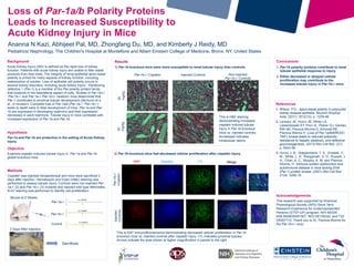
2) Par-1b knockout mice had decreased cellular proliferation after cisplatin injury.
Conclusions
1. Par-1b polarity proteins contribute to renal
tubular epithelial response to injury.
2. Either decreased or delayed cellular
proliferation may contribute to the
increased tubular injury in Par-1b-/- mice.
Par-1b-/- Cisplatin Injected Controls Non-injected
Par-1b-/- Controls
20
mg/kg
30
mg/kg
This is H&E staining
demonstrating increased
cisplatin induced tubular
injury in Par-1b knockout
mice vs. injected controls.
Arrowhead indicates
intratubular debris.
Mouse at 5 Weeks
3 Days After Injection
Sacrificed
Par-1a-/-
Par-1b-/-
Control
Methods
Cisplatin was injected intraperitoneal and mice were sacrificed 3
days after injection. Hematoxylin and Eosin (H&E) staining was
performed to assess tubular injury. Controls were non-injected Par-
1a-/- (2) and Par-1b-/- (3) mutants and injected wild type littermates.
Ki-67 staining was performed to identify cell proliferation.
Ki67 Hoechst LTL Merge
Par-1b-/-
Cisplatin
Injected
Controls
This is Ki67 immunofluorescence demonstrating decreased cellular proliferation in Par-1b
knockout mice vs. injected controls after cisplatin injury. LTL indicates proximal tubules.
Arrows indicate the area shown at higher magnification in panels to the right.