International Journal of Cardiovascular Diseases & Diagnosis
Dysregulation of ferroportin 1 interferes with spleen organogenesis in polycythaemia mice
1. 4871
Introduction
As a cofactor mediating oxidation-reduction chemistry (for a
review, see Aisen et al., 2001), iron governs a plethora of
pathways involved in cellular metabolism. For example, iron
is essential for hemoglobin synthesis, which comprises the
majority of daily iron utilization in humans. Organismal iron
levels must be tightly controlled, because both iron deficiency
and overload lead to severe consequences ranging from anemia
to iron-induced tissue damage affecting multiple organs (for a
review, see Andrews, 2000). Mammalian iron homeostasis is
achieved via the organismal interplay of multiple proteins that
are regulated by both transcriptional and post-transcriptional
mechanisms (for a review, see Hentze et al., 2004). In the
duodenum, the pivotal site of organismal iron uptake,
ferroportin 1 (Fpn1; also known as MTP1, IREG1, SLC11A3,
SLC40A1) functions as the principal basolateral iron
transporter in enterocytes (Abboud and Haile, 2000; Donovan
et al., 2000; McKie et al., 2000). Furthermore, Fpn1 expression
has been detected at other sites involved in iron homeostasis,
including hepatocytes, reticuloendothelial macrophages of the
liver and spleen, and placental syncytiotrophoblast cells (for a
review, see McKie and Barlow, 2004).
We have recently identified a 58-base pair microdeletion in
the promoter region of the Fpn1 locus in radiation-induced
polycythaemia (Pcm) mice (Mok et al., 2004). This deletion,
located four nucleotides upstream of the TATA box, conferred
aberrant transcription initiation at the Fpn1 locus, which
resulted in the absence of the iron-responsive element (IRE) in
the 5′ untranslated region (UTR) of the vast majority of hepatic
Fpn1 transcripts in Pcm homozygotes. Consistent with a
critical in vivo role for the IRE in translational regulation of
the Fpn1 mRNA in response to cellular iron levels, Pcm mutant
mice displayed significant elevations in Fpn1 protein levels in
liver and duodenum, as well as organismal iron overload during
early postnatal development. Strikingly, an erythropoietin
(Epo)-dependent polycythemia was evident at 7 weeks of age
in Pcm heterozygotes. Abrogation of the iron accumulation and
polycythemia in adult Pcm mutant animals implicated hepcidin
(Hamp), the hormonal regulator of iron homeostasis, in a
superimposed regulatory mechanism on Fpn1 protein levels
(Mok et al., 2004). Unexpectedly, Pcm mutant mice
demonstrated a profound iron deficiency at birth, with a severe
hypochromic, microcytic anemia in homozygotes. In the
present study, we have identified dysregulated expression of
placental Fpn1 during late gestation as the basis for neonatal
iron deficiency in Pcm mutants. Furthermore, we have
uncovered evidence for tissue specificity of developmental
Fpn1 dysregulation, which associates with apoptotic cell death
and semidominant defects in Pcm spleen organogenesis.
Surprisingly little is known about the development of the
Regulatory interferences at the iron transporter
ferroportin 1 (Fpn1) cause transient defects in iron
homeostasis and erythropoiesis in polycythaemia (Pcm)
mutant mice. The present study identified decreased Fpn1
expression in placental syncytiotrophoblast cells at late
gestation as the mechanism of neonatal iron deficiency
in Pcm mutants. Tissue specificity of embryonic Fpn1
dysregulation was evident from concomitant decreases in
Fpn1 mRNA and protein expression in placenta and liver,
as opposed to upregulation of Fpn1 protein despite
decreased transcript levels in spleen, implicating post-
transcriptional regulation of Fpn1. Dysregulation of
Fpn1 and decreased iron levels in Pcm mutant spleens
correlated with apoptotic cell death in the stroma,
resulting in a semidominant spleen regression. At 7 weeks
of age, a transient increase in spleen size in Pcm
heterozygotes reflected a transient erythropoietin-mediated
polycythemia. Structurally, Pcm mutant spleens displayed
a severe defect in red pulp formation, including disruption
of the sinusoidal endothelium, as well as discrete defects in
white pulp organization during postnatal development.
Reduced functional competence of the Pcm mutant spleen
was manifested by an impaired response to chemically
induced hemolytic anemia. Thus, aberrant Fpn1 regulation
and iron homeostasis interferes with development of the
spleen stroma during embryogenesis, resulting in a novel
defect in spleen architecture postnatally.
Key words: Ferroportin 1, Spleen, Iron homeostasis, Polycythaemia
mutant, Apoptosis
Summary
Dysregulation of ferroportin 1 interferes with spleen organogenesis
in polycythaemia mice
Henry Mok1, Miriam Mendoza1, Josef T. Prchal2, Péter Balogh3 and Armin Schumacher1,*
1Department of Molecular and Human Genetics, Baylor College of Medicine, Houston, TX 77030, USA
2Division of Hematology, Baylor College of Medicine and Michael DeBakey VAH Medical Center, Houston, TX 77030, USA
3Department of Immunology and Biotechnology, University of Pécs, Pécs, Hungary
*Author for correspondence (e-mail: armins@bcm.tmc.edu)
Accepted 6 July 2004
Development 131, 4871-4881
Published by The Company of Biologists 2004
doi:10.1242/dev.01342
Research article Development and disease
2. 4872
spleen, which is derived from the coelomic epithelium and
mesenchyme of the dorsal mesogastrium (Green, 1967). The
spleen subserves important organismal functions, which can be
attributed to distinct anatomical regions within the organ. It
represents the largest single lymphoid organ and performs a
critical immunological role reflected in the highly organized
arrangement of lymphoid and accessory cells within the white
pulp (for a review, see Fu and Chaplin, 1999). The lymphocytic
composition of the white pulp is different from the red pulp,
which also hosts a sizeable plasma cell population (Nolte et al.,
2000; Garcia De Vinuesa et al., 1999). Whereas the white pulp
is primarily engaged in mounting adaptive immune responses,
the red pulp, composed of a stromal reticular network of
endothelial sinusoids, macrophages, erythroid and accessory
cells, performs a digestive function, clearing damaged and
senescent red blood cells from the circulation. This composite
function of the spleen is a direct reflection of embryonic spleen
development, and results from colonization of a stromal
infrastructure, consisting of intrinsic mesenchyme, by extrinsic
hematopoietic cells at approximately embryonic (E) day 15 of
murine embryogenesis (Sasaki and Matsumura, 1988).
Aside from the relative dearth of well-characterized splenic
markers, insight into the mechanisms regulating spleen
development has been hampered by a limited number of mutant
alleles. Over the past decades, several murine models of
aberrant spleen development have been characterized,
including dominant hemimelia (Searle, 1959; Green, 1967),
and null mutants for transcription factors Hox11 (Roberts et
al., 1994; Dear et al., 1995; Koehler et al., 2000; Kanzler and
Dear, 2001), Wt1 (Herzer et al., 1999), Bapx1 (Lettice et al.,
1999; Tribioli and Lufkin, 1999; Akazawa et al., 2000), and
capsulin (Lu et al., 2000). Following specification of the spleen
primordium around E11.5 (Dear et al., 1995), these mutants
display progressive spleen regression, resulting in its complete
absence as early as E13.5, but no later than E15.5. Therefore,
based on the early regression of the spleen, these models are
ill-suited to address hematopoietic cell colonization and
potential cellular interactions between intrinsic and extrinsic
cell populations required for structural and functional
competence of the spleen. Here, we report a novel defect in
late embryonic spleen development in the context of aberrant
fetal iron homeostasis in Pcm mutant mice, which manifests as
a severe disruption of the red pulp sinusoidal endothelium in
the postnatal spleen.
Materials and methods
Mice and genotyping
Pcm mice originated from radiation mutagenesis of
(101/HeH×C3H/HeH) F1 hybrids (Cattanach, 1995), and were
crossed onto the A/J inbred background to generate partial congenics
(Mok et al., 2004). In the present study, analyses were performed on
animals from N5 and subsequent backcross generations. Age-matched
embryos were generated by timed matings; noon of the day of vaginal
plug detection was considered E0.5. PCR-based genotyping was
performed using primers flanking the Pcm deletion, as described
previously (Mok et al., 2004). All animal experiments in this study
were approved by the Institutional Animal Care and Use Committee
of Baylor College of Medicine.
Western blot analysis
Protein lysates from embryonic spleen, placenta and liver were
prepared and quantified as described previously (Mok et al., 2004).
Five µg of unboiled protein lysates from liver and placenta and 2.5
µg of unboiled spleen lysate were separated by electrophoresis on 8%
SDS polyacrylamide gels and transferred to PVDF membrane
(BioRad). Blocking was achieved by incubation in TRIS-buffered
saline containing 5% BSA and 0.1% Tween 20. Membranes were
incubated overnight at 4°C using primary antibodies against mouse
Fpn1 (kindly provided by D. Haile) at 1:2000 and mouse actin (Santa
Cruz Biotechnology) at 1:5000. Following washing, membranes were
incubated with horseradish peroxidase-conjugated secondary
antibodies (1:5000) against rabbit (for Fpn1) or goat (for actin), and
signals were detected using luminol reagent (Santa Cruz
Biotechnology).
mRNA analyses
Semi-quantitative RT-PCR for Hamp expression on E16.5 liver
mRNA, and 5′ rapid amplification of cDNA ends (5′RACE) from
E16.5 placenta and liver as well as E15.5 spleen were performed as
described previously (Mok et al., 2004). Fpn1 mRNA expression was
quantified via real-time RT-PCR on total RNA samples isolated from
E16.5 placental and liver as well as E15.5 spleen. Reactions and signal
detection were performed on an ABI Prism 7000 Sequence Detection
System (Applied Biosystems). Commercially available Fpn1 primers
and probe were employed (Mm00489837_m1, Applied Biosystems).
An 18S rRNA assay was performed using primers 5′-TCGAG-
GCCCTGTAATTGGAA-3′ (forward), 5′-CCCTCCAATGGATCC-
TCGTT-3′ (reverse), and TaqMan MGB probe 5′-AGTCCACT-
TTAAATCCTT-3′ labeled with VIC (Applied Biosystems). Relative
amounts of mRNA were expressed as a ratio to 18S rRNA levels, and
normalized to an arbitrary wild-type (WT) ratio of 1.
Histology and immunohistochemistry
For histology and immunohistochemistry for Fpn1, F4/80 and Wt1,
spleen, placenta and liver were fixed in Bouin’s reagent (postnatal
stages) or 4% PFA/PBS (embryonic stages), dehydrated in a graded
series of ethanol, embedded in paraffin and sectioned at 5-10 µm.
Prussian blue staining for iron was performed using the Accustain iron
staining kit, according to the manufacturer (Sigma). For Fpn1 and
F4/80 antigen retrieval, sections were treated with 3% H2O2 in
methanol, whereas for Wt1, sections were boiled in 0.01 M citric acid,
pH 6.0. Blocking was achieved with goat (Fpn1 and Wt1) or rabbit
serum (F4/80). Primary antibody incubations were performed
overnight at 4°C using antibodies against Fpn1 at 1:100, F4/80 (clone
CI:A3-1, Serotec) at 1:100, or Wt1 (C-19, Santa Cruz Biotechnology)
at 1:1000 dilution. Secondary antibodies were peroxidase-conjugated
with the Vectastain Elite ABC Kit (Vector Laboratories), followed
by signal detection with Vector NovaRED substrate (Vector
Laboratories). Immunohistochemistry for B220, CD3, Ter119,
MAdCAM-1, IBL-7/1, IBL-9/2 and IBL-7/22 was performed on
frozen sections of 12-week-old spleens. Cryostat sections were
stained with rat hybridoma supernatants containing IBL-7/1, IBL-9/2
and IBL-7/22 antibodies followed by biotin-amplified alkaline
phosphatase detection as described (Balázs et al., 2001). Briefly, the
sections were fixed in chilled acetone for 10 minutes, dried, and were
encircled with water-repellent wax pen. Following rehydration with
PBS containing 10% BSA for 20 minutes and removal of excess
buffer, the sections were incubated with undiluted hybridoma
supernatants against lymphocyte subsets and endothelial markers for
45 minutes. The Ly-76 erythroid antigen was detected by the
monoclonal antibody Ter119 (BD Pharmingen) at 1 µg/ml
concentration in PBS. The reaction was developed with biotinylated
mouse anti-rat kappa chain reagent (clone MRK-1, BD Pharmingen)
at 1 µg/ml, and ExtraAvidine-alkaline phosphatase conjugate (Sigma-
Aldrich) using NBT/BCIP in the presence of 1 mg/ml levamisole.
Sections were counterstained with methylene green. Rat monoclonal
antibodies used: anti-B220 (clone RA3-6B2); anti-CD3 (clone KT-3);
anti-MAdCAM-1 (clone MECA-367); IBL-7/1, IBL-7/22 and IBL-
Development 131 (19) Research article
3. 4873Defects in spleen development in Pcm miceDevelopment and disease
9/2 monoclonal antibodies were produced in the Department of
Immunology and Biotechnology, University of Pécs, Pécs, Hungary
(Balázs et al., 2001).
Determination of iron levels in embryonic tissues
The non-heme iron content of individual E15.5 liver samples was
determined as described previously (Mok et al., 2004). Because of the
small amount of tissue, E15.5 spleens were pooled by genotype, dried,
and quantified similar to liver samples.
TUNEL analysis
Embryonic spleen and liver were dissected out from E15.5 and E16.5
embryos, fixed in 4% PFA/PBS for 2 hours at 4°C, dehydrated in a
graded series of ethanol, embedded in paraffin and sectioned at 5 µm.
Sections were processed for detection of TUNEL-positive cells using
the In Situ Cell Death Detection Kit (Roche), and mounted with the
Vectashield Hard Set mounting medium, containing DAPI (Vector
Laboratories). Images were acquired via fluorescence microscopy for
fluorescein (TUNEL) and DAPI signals using separate channels, and
merged using Adobe Photoshop version 6.0.
Phenylhydrazine treatment of mice
To induce hemolytic anemia in 3- and 7-week-old wild-type and Pcm
mutant mice, phenylhydrazine hydrochloride dissolved in sterile
saline was administered intraperitoneally at a dose of 60 mg/kg body
weight twice at a 24-hour interval on day 0 and day 1. Mice were
euthanized on day 4, and spleens were dissected out and weighed.
Baseline spleen values were obtained by comparison of spleen
weights from untreated wild-type and heterozygous mutant mice at 3
and 7 weeks of age.
Statistical analyses
All data are reported as the mean±s.d. All comparisons were
performed versus wild-type cohorts, and analyzed for statistically
significant differences using the Student’s unpaired t-test.
Results
Decreased placental and liver Fpn1 protein
expression in Pcm mutant embryos
We have previously demonstrated that Pcm mutant animals are
iron deficient at birth (Mok et al., 2004), consistent with the
putative role of placental Fpn1 in maternal-to-fetal iron
transport (Donovan et al., 2000; McKie et al., 2000). In support
of this hypothesis, we observed a marked decrease in placental
Fpn1 expression in Pcm mutants at E16.5 by western blot
analysis (Fig. 1A). Likewise, Pcm mutant liver showed a
similar decrease in Fpn1 protein levels (Fig. 1B). Fpn1
expression has been demonstrated at the basolateral surface of
syncytiotrophoblast cells in human placenta, where it is
Fig. 1. Decreased Fpn1 protein expression in Pcm mutant placenta
and embryonic liver is consistent with fetal iron deficiency, and
contrasts with increased Fpn1 expression in embryonic spleen. At
E16.5, placenta (A) and liver (B) show a graded decrease in Fpn1
protein expression on western blot. Approximate molecular mass:
Fpn1, 68 kDa; actin, 41 kDa. (C) Western blot analysis reveals
increased Fpn1 protein expression in E15.5 spleen.
(D) Immunohistochemistry demonstrates a significant reduction of
labyrinth cell expression of Fpn1 in E16.5 placenta from Pcm
homozygotes. Original magnification 400×.
(E) Immunohistochemistry reveals a widespread increase in Fpn1
expression in E15.5 Pcm mutant spleens. Original magnification
400×. (F) Analysis of non-heme iron demonstrates significant
reduction of iron content in E15.5 Pcm livers, more severe in
homozygotes. *, P<0.0001. (G) Semiquantitative RT-PCR of E16.5
liver indicates lower Hamp mRNA expression in Pcm mutants. Band
sizes: Hamp, 171 bp; β-actin, 250 bp.
4. 4874
implicated in iron transport to the embryonic circulation
(Donovan et al., 2000). Indeed, by immunohistochemistry,
high levels of Fpn1 expression were detected in
syncytiotrophoblast cells in the labyrinth cell layer in wild-type
mouse placenta (Fig. 1D). In contrast, Fpn1 expression was
decreased to near background levels in Pcm homozygous
placenta at E16.5 (Fig. 1D), consistent with western analysis
(Fig. 1A). Furthermore, the architecture of the labyrinth cell
layer appeared to be preserved in mutant placenta (data not
shown).
Iron balance in Pcm mutant embryos
Given the maximal maternal iron transport to the fetus during
late gestation (for a review, see Srai et al., 2002), decreased
placental expression of Fpn1 should result in iron deficiency
in Pcm mutant embryos. Indeed, a significant reduction of
hepatic iron content in Pcm mutant embryos was observed,
more severe in homozygotes (Fig. 1F). Corollary with the
decreased hepatic iron content (Fig. 1F), Pcm mutant spleens
exhibited a graded decrease in spleen iron levels at E15.5.
Because of the small size of the spleens, only multiple
samples pooled by genotype yielded readings above
background level (n=4 +/+, 13.3 ng iron/spleen; n=5, Pcm/+,
9.9 ng iron/spleen; n=5, Pcm/Pcm, 4.9 ng iron/spleen).
Nonetheless, it appears likely that, despite pooling of
samples, the colorimetric assay may underestimate the
degree of iron deficiency in Pcm mutant spleens.
Furthermore, at all stages analyzed, i.e. E15.5 until birth,
homozygous mutant embryos exhibited marked pallor (data
not shown). Thus, decreased placental expression of Fpn1
probably represents the major contributing factor to neonatal
iron deficiency and anemia in Pcm mutant mice (Mok et al.,
2004).
Interestingly, Hamp mRNA levels were significantly
reduced in E16.5 Pcm mutant liver by semi-quantitative RT-
PCR (Fig. 1G), indicating that Hamp, the principal negative
hormonal regulator of iron balance (Park et al., 2001; Pigeon
et al., 2001) (for a review, see Ganz, 2003), is also responsive
to iron deficiency during embryogenesis.
Increased Fpn1 protein expression in the spleen of
Pcm mutant embryos
In striking contrast to placenta and liver, Fpn1 protein levels
were higher in embryonic spleen from Pcm mutant embryos
(Fig. 1C). Importantly, immunohistochemistry detected high
levels of Fpn1 expression throughout the spleen of Pcm
homozygous embryos, consistent with upregulation of Fpn1
expression in stromal cells (Fig. 1E). Thus, the present study
uncovered evidence for tissue-specific regulation of embryonic
Fpn1 expression by virtue of differential protein levels in Pcm
mutant placenta and liver compared with spleen.
Decreased Fpn1 mRNA levels in placenta, liver and
spleen
To determine whether transcript abundance caused the
observed changes in embryonic Fpn1 protein levels, we
performed real-time RT-PCR analysis for Fpn1 mRNA
expression. Fpn1 mRNA levels were statistically significantly
reduced in both placenta and liver from Pcm mutants (Fig.
2A,B), consistent with the decreased Fpn1 protein expression
(Fig. 1A,B). Surprisingly, Fpn1 mRNA levels were also
significantly reduced in Pcm homozygous mutant spleen (Fig.
2C), despite increased Fpn1 protein levels (Fig. 1C,E). This
discordance between Fpn1 mRNA and protein expression in
the spleen indicated that the upregulation of Fpn1 resulted from
a post-transcriptional mechanism.
Aberrant transcription initiation at the Fpn1 locus leads to
disruption of the IRE-mediated post-transcriptional regulation
during postnatal development in Pcm mutant mice (Mok et al.,
2004). Similar to postnatal liver, analysis of Fpn1 transcripts
by 5′RACE from wild-type E15.5 spleen and E16.5 placenta
showed a single, predominant band, reflecting homogeneous
transcription start sites (Fig. 2D,E). However, analysis of
Pcm/Pcm placenta and embryonic spleen revealed a significant
Development 131 (19) Research article
Fig. 2. Embryonic Fpn1 mRNA expression is decreased in Pcm
mutant mice. (A) Real-time RT-PCR analysis of E15.5 spleen
shows reduction of Fpn1 mRNA in Pcm homozygous mutants.
**, P<0.01. Decreased mRNA levels in placenta (B) and liver
(C) in E16.5 Pcm mutants. *, P<0.05; **, P<0.01;
***, P<0.001. 5′RACE PCR of E15.5 spleen (D) and E16.5
placenta mRNA (E) demonstrates aberrant transcripts in Pcm
homozygous mutants. Wild-type band, 630 bp.
5. 4875Defects in spleen development in Pcm miceDevelopment and disease
reduction in the intensity of this band, as well as multiple,
additional transcripts (Fig. 2D,E). To test whether transcript
identity could explain the divergent Fpn1 protein expression
levels in placenta and spleen, we performed DNA sequence
analysis of the 5′RACE clones. Sequence data from wild-type
transcripts from both the placenta and spleen reiterated
published transcription start sites (Liu et al., 2002; Mok et al.,
2004). In contrast, cDNAs recovered from Pcm mutant tissues
demonstrated significant transcript heterogeneity and were
aberrant with respect to transcription start site and mRNA
splicing. Fpn1 transcripts devoid of the IRE, which were
predominant in homozygous mutant tissue postnatally (Mok et
al., 2004), comprised less than half of sequenced clones from
mutant placenta and embryonic spleen (data not shown).
Importantly, despite transcript heterogeneity, no evidence for
aberrant Fpn1 protein isoforms was observed by western
analysis (data not shown). Therefore, neither Fpn1 transcript
abundance nor sequence appeared sufficient to explain the
increased Fpn1 protein levels in Pcm mutant embryonic spleen,
indicating an unknown, post-transcriptional regulatory
mechanism.
Semidominant defect in Pcm mutant spleens during
late embryogenesis
To ascertain whether aberrant Fpn1 protein expression in E15.5
spleen associated with an organ phenotype, we conducted a
time-course analysis of spleen development. At E15.5, the
spleen appeared to be consistent across all genotypes with
regard to organ size and gross morphology (Fig. 3A). However,
beginning at E16.5 (data not shown) and increasingly evident
at E17.5, Pcm mutant spleens were reduced in size relative
to wild type, more severe in homozygotes (Fig. 3B). It is
noteworthy that the spleen size in homozygotes effectively
regressed, rather than merely stalled, as evident from
comparison of spleen size at E17.5 and E15.5 (Fig. 3A,B). At
birth, a completely penetrant, semidominant defect in spleen
size was observed in Pcm mutant pups (Fig. 3C). Thus, unlike
previous models of defective splenogenesis (Searle, 1959;
Roberts et al., 1994; Herzer et al., 1999; Lettice et al., 1999;
Lu et al., 2000), we observed grossly intact spleens in Pcm
mutants at E15.5 (Fig. 3A), indicating that Pcm represents a
novel murine model for disrupted splenogenesis.
Increased apoptosis in spleens from Pcm mutant
embryos
The regression of spleen size in Pcm homozygous mutants
during late gestation (Fig. 3A,B) implicated cell death by
apoptosis as the likely mechanism. Indeed, a significant
increase in TUNEL-positive foci was detected in E15.5 and
E16.5 homozygous mutant spleens (Fig. 4A,B). In addition,
heterozygous mutants exhibited moderately increased TUNEL
foci relative to wild-type spleens, which demonstrated minimal
baseline apoptosis. Importantly, significantly increased
apoptotic cell death appeared to be restricted to Pcm mutant
spleens, as the liver displayed similar levels of TUNEL-
positive foci across all genotypes at these stages (data not
shown).
At E15, the spleen constitutes a pre-hematopoietic organ,
consisting of predominantly mesenchymal cells and few,
scattered mononuclear cells (Sasaki and Matsumura, 1988).
Using a monoclonal antibody against a macrophage/monocyte-
associated surface antigen, F4/80 (Morris et al., 1991), we
observed no difference in the number of macrophage/monocyte
lineage cells among the genotypes in E15.5 spleens (Fig. 4C).
Strikingly, F4/80-positive cells appeared to be less frequent
than TUNEL-positive foci in homozygous mutant spleens (Fig.
4A,B). Furthermore, macrophage/monocyte lineage cells were
observed throughout all later stages of embryonic spleen
Fig. 3. Pcm mutant mice show a semidominant defect in spleen
development during late embryogenesis. (A) Spleen development
and size appear consistent across all genotypes at E15.5. Original
magnification 40×. (B) By E17.5, significant reduction in spleen size
is observed in Pcm mutants, more severe in homozygotes. Compare
with A and note that spleen size in homozygotes regresses. Original
magnification 40×. At P0 (C) and 7 weeks (D), spleens (arrowheads)
of homozygous mutants maintain consistent decrease in size,
whereas heterozygotes undergo a significant increase in size from P0
to 7 weeks. Original magnification (C) 16× and (D) 6×. H&E
staining of spleen sections at P0 (E) and 7 weeks (F) reveals red and
white pulp compartmentalization of wild-type and mutant spleens;
arrowhead indicates white pulp follicle. Original magnification
(E) 200× and (F) 50×.
6. 4876
development at a similar frequency and distribution across all
genotypes (data not shown).
The tumor suppressor Wt1 plays an essential role in organ
development, including that of the kidney, gonad and
mesothelial structures (Kreidberg et al., 1993). In addition, Wt1
regulates spleen development, as Wt1 null mice exhibit
complete regression of the spleen by E15.5 (Herzer et al.,
1999). Using Wt1 as a marker for splenic stromal cells,
analysis at E15.5 demonstrated a generalized pattern of Wt1
expression encompassing the majority of cells present at this
stage in both wild-type and Pcm mutant spleens (Fig. 4D). At
birth, similar to other putative stromal cell markers in the
spleen, such as Hox11 (Dear et al., 1995; Kanzler and Dear,
2001) and Bapx1 (Akazawa et al., 2000), Wt1 expression levels
appeared significantly reduced and were primarily restricted to
the capsule and subcapsular region in wild type (data not
shown). Because of the low levels of Wt1 expression and
abnormal spleen structure in postnatal day 0 (P0) Pcm
homozygotes (Fig. 3C), Wt1 analysis at this stage was not
informative (data not shown). Importantly, at E15.5, the pattern
of Wt1 expression (Fig. 4D) correlated with the Fpn1
expression pattern in Pcm mutant spleens (Fig. 1E).
Taken together, the regression of Pcm spleens could not be
reconciled with primarily macrophage cell death during spleen
development. Rather, increased Fpn1 protein expression in
stromal cells, in the context of organismal iron deficiency,
associates with apoptotic death of stromal cells, resulting in the
semidominant defect in spleen size at birth.
Postnatal changes in Pcm mutant spleens
Homozygous Pcm mutants maintained a decreased spleen
size, as observed at 7 weeks of age (Fig. 3D). Conversely,
heterozygous spleens gradually increased in size from birth,
reaching wild-type size at 7 weeks of age (Fig. 3D).
Histologically, at P0, Pcm heterozygous spleens appeared
comparable to wild type, whereas homozygous spleens
displayed discrete alterations in cellular composition, such as
a decreased prevalence of basophilic cells (Fig. 3E). Postnatal
organization and development of the white pulp appeared to
occur to a significant extent in both heterozygous and
homozygous mutant spleens, as distinct white pulp follicles
were clearly identifiable at 7 weeks (Fig. 3F). In addition,
heterozygous spleens were notable for increased eosinophilic
cells within the red pulp as well as the marginal sinus
surrounding the white pulp follicles. Seven-week-old
homozygous spleens exhibited an increased white pulp to red
pulp ratio relative to wild type (Fig. 3F). Given the increased
rate of red blood cell production characteristic of Pcm
heterozygotes at 7 weeks of age (Mok et al., 2004), the
histological changes observed in the red pulp were consistent
with increased erythropoiesis in Pcm heterozygous spleens.
Impaired red pulp function in Pcm heterozygous
spleen
Given the regression in spleen size during late gestation (Fig.
3C,D), Pcm homozygotes are likely to become functionally
asplenic. In contrast, Pcm heterozygotes retained a significant
amount of spleen stroma, and exhibited a significant increase
in relative spleen size from during postnatal development (Fig.
3C,D). Consistent with its reduced size at P0 (Fig. 3C),
heterozygous mutant spleens demonstrated a significantly
lower weight at 3 weeks of age as compared with wild type
(Fig. 5A). However, by 7 weeks of age, and correlating with
the gross appearance (Fig. 3D), similar spleen weights were
measured in wild-type and heterozygous mutant mice (Fig.
5A). This increase in relative spleen size was transient in
nature, as by 12 weeks of age, heterozygous spleen weights
were again statistically significantly reduced as compared with
wild type (Fig. 5A).
Recently, we showed that Pcm heterozygotes displayed a
transient, Epo-stimulated polycythemia at 7 weeks of age (Mok
et al., 2004). Concordant with the normalization of both Epo
expression and hematocrit in 12-week old Pcm mutants (Mok
Development 131 (19) Research article
Fig. 4. Increased apoptosis detected in Pcm mutant spleens. TUNEL
analysis at E15.5 (A) and E16.5 (B) indicates a significant increase in
TUNEL-positive foci in Pcm mutant spleens, higher in homozygotes.
Images represent merged composites of TUNEL (fluorescein) and
DAPI-counterstained sections, original magnification 200×.
(C) Immunohistochemistry for F4/80 at E15.5 reveals similar
distribution of positive cells within spleens across all genotypes.
Original magnification, 400×. (D) Immunohistochemistry for Wt1 at
E15.5 demonstrates widespread stromal cell staining within the
spleen. Original magnification, 400×.
7. 4877Defects in spleen development in Pcm miceDevelopment and disease
et al., 2004), the splenic weight declined at this age (Fig. 5A).
Furthermore, the prevalence of eosinophilic cells within the red
pulp and marginal sinus, which was prominent at 7 weeks of
age, restored to wild-type pattern (data not shown). Therefore,
in agreement with the significant function of the postnatal
spleen as an erythropoietic organ in the mouse (Brodsky et al.,
1966; Bozzini et al., 1970), the transient nature of the increase
in spleen size and histological changes in Pcm heterozygotes
probably reflects augmented splenic erythropoiesis because of
Epo stimulation.
To quantify the functional erythropoietic capacity of Pcm
heterozygous spleens, we performed phenylhydrazine
treatment of mice to induce hemolytic anemia (Itano et al.,
1975). Strikingly, Pcm heterozygous spleens were notable for
a significantly reduced hyperplastic response at both 3 and 7
weeks of age (Fig. 5B,C), indicating a decreased functional
capacity to foster erythroid progenitor and precursor
proliferation. No increase in spleen size was evident in
phenylhydrazine-treated Pcm homozygotes at 3 weeks of age,
consistent with functional asplenia (data not shown).
Additional evidence for the functional capacity of Pcm
heterozygous spleens was gleaned from the analysis of iron
distribution in the context of erythrophagocytosis. At P0, no
iron staining was observed within the Pcm heterozygous
spleen, reflecting the perinatal iron deficiency in Pcm mutants
(Fig. 5D). However, by 12 weeks of age, Pcm heterozygotes
displayed a significant, punctate pattern of iron accumulation
confined to the red pulp region (Fig. 5E). Given the
polycythemia at 7 weeks of age and hematocrit normalization
at 12 weeks of age in Pcm heterozygotes, this staining pattern
probably reflects erythrophagocytosis followed by iron
scavenging within the reticuloendothelial macrophages of the
red pulp.
Taken together, the Pcm heterozygous mutant spleens appear
to retain red pulp function with regard to erythrophagocytosis,
as well as extramedullary hematopoiesis in response to
stimulation, albeit at a significantly reduced level relative to
wild type.
Characterization of hematopoietic cell lineages in
postnatal Pcm mutant spleens
Ter119 represents a marker for late erythroid lineage cells
(Kina et al., 2000). Within the red pulp, Ter119-positive
erythroid cells were observed at a similar density and
distribution in wild-type and heterozygous spleens at 12 weeks
of age (Fig. 6A). Homozygotes displayed a moderately reduced
density of Ter119-positive cells in red pulp regions (Fig. 6A).
The mature white pulp follicle consists of a T-cell rich,
periarteriolar lymphoid sheath (PALS) region, which is
surrounded by primary follicles comprised of B cells, the
predominant lymphoid cell population of the spleen (Veerman
and van Ewijk, 1975) (for a review, see Fu and Chaplin, 1999).
Pcm heterozygous spleens displayed a similar appearance of
B- and T-cell regions, as demonstrated by B220 and CD3
expression, respectively (Fig. 6B,C). Interestingly, Pcm
homozygotes exhibited an increased ratio of B to T cells. In
addition, the central arteriole, normally positioned within a
central location of the T-cell PALS region, was located more
peripherally in heterozygotes, and appeared within the B-cell
region in Pcm homozygotes (Fig. 6B,C). Thus, although the
overall follicular structure of the white pulp appeared largely
intact, lymphocyte composition and positioning of the central
arteriole were abnormal in Pcm mutant spleens, more severe
in homozygotes.
Red pulp sinusoid abnormalities in Pcm mutant
spleens
The marginal sinus forms a discontinuous endothelial network
surrounding the white pulp, which, in conjunction with the
marginal zone, represents the site of immunological
interactions and cellular extravasation from peripheral blood
into the white pulp (Schmidt et al., 1993) (for a review, see Fu
and Chaplin, 1999). The expression of cellular adhesion
molecule MAdCAM-1 marks endothelial cells of the marginal
Fig. 5. Pcm heterozygous mutant spleens exhibit functional
competence for red pulp hyperplasia and red blood cell clearance.
(A) Spleen weights demonstrate a transient increase in Pcm
heterozygotes at 7 weeks of age. *, P<0.01. Phenylhydrazine
treatment of 3-week (B) and 7-week-old mice (C) demonstrates
reduced splenic hyperplasia in Pcm heterozygotes. **, P<0.0001.
(D) Prussian blue staining of P0 spleen reveals no iron accumulation
in wild-type or Pcm heterozygous mutants. Original magnification,
200×. (E) At 12 weeks of age, Pcm heterozygotes display significant
red pulp iron accumulation. Original magnification, 100×.
8. 4878
sinus in the spleen (Kraal et al., 1995). We observed
MAdCAM-1 expression surrounding white pulp follicles in
Pcm mutant spleens at 12 weeks of age, delineating the
presence of the marginal sinus (Fig. 6D). IBL-7/1, which also
marks the marginal sinus (Balazs et al., 1999), similarly
encompassed the white pulp follicles (Fig. 6E). Thus, the
marginal sinus appears to be largely intact in Pcm mutant
spleens. Immunoreactivity to IBL-7/1 has also been
demonstrated in a heterogeneous pattern within the red pulp
sinuses (Balazs et al., 2001), as observed in wild-type spleens
(Fig. 6E). Strikingly, this red pulp staining pattern was
markedly reduced in both heterozygous and homozygous
mutant spleens, indicating an abnormality of the red pulp
sinusoids. The IBL-9/2 antibody identifies a complementary
subset of red pulp sinusoidal endothelium (Balazs et al., 2001).
Likewise, the orderly distribution of IBL-9/2 staining
endothelium was severely disrupted in Pcm mutant spleens
(Fig. 6F). The IBL-7/22 antibody recognizes sinusoidal
components in a pan-endothelial pattern, as well as reticular
fibroblasts of the PALS and red pulp (Balazs et al., 1999).
Heterozygous spleens displayed a decrease in IBL-7/22
reactivity (Fig. 6G), consistent with the reduced IBL-7/1- and
IBL-9/2-positive red pulp sinusoid endothelial subpopulations
(Fig. 6E,F). Interestingly, Pcm homozygotes displayed a higher
level of IBL-7/22 reactivity than heterozygotes, but were
disorganized relative to wild type (Fig. 6G). Given the
reduction in IBL/7-1 and IBL-9/2 reactivity in Pcm
homozygotes (Fig. 6E,F), this probably reflects an increased
prevalence of reticular fibroblasts within the sinusoidal
meshwork of the red pulp.
In summary, significant abnormalities in red pulp sinusoidal
architecture were observed in Pcm mutant spleens, which are
consistent with the reduced functional competence of the red
pulp during Epo-stimulated erythropoiesis in Pcm
heterozygous spleens.
Discussion
The hypermorphic Pcm mutation causes upregulation of Fpn1
expression, resulting in increased organismal iron uptake
during postnatal development (Mok et al., 2004). Because
mutant Fpn1 transcripts lacked an IRE, which inhibits mRNA
translation in response to cellular iron levels (Abboud and
Haile, 2000; McKie et al., 2000; Liu et al., 2002), our data
provided the first in vivo evidence for the importance of
posttranscriptional regulation of Fpn1 expression. Surprisingly,
Pcm mutant animals exhibited a severe iron deficiency at birth,
more severe in homozygotes (Mok et al., 2004). Because fetal
iron and other nutrient requirements are met by placental
transport from the maternal to fetal blood circulation, maternal
Development 131 (19) Research article
Fig. 6. Intact white pulp follicles but aberrant red pulp sinusoidal
architecture in Pcm mutant spleens. Immunohistochemistry of
hematopoietic lineage markers Ter119 (A), B220 (B), and CD3 (C)
within the spleen at 12 weeks of age. Arrowheads denote central
arteriole. (D) MAdCAM-1 immunohistochemistry indicates the
presence of marginal sinuses in Pcm mutant spleens at 12 weeks of
age. Immunohistochemistry of IBL-7/1 (E), IBL-9/2 (F), and IBL-
7/22 (G) demonstrates abnormal arrangement of the
microvasculature endothelial components of the red pulp in Pcm mutant spleens at 12 weeks of age. Original magnification, 200×.
9. 4879Defects in spleen development in Pcm miceDevelopment and disease
iron deficiency exerts profound effects on fetal as well as
postnatal development (Crowe et al., 1995) (for a review, see
McArdle et al., 2003). Interestingly, fetuses exhibit decreased
severity of iron deficiency in comparison to the mother,
indicating the presence of regulatory mechanisms to sustain an
adequate supply of iron to the fetus (Gambling et al., 2001).
Characterization of an Fpn1 mutation in zebrafish identified a
role for this iron transporter in maternal-to-fetal efflux of iron
via the placenta (Donovan et al., 2000). Our data confirm that
decreased placental expression of Fpn1 mediates the fetal iron
deficiency, as Pcm homozygous mutants displayed greatly
diminished syncytiotrophoblast cell expression of Fpn1 during
late gestation (for a model, see Fig. 7). Furthermore, transgenic
overexpression studies implicated Hamp in the upregulation of
placental iron transport (Nicolas et al., 2002). Conversely, the
present results provide evidence that Hamp expression is
responsive to the fetal iron status, as Pcm mutant liver exhibited
decreased Hamp mRNA levels. Thus, the Hamp-Fpn1
regulatory axis appears to govern iron homeostasis during both
embryonic (the present study) and postnatal development (Mok
et al., 2004) via its function in placenta, liver and duodenum.
Placental Fpn1 expression has been hypothesized to be
developmentally regulated in the mouse, with highest levels
during late gestation (McKie et al., 2000). At this critical
developmental phase, placental and liver Fpn1 transcript levels
are significantly reduced in Pcm mutants, correlating with
reduced Fpn1 protein expression. These data strongly suggest
that a transcriptional mechanism governs developmental Fpn1
regulation in these tissues. Given the proximity of the Pcm
microdeletion to a putative TATA box (Mok et al., 2004), it is
conceivable that aberrant transcript levels result from a
reduction of transcriptional efficiency and/or basal promoter
activity. Alternatively, the microdeletion could disrupt putative
transcription factor binding sites. Clearly, further investigation
is warranted into the precise mechanisms by which the Pcm
microdeletion dysregulates Fpn1 transcription.
In striking contrast to placenta and liver, Fpn1 protein levels
were increased in Pcm mutant spleen, despite significantly
decreased Fpn1 mRNA levels. Based on Fpn1 transcript levels
and sequence, it appears unlikely that the increased Fpn1
protein levels result from a transcriptional mechanism.
Furthermore, although decreased Hamp expression is
consistent with increased Fpn1 protein levels in the spleen,
decreased Fpn1 protein expression in the placenta and liver
renders a systemic regulatory effect unlikely. Thus, similar to
postnatal development (Mok et al., 2004), an unknown post-
transcriptional mechanism appears to govern Fpn1 protein
levels during late gestation, which warrants additional studies.
In this context, it would be of significant interest to characterize
further the basis for decreased Fpn1 protein expression in
embryonic liver, which, strikingly, transitions to increased
hepatic Fpn1 protein levels at birth (Mok et al., 2004).
Regardless of the mechanisms of tissue-specific Fpn1
regulation, increased Fpn1-mediated iron efflux in stromal
cells should potentiate cellular iron deficiency in the spleen
resulting from decreased embryonic iron levels. This
pathophysiology correlates with dramatic consequences at the
cellular and organ level, which become manifest as a
semidominant defect in spleen development during late
gestation (for a model, see Fig. 7).
Consistent with its pleiotropic role in cellular metabolism,
iron has long been known to modulate pathways that regulate
proliferation and cell death (for a review, see Le and
Richardson, 2002). For example, iron chelation has been
demonstrated to induce apoptosis (Fukuchi et al., 1994; Haq et
al., 1995) and promote cell-cycle arrest (Lederman et al.,
1984). Similarly, cellular hypoxia represents a potent stimulus
for the apoptotic cascade (for a review, see Brunelle and
Chandel, 2002). The effects of iron chelation mimic cellular
hypoxia (Wang and Semenza, 1993), presumably via iron-
dependent regulators of the hypoxia signaling cascade, such as
a prolyl hydroxylase (Ivan et al., 2001; Jaakkola et al., 2001),
which regulates hypoxia-inducible factor-1α (HIF-1α) (for a
review, see Semenza, 2003). Therefore, it is conceivable that
apoptotic cell death detected in Pcm mutant spleens results
from a synergistic effect of iron deficiency and anemia during
development. Recently, upregulation of the pleomorphic
adenomas gene-like 2 (PLAGL2) protein, a putative zinc-finger
transcription factor, has been demonstrated in response to
hypoxia and iron chelation (Furukawa et al., 2001). In turn,
PLAGL2 induces apoptosis and upregulation of a proapoptotic
factor BNip3 (Mizutani et al., 2002). Interestingly, BNip3 is
itself induced by hypoxia, a unique feature of regulation among
the Bcl-2 family of apoptotic factors (Bruick, 2000). Thus,
given the relative specificity of these factors in response to iron
and hypoxia stimuli, we are currently assessing their potential
involvement in the stromal cell apoptosis in Pcm mutant
spleens. Furthermore, to the best of our knowledge, defects in
spleen development have not been reported in extant mouse
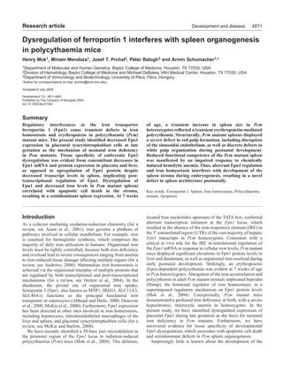
Fig. 7. Model for Fpn1-mediated embryonic iron deficiency and
spleen stromal defects in Pcm mice. Decreased Fpn1 mRNA and
protein expression in placental syncytiotrophoblast cells leads to
decreased maternal-to-fetal iron transport and embryonic iron
deficiency. Although the spleen exhibits decreased Fpn1 mRNA,
increased Fpn1 protein is observed, which should mediate cellular
iron efflux from stromal cells. Under the influence of iron deficiency
and/or hypoxia, spleen stromal cell death translates to decreased
organ size and defects of the red pulp vascular endothelium. The
broken line indicates the uncertain relationship between decreased
Fpn1 expression in the fetal liver and the functional consequences,
for instance with regard to fetal erythropoiesis.
10. 4880
models of iron deficiency and anemia. A detailed analysis of
these mutants would corroborate whether iron deficiency
and/or Fpn1 protein upregulation induce apoptosis in spleen
stromal cells.
The defects in spleen organogenesis in Pcm mutant mice are
distinct from existing genetic mouse models of aberrant spleen
development. Complete asplenia is observed in dominant
hemimelia (Searle, 1959; Green, 1967), Hox11 (Roberts et al.,
1994; Dear et al., 1995; Koehler et al., 2000; Kanzler and Dear,
2001), Wt1 (Herzer et al., 1999), Bapx1 (Lettice et al., 1999;
Tribioli and Lufkin, 1999; Akazawa et al., 2000) and capsulin
mutant mice (Lu et al., 2000). In these mutants, primary
induction of the splenic primordium is followed by complete
involution of the organ before E15.5. Evidence for apoptotic
cell death within the spleen has been observed in Hox11 (Dear
et al., 1995), Wt1 (Herzer et al., 1999) and capsulin mutant
animals (Lu et al., 2000). Because Pcm mutant spleens appear
intact at E15.5, this implicates Fpn1 and iron homeostasis in
the disruption of a distinct, subsequent developmental phase
in spleen organogenesis. Attempts to determine genetic
interaction of Hox11 with Wt1 (Koehler et al., 2000), Bapx1
(Akazawa et al., 2000) and capsulin (Lu et al., 2000) provided
evidence that Wt1 functions downstream of Hox11 (Koehler et
al., 2000). Interestingly, Pcm mutant spleens at E15.5 exhibit
similar Wt1 expression levels and patterns compared with wild
type, suggesting that the defects in Pcm mice are independent
of Hox11 and Wt1 function. As Pcm heterozygotes display a
reduced severity of splenic disruption and homozygotes retain
residual spleen tissue, Pcm mice represent a complementary
resource to the existing mutants that will enable a more
comprehensive understanding of the mechanisms and
pathways of spleen organogenesis throughout development.
In the mouse, the red pulp constitutes a significant
erythropoietic organ during normal development as well as in
response to hypoxia, phlebotomy, or exogenous Epo
administration (Brodsky et al., 1966; Bozzini et al., 1970). Pcm
heterozygotes demonstrate an Epo-dependent polycythemia at
7 weeks of age (Mok et al., 2004). Here, we show that Pcm
heterozygous spleens demonstrate changes consistent with
elevated Epo levels, including a transient increase in spleen
weight and red pulp hyperplasia, which temporally coincide
with the transient polycythemia. In contrast, similar to
genetically asplenic or splenectomized mice, the spleen
rudiment in Pcm homozygotes should not respond to factors
stimulating erythropoiesis (Bozzini et al., 1970; Lozzio, 1972).
Therefore, despite elevated Epo levels, functional asplenia and
severe perinatal iron deficiency probably represent the limiting
factors toward the diminished rate of productive erythropoiesis
during postnatal development in Pcm homozygotes, resulting
in the lower peak hematocrit as compared with heterozygous
mutants.
The stromal defects during organogenesis of Pcm mutant
spleens correlate well with the severe red pulp abnormalities
postnatally, such as aberrant sinusoidal endothelial cell
populations. In turn, these defects lead to decreased functional
competence of Pcm mutant spleens, as reflected by impaired
splenic hyperplasia in response to phenylhydrazine treatment.
Furthermore, discrete abnormalities of the white pulp, as well
as the marginal zone, have also been detected. Thus, it is likely
that interactions between the intrinsic, stromal cell population,
and extrinsic, hematopoietic cell lineages mediate proper
structural and functional organization of the spleen during
postnatal development. Therefore, further characterization of
the patterning and organization of the white pulp in Pcm
mutant spleens should yield additional insight into the
mechanisms of spleen organogenesis in mammals.
We thank David Haile for generously providing the Fpn1 antibody
reagent used in this study; we are grateful to Jaroslav Jelinek and
William Craigen, as well as members of our laboratory, for helpful
discussions and critical reading of the manuscript. We acknowledge
Sonia Pai and Salomon Durrani for excellent technical assistance, and
Gerard Karsenty for generous microscope access. H.M. is supported
by an individual National Research Service Award from the National
Institute of Environmental Health Sciences, NIH; has received NIH
training grant support through the Department of Molecular and
Human Genetics; and is a member of the Medical Scientist Training
Program of Baylor College of Medicine funded by the NIH. P.B. was
supported by the Széchenyi István Postdoctoral Fellowship of the
Hungarian Academy of Sciences and ETT grant No. 592/2003. This
work was supported by a research grant from the NIH to A.S.
References
Abboud, S. and Haile, D. J. (2000). A novel mammalian iron-regulated
protein involved in intracellular iron metabolism. J. Biol. Chem. 275, 19906-
19912.
Aisen, P., Enns, C. and Wessling-Resnick, M. (2001). Chemistry and biology
of eukaryotic iron metabolism. Int. J. Biochem. Cell. Biol. 33, 940-959.
Akazawa, H., Komuro, I., Sugitani, Y., Yazaki, Y., Nagai, R. and Noda, T.
(2000). Targeted disruption of the homeobox transcription factor Bapx1
results in lethal skeletal dysplasia with asplenia and gastroduodenal
malformation. Genes Cells 5, 499-513.
Andrews, N. C. (2000). Iron metabolism: iron deficiency and iron overload.
Annu. Rev. Genomics Hum. Genet. 1, 75-98.
Balazs, M., Grama, L. and Balogh, P. (1999). Detection of phenotypic
heterogeneity within the murine splenic vasculature using rat monoclonal
antibodies IBL-7/1 and IBL-7/22. Hybridoma 18, 177-182.
Balazs, M., Horvath, G., Grama, L. and Balogh, P. (2001). Phenotypic
identification and development of distinct microvascular compartments in
the postnatal mouse spleen. Cell. Immunol. 212, 126-137.
Bozzini, C. E., Barrio Rendo, M. E., Devoto, F. C. and Epper, C. E. (1970).
Studies on medullary and extramedullary erythropoiesis in the adult mouse.
Am. J. Physiol. 219, 724-728.
Brodsky, I., Dennis, L. H., Kahn, S. B. and Brady, L. W. (1966). Normal
mouse erythropoiesis. I. The role of the spleen in mouse erythropoiesis.
Cancer Res. 26, 198-201.
Bruick, R. K. (2000). Expression of the gene encoding the proapoptotic Nip3
protein is induced by hypoxia. Proc. Natl. Acad. Sci. USA 97, 9082-9087.
Brunelle, J. K. and Chandel, N. S. (2002). Oxygen deprivation induced cell
death: an update. Apoptosis 7, 475-482.
Cattanach, B. M. (1995). A dominant polycythaemia. Mouse Genome 93,
1027-1028.
Crowe, C., Dandekar, P., Fox, M., Dhingra, K., Bennet, L. and Hanson,
M. A. (1995). The effects of anaemia on heart, placenta and body weight,
and blood pressure in fetal and neonatal rats. J. Physiol. 488, 515-519.
Dear, T. N., Colledge, W. H., Carlton, M. B., Lavenir, I., Larson, T., Smith,
A. J., Warren, A. J., Evans, M. J., Sofroniew, M. V. and Rabbitts, T. H.
(1995). The Hox11 gene is essential for cell survival during spleen
development. Development 121, 2909-2915.
Donovan, A., Brownlie, A., Zhou, Y., Shepard, J., Pratt, S. J., Moynihan,
J., Paw, B. H., Drejer, A., Barut, B., Zapata, A. et al. (2000). Positional
cloning of zebrafish ferroportin1 identifies a conserved vertebrate iron
exporter. Nature 403, 776-781.
Fu, Y. X. and Chaplin, D. D. (1999). Development and maturation of
secondary lymphoid tissues. Annu. Rev. Immunol. 17, 399-433.
Fukuchi, K., Tomoyasu, S., Tsuruoka, N. and Gomi, K. (1994). Iron
deprivation-induced apoptosis in HL-60 cells. FEBS Lett. 350, 139-142.
Furukawa, T., Adachi, Y., Fujisawa, J., Kambe, T., Yamaguchi-Iwai, Y.,
Sasaki, R., Kuwahara, J., Ikehara, S., Tokunaga, R. and Taketani, S.
(2001). Involvement of PLAGL2 in activation of iron deficient- and hypoxia-
induced gene expression in mouse cell lines. Oncogene 20, 4718-4727.
Development 131 (19) Research article
11. 4881Defects in spleen development in Pcm miceDevelopment and disease
Gambling, L., Danzeisen, R., Gair, S., Lea, R. G., Charania, Z., Solanky,
N., Joory, K. D., Srai, S. K. and McArdle, H. J. (2001). Effect of iron
deficiency on placental transfer of iron and expression of iron transport
proteins in vivo and in vitro. Biochem. J. 356, 883-889.
Ganz, T. (2003). Hepcidin, a key regulator of iron metabolism and mediator
of anemia of inflammation. Blood 102, 783-788.
Garcia De Vinuesa, C., Gulbranson-Judge, A., Khan, M., O’Leary, P.,
Cascalho, M., Wabl, M., Klaus, G. G., Owen, M. J. and MacLennan, I.
C. (1999). Dendritic cells associated with plasmablast survival. Eur. J.
Immunol. 29, 3712-3721.
Green, M. C. (1967). A defect of the splanchnic mesoderm caused by the
mutant gene dominant hemimelia in the mouse. Dev. Biol. 15, 62-89.
Haq, R. U., Wereley, J. P. and Chitambar, C. R. (1995). Induction of
apoptosis by iron deprivation in human leukemic CCRF-CEM cells. Exp.
Hematol. 23, 428-432.
Hentze, M. W., Muckenthaler, M. U. and Andrews, N. C. (2004). Balancing
acts: molecular control of mammalian iron metabolism. Cell 117, 285-297.
Herzer, U., Crocoll, A., Barton, D., Howells, N. and Englert, C. (1999). The
Wilms tumor suppressor gene wt1 is required for development of the spleen.
Curr. Biol. 9, 837-840.
Itano, H. A., Hirota, K. and Hosokawa, K. (1975). Mechanism of induction
of haemolytic anaemia by phenylhydrazine. Nature 256, 665-667.
Ivan, M., Kondo, K., Yang, H., Kim, W., Valiando, J., Ohh, M., Salic, A.,
Asara, J. M., Lane, W. S. and Kaelin, W. G., Jr (2001). HIFalpha targeted
for VHL-mediated destruction by proline hydroxylation: implications for
O2 sensing. Science 292, 464-468.
Jaakkola, P., Mole, D. R., Tian, Y. M., Wilson, M. I., Gielbert, J., Gaskell,
S. J., Kriegsheim, A. V., Hebestreit, H. F., Mukherji, M., Schofield, C.
J. et al. (2001). Targeting of HIF-alpha to the von Hippel-Lindau
ubiquitylation complex by O2-regulated prolyl hydroxylation. Science 292,
468-472.
Kanzler, B. and Dear, T. N. (2001). Hox11 acts cell autonomously in spleen
development and its absence results in altered cell fate of mesenchymal
spleen precursors. Dev. Biol. 234, 231-243.
Kina, T., Ikuta, K., Takayama, E., Wada, K., Majumdar, A. S., Weissman,
I. L. and Katsura, Y. (2000). The monoclonal antibody TER-119
recognizes a molecule associated with glycophorin A and specifically marks
the late stages of murine erythroid lineage. Br. J. Haematol. 109, 280-287.
Koehler, K., Franz, T. and Dear, T. N. (2000). Hox11 is required to maintain
normal Wt1 mRNA levels in the developing spleen. Dev. Dyn. 218, 201-
206.
Kraal, G., Schornagel, K., Streeter, P. R., Holzmann, B. and Butcher, E.
C. (1995). Expression of the mucosal vascular addressin, MAdCAM-1, on
sinus-lining cells in the spleen. Am. J. Pathol. 147, 763-771.
Kreidberg, J. A., Sariola, H., Loring, J. M., Maeda, M., Pelletier, J.,
Housman, D. and Jaenisch, R. (1993). WT-1 is required for early kidney
development. Cell 74, 679-691.
Le, N. T. and Richardson, D. R. (2002). The role of iron in cell cycle
progression and the proliferation of neoplastic cells. Biochim. Biophys. Acta.
1603, 31-46.
Lederman, H. M., Cohen, A., Lee, J. W., Freedman, M. H. and Gelfand,
E. W. (1984). Deferoxamine: a reversible S-phase inhibitor of human
lymphocyte proliferation. Blood 64, 748-753.
Lettice, L. A., Purdie, L. A., Carlson, G. J., Kilanowski, F., Dorin, J. and
Hill, R. E. (1999). The mouse bagpipe gene controls development of axial
skeleton, skull, and spleen. Proc. Natl. Acad. Sci. USA 96, 9695-9700.
Liu, X., Hill, P. and Haile, D. J. (2002). Role of the ferroportin iron-
responsive element in iron and nitric oxide dependent gene regulation. Blood
Cells Mol. Dis. 29, 315-326.
Lozzio, B. B. (1972). Hematopoiesis in congenitally asplenic mice. Am. J.
Physiol. 222, 290-295.
Lu, J., Chang, P., Richardson, J. A., Gan, L., Weiler, H. and Olson, E. N.
(2000). The basic helix-loop-helix transcription factor capsulin controls
spleen organogenesis. Proc. Natl. Acad. Sci. USA 97, 9525-9530.
McArdle, H. J., Danzeisen, R., Fosset, C. and Gambling, L. (2003). The
role of the placenta in iron transfer from mother to fetus and the relationship
between iron status and fetal outcome. Biometals 16, 161-167.
McKie, A. T. and Barlow, D. J. (2004). The SLC40 basolateral iron
transporter family (IREG1/ferroportin/MTP1). Pflügers Arch. 447, 801-806.
McKie, A. T., Marciani, P., Rolfs, A., Brennan, K., Wehr, K., Barrow, D.,
Miret, S., Bomford, A., Peters, T. J., Farzaneh, F. et al. (2000). A novel
duodenal iron-regulated transporter, IREG1, implicated in the basolateral
transfer of iron to the circulation. Mol. Cell. 5, 299-309.
Mizutani, A., Furukawa, T., Adachi, Y., Ikehara, S. and Taketani, S.
(2002). A zinc-finger protein, PLAGL2, induces the expression of a
proapoptotic protein Nip3, leading to cellular apoptosis. J. Biol. Chem. 277,
15851-15858.
Mok, H., Jelinek, J., Pai, S., Cattanach, B. M., Prchal, J. T., Youssoufian,
H. and Schumacher, A. (2004). Disruption of ferroportin 1 regulation
causes dynamic alterations in iron homeostasis and erythropoiesis in
polycythaemia mice. Development 131, 1859-1868.
Morris, L., Graham, C. F. and Gordon, S. (1991). Macrophages in
haemopoietic and other tissues of the developing mouse detected by the
monoclonal antibody F4/80. Development 112, 517-526.
Nicolas, G., Bennoun, M., Porteu, A., Mativet, S., Beaumont, C.,
Grandchamp, B., Sirito, M., Sawadogo, M., Kahn, A. and Vaulont, S.
(2002). Severe iron deficiency anemia in transgenic mice expressing liver
hepcidin. Proc. Natl. Acad. Sci. USA 99, 4596-4601.
Nolte, M. A., Hoen, E. N., van Stijn, A., Kraal, G. and Mebius, R. E. (2000).
Isolation of the intact white pulp. Quantitative and qualitative analysis of
the cellular composition of the splenic compartments. Eur. J. Immunol. 30,
626-634.
Park, C. H., Valore, E. V., Waring, A. J. and Ganz, T. (2001). Hepcidin, a
urinary antimicrobial peptide synthesized in the liver. J. Biol. Chem. 276,
7806-7810.
Pigeon, C., Ilyin, G., Courselaud, B., Leroyer, P., Turlin, B., Brissot, P. and
Loreal, O. (2001). A new mouse liver-specific gene, encoding a protein
homologous to human antimicrobial peptide hepcidin, is overexpressed
during iron overload. J. Biol. Chem. 276, 7811-7819.
Roberts, C. W., Shutter, J. R. and Korsmeyer, S. J. (1994). Hox11 controls
the genesis of the spleen. Nature 368, 747-749.
Sasaki, K. and Matsumura, G. (1988). Spleen lymphocytes and
haemopoiesis in the mouse embryo. J. Anat. 160, 27-37.
Schmidt, E. E., MacDonald, I. C. and Groom, A. C. (1993). Comparative
aspects of splenic microcirculatory pathways in mammals: the region
bordering the white pulp. Scanning Microsc. 7, 613-628.
Searle, A. G. (1959). Hereditary absence of spleen in the mouse. Nature 184,
1419-1420.
Semenza, G. L. (2003). Targeting HIF-1 for cancer therapy. Nat. Rev. Cancer
3, 721-732.
Srai, S. K., Bomford, A. and McArdle, H. J. (2002). Iron transport across
cell membranes: molecular understanding of duodenal and placental iron
uptake. Best Pract. Res. Clin. Haematol. 15, 243-259.
Tribioli, C. and Lufkin, T. (1999). The murine Bapx1 homeobox gene plays
a critical role in embryonic development of the axial skeleton and spleen.
Development 126, 5699-5711.
Veerman, A. J. and van Ewijk, W. (1975). White pulp compartments in the
spleen of rats and mice. A light and electron microscopic study of lymphoid
and non-lymphoid celltypes in T- and B-areas. Cell Tissue Res. 156, 417-
441.
Wang, G. L. and Semenza, G. L. (1993). Desferrioxamine induces
erythropoietin gene expression and hypoxia-inducible factor 1 DNA-binding
activity: implications for models of hypoxia signal transduction. Blood 82,
3610-3615.