
Recommended
More Related Content
What's hot
What's hot (18)
Similar to Role of PINK1 and Parkin in Parkinson's Disease
Similar to Role of PINK1 and Parkin in Parkinson's Disease (20)
Role of PINK1 and Parkin in Parkinson's Disease
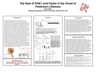
- 1. The Role of PINK1 and Parkin in the Onset of Parkinson’s Disease Ryan Elido Biology Department, Pacific University, Forest Grove, OR Background Parkinson’s disease is the second most common type of neurodegenerative disorder in humans (Chen & Chan, 2009). According to the Parkinson’s Disease Foundation website, it is estimated that seven to ten million people worldwide are living with Parkinson’s disease. Most individuals are diagnosed with Parkinson’s at the age of 60 years old. Parkinson’s is characterized by tremors of the limbs while resting, “cogwheel” movement in limbs, a slow and disturbed gait, and muscle weakness in the face and throat. Degeneration of dopaminergic neurons in the substantia nigra was seen in post-mortem autopsies in people who died with Parkinson’s (Chen & Chan, 2009; Gispert et al., 2009; Chaturvedi & Beal, 2013; Janezic et al., 2013). The substantia nigra is a structure located in the mesencephalon of the brain and is associated with the reward pathway and movement. It is characterized by a dark stripe due to a high concentration of neuromelanin in this region and primarily contains dopaminergic neurons. While the causes of the majority of Parkinson’s cases are idiopathic or unknown, about 10% of cases are familial and linked to defective genes (Chen & Chan, 2009). Treatment options are limited to physical and occupational therapy and therapeutic drugs designed to alleviate the symptoms associated with Parkinson’s. Mitochondria are organelles that are responsible for many cellular functions such as producing energy-rich molecules (ATP and NAD+) and apoptosis. Both PTEN induced putative kinase 1 (PINK1) and parkin are involved in maintaining mitochondrial integrity and degradation (mitophagy) (Chaturvedi & Beal, 2013; Gispert et al., 2009; Chen & Chan, 2009). A decrease in the mitochondrial membrane potential (ΔΨm) results in the inhibition of PINK1 degradation, therefore causing an accumulation of PINK1 proteins (Fedorowicz et al., 2013; Jones, 2010). Evidence Conclusions Evidence suggests that removing the PINK1 gene affects the regulation of mitochondrial turnover and degradation. Parkin also plays an important role in maintaining mitochondrial integrity, which is evident in the findings of Clark et al. (2006). In addition, parkin is shown to be involved in mitochondrial localization and PINK1 activity (Koyano et al., 2013). PINK1 acts upstream from parkin, meaning that these two genes are located in the same biochemical pathway. Mutations in either PINK1 or parkin lead to the decrease in mitochondrial activity, increased levels of ROS, and decreased levels of antioxidants like glutathione. This series of events is what is thought to lead to neurodegeneration and the cause of Parkinson’s disease. New questions that have developed include: Is there a connection between idiopathic and familial forms of Parkinson’s? Are there other genes involved in the onset of Parkinson’s disease? Future directions to this analysis include the use of human dopaminergic neurons to study the effects of PINK1/parkin mutations. Literature Cited Anichtchik, O., Diekmann, H., Fleming, A., Roach, A., Goldsmith, P., and Rubinsztein, D. C. “Loss of PINK1 Function Affects Development and results in Neurodegeneration in Zebrafish.” J. Neurosci. 28.33 (2008): 8199-8207. Chaturvedi, R., and Beal, M. "Mitochondrial Diseases of the Brain." Free Radical Biology and Medicine. (2013): 2-9. Chen , H., and Chan, D. "Mitochondrial dynamics - fusion, fission, movement, and mitophagy - in neurodegenerative diseases." Human Molecular Genetics. 18.2 (2009): 169-176. Clark, I. E., Dodson, M. W., Jiang, C., Cao, J., Huh, J. R., Hong Seol, J., Ji Yoo, S., Hay, B. A., and Guo, M. “Drosophila PINK1 is Required for Mitochondrial Function and Interacts Genetically with Parkin.” Nature. 441. (2006): 1162-1166. Fedorowicz, M.A., de Vries-Schneider, R., Rüb, C., Becker, D., Huang, Y., Zhou, C., Wolken, D., Voos, W., Liu, Y., and Przedborski, S. “Cytosolic Cleaved PINK1 represses Parkin Translocation to Mitochondria and Mitophagy.” EMBO Reports. 15.1 (2013): 86-93 Gispert, S., Ricciardi F., et al. "Parkinson Phenotype in Aged PINK1-Deficient Mice is Accompanied by Progressive Mitochondrial Dysfunction in Absence of Neurodegeneration." PLoS ONE. 4.6 (2009): 1-15. Janezic, S., Threlfell S., et al. "Deficits in dopaminergic transmission precede neuron loss and dysfunction in a new Parkinson model." PNAS. (2013): 1-10. Jones, R. “The Roles of PINK1 and Parkin in Parkinson’s Disease.” PLoS Biol. 8.1 (2010): 1-2. Koyano, F., Okatsu, K., Ishigaki, S., Fujioka, Y., Kimura, M., Sobue, G., Tanaka, K., and Matsuda, N. “The principal PINK1 and Parkin cellular events triggered in response to dissipation of mitochondrial membrane potential occur in primary neurons.” Genes to Cells. 18 (2013): 672-681. Parkinson's Disease Foundation (PDF). n.p., n.d. Web. 20 Apr. 2014. <http://www.pdf.org>. Trancikova, A., Tsika, E., Moore, D. J. “Mitochondrial Dysfunction in Genetic Animal Models of Parkinson’s Disease.” Antioxidants & Redox Signaling. 16.9 (2012): 896-919. * * Specific Question How do mutations in PINK1 and parkin affect mitochondrial function and lead to Parkinson's disease? Broader Implications Parkinson’s disease is the second most common form of neurodegenerative disorder in humans. Treatment options remain limited to physical therapy and medication (e.g. L-DOPA). Although 90% of Parkinson's cases are of unknown origins, understanding the development of familial forms of Parkinson’s can aid in the development in preventative treatments. The option of gene therapy is possible if the mechanism of the PINK1/parkin pathway is better understood. New drugs can be developed to counter the effects of this mutation and prevent the onset of this disease. In the meantime, the goal is to prevent the development of this disease by refraining from drug consumption (e.g. opioids), as well as living a healthy lifestyle to postpone the development of this disease. Combined costs for treatments and government assistance can cost up to $25 billion per year in the United States alone (pdf.org). Once new forms to prevent the onset of this disease have been developed, overall costs to care for patients with Parkinson’s will decrease, which benefits the public’s health and our economy. Figure 2. Survival rates of wild type versus PINK1 mutant Drosophila after exposure to different stressors (a-d) and ATP concentration in muscle (f) Graph shows a decrease in survival rates among PINK1 mutant flies after each treatment. (f) shows an overall lower concentration of ATP in PINK1 mutants compared to WT flies. Dysfunctional PINK1 genes decreases the organism’s resistance towards oxidative stress and damage due to the presence of reactive oxygen species (ROS). According to the data, PINK1 mutations affect electron transport chain due to low yield of ATP in PINK1 mutants (f). Increased sensitivity to oxidative stress coupled with a low yield in ATP production can result in damaged to affected cells and cell death. (Clark et al., 2006). Table 1. Mitochondrial dysfunction due to PINK1 and parkin gene knockouts. Missing or non- functional PINK1 and parkin genes results in a change of mitochondrial activity; a missing parkin gene results in a decrease in respiration (low ATP production). In addition, missing both PINK1 and parkin results in a decreased mitochondrial membrane potential, affecting PINK1/parkin pathway, as well as increased ROS levels and decreased antioxidant levels (i.e. glutathione). (Trancikova et al., 2013). Figure 1. Parkin mutations impair mitochondrial localization and E3 activity after carbonyl cyanide m-chlorophenylhydrazone (CCCP) treatment for 3 hours at 30 uM in mouse neurons. (B) Number of neurons with GFP-parkin on mitochondria. (C)E3 activity of parkin with parkin mutations shown using immunoblotting with an anti-parkin antibody. With the exception of the R275W cell line, presence of ubiquitin-GFP-parkin not present in CCCP-treated cells with mutated parkin. Mutations in parkin affect autophagy of mitochondria due to the impairment of PINK1/parkin function. Without the degradation of damaged mitochondria, abnormal cell function can result, leading to the onset of disease like Parkinson’s (Koyano et al., 2013). (Chen & Chan, 2009) (Fedorowicz et al., 2013)