2. Four mechanisms by which small molecules and
ions are transported across cellular membranes
3. Membrane Proteins Mediate Transport of Most
Molecules and All Ions Across Biomembranes
Overview of membrane transport proteins. Gradients are indicated by
triangles with the tip pointing toward lower concentration, electrical
potential, or both.
4. 1. Ion channels
1.1. Concept of ion channel
can transport ions across
membrane down electrochemical gradients
5. 1. Ion channels
1.2. Property of ion channel
Ion Channel proteins form a hydrophilic passageway across the
membrane through which multiple ions move simultaneously,
single file at a very rapid rate.
1 - channel domains (typically four per
channel), 2 - outer vestibule, 3 - selectivity
filter, 4 - diameter of selectivity filter, 5 -
phosphorylation site, 6 - cell membrane.
6. 1. Ion channels
1.3. Diversity of ion channel
Classification by gating
Ligand-gated
Voltage-gated
Stress gating
Classification by type of ions
Potassium channels, Sodium channels, Calcium channels
Classification by cellular localization
Plasma membrane channels, Endoplasmic reticulum channels
Mitochondrial channels
9. Opening of Acetylcholine-Gated Cation Channels
Leads to Muscle Contraction
Sequential activation of gated ion channels at a neuromuscular
junction(synapse).
10. 2. Transporters (carrier protein)
2.1. Concept of Transporter
carrier proteins is the integral transmembrane proteins
embedded in the cell membrane would have a high
affinity for specific substances on the cell exterior and
would next undergo a conformational change to facilitate
the passage of these substances to the cell interior across
the membrane barriers, mediated both passive and active
transport
12. 2. Transporters (carrier protein)
2.2. Diversity of Transporter
Uniporters: transport a single type of molecule down its concentration
gradient via facilitated diffusion. Glucose and amino acids
cross the plasma membrane into most mammalian cells
with the aid of uniporters.
Uniporter carrier proteins work by binding
to one molecule of substrate at a time and
transporting it with its concentration gradient.
13. GLUT1 Uniporter Transports Glucose into Most Mammalian
Cells Glucose transporter 1
Most mammalian cells use blood glucose
as the major source of cellular energy and
express GLUT1. Since the glucose
concentration usually is higher in the
extracellular medium (blood in the case of
erythrocytes) than in the cell, GLUT1
generally catalyzes the net import of glucose
from the extracellular medium into the cell.
1.3.1. Uniporters
14. Model of uniport transport by GLUT1. In one conformation, the
glucose-binding site faces outward; in the other, the binding site faces
inward. Binding of glucose to the outward-facing site (step 1 ) triggers a
conformational change in the transporter that results in the binding
site’s facing inward toward the cytosol (step 2 ). Glucose then is
released to the inside of the cell (step 3 ). Finally, the transporter
undergoes the reverse conformational change, regenerating the outward-
facing binding site (step 4 ). If the concentration of glucose is higher
inside the cell than outside, the cycle will work in reverse (step 4→step
1 ), resulting in net movement of glucose from inside to out.
15. 2. Transporters (carrier protein)
2.2. Diversity of Transporter
Cotransporters: are a subcategory of membrane transport proteins
(transporters) that couple the favorable movement
of one molecule with its concentration gradient and
unfavorable movement of another molecule against
its concentration gradient.
utilize electric potential
and/or chemical gradients
to move protons and ions
16. 2.2.1.Symporters
Na+-linked symporters import amino acids and glucose into
animal cells against high concentration gradients
The two-Na+/one-glucose symporter, a protein that couples import
of one glucose molecule to the import of two Na+ ions:
2 Na+
out + glucoseout →2 Na+
in + glucosein
18. 2.2.2.Antiporters
Na+-linked antiporter exports Ca2+ from cardiac muscle cells.
In cardiac muscle cells a three-Na+one-Ca2+ antiporter plays the principal role
in maintaining a low concentration of Ca2+in the cytosol. The transport
reaction mediated by this cation antiporter can be written:
3 Na+
out + Ca2+
in →3Na+
in + Ca2+
out
19. Several Cotransporters Regulate Cytosolic pH
The anaerobic metabolism of glucose yields
lactic acid, and aerobic metabolism yields CO2,
which adds water to form carbonic acid (H2CO3).
These weak acids dissociate, yielding H+ ions
(protons); if these excess protons were not removed
from cells, the cytosolic pH would drop
precipitously, endangering cellular functions. Two
types of cotransport proteins help remove some of
the “excess” protons generated during metabolism
in animal cells.
20. The activity of membrane transport proteins that
regulate the cytosolic pH of mammalian cells changes
with pH.
21. Multiple Transport Proteins Are Needed to Move
Glucose and Amino Acids Across Epithelia
Transcellular transport of glucose from the
intestinal lumen into the blood.
epithelia
22. 1. The macrophage internalizes pathogens through facilitated
diffusion. ( )
2. Steroid hormones cross cell membrane through active
transport. ( )
3. When the presynaptic neuron is stimulated, the flow of Na+
enters the cell membrane of postsynaptic neuron through
facilitated diffusion. ( )
4. Glycerin in cosmetics cross the membrane of skin cell through
active transport. ( )
True or False
23. Summary
Difference between channels and carriers
1. A carrier is not open simultaneously to both the extracellular and
intracellular environments. In contrast, a channel can be open to
both environments at the same time, allowing the solutes it
transports to diffuse without interruption;
2. Carriers have binding sites, but pores and channels do not;
3. Each carrier protein is designed to recognize only one substance or
one group of very similar substances
24. Homework
How does a glucose molecule transport from the
intestinal lumen into the blood?
Thanks for your attention!
Editor's Notes
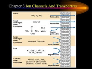
From the previous chapter, we have known the composition, structure and functions of Biomembrane. The most important function of Biomembrane is that it works as a barrier between inside and outside of the cell or organelle, and control the entry and exit of cell.(讲气体如何进入细胞、乙醇如何进入细胞)because the biomembrane is a impermeable membrane, not all of molecule pass through the biomembrane freely. Only gases and small polar molecules like ethanol pass through the biomembrane freely. a small amount of water or Urea transport through the membrane freely.in this chapter, we will learn two types of membrane protein which facilitate the small molecules moving through the biomembrane.
passive diffusion is the simplest way of passive transport. Special type
Next, lets see the overview of membrane transport proteins. Because different cell types require different mixtures of low-molecular-weight compounds, the plasma membrane of each cell type contains a specific set of transport proteins that allow only certain ions and molecules to cross. Similarly, organelles within the cell often have a different internal environment from that of the surrounding cytosol, and organelle membranes contain specific transport proteins that maintain this difference.(分别描述三种)Pumps utilize the energy released by ATP hydrolysis to power movement of specific ions (red circles) or small molecules against their electrochemical gradient. Channels permit movement of specific ions (or water) down their electrochemical gradient. Transporters, which fall into three groups, facilitate movement of specific small molecules or ions.
Channel proteins transport water or specific types of ions down their concentration or electric potential gradients.Today, we focus on the ion channel,a Channel protein that transport ions.(读PPT第一条),Their functions include establishing a resting membrane potential, shaping action potentials and other electrical signals by gating the flow of ions across the cell membrane, that means ion channel control the membrane potential of a biomembrane. In electrically excitable cells such as neurons and muscle cells, the membrane potential is used for transmitting signals between different parts of a cell, so ion channels are the excitable compoents of excitable cells and very important for excitable cells.
so ion channels are very important for excitable cells, like neurons, muscle and synapses.
1.Ion channels are integral membrane proteins with a pore through which ions can travel between extracellular space and cell interior.(读PPT标题下一段)描述图片,in most case, a ion channel are composed of several parts.(1) channel domain, have several subunits, typically four per channel,forming a single aqueous pore at the axial intersection.讲到结构3时接下面一段。
Because of selectivity filter,the Most channels are specific (selective) for one ion; for example, most potassium channels are characterized by 1000:1 selectivity ratio for potassium over sodium, though potassium and sodium ions have the same charge and differ only slightly in their radius.
There are over 300 types of ion channels,they may be classified by the nature of their gating, the species of ions passing through those gates, the number of gates (pores) and localization of proteins. Depending on the gating ,Ion channels may be classified to(1/2/3),
Voltage – Regulated by the difference in voltage across the membrane;
Stress – Regulated by physical pressure on the transporter (as in the cochlea of the ear);
Ligand – Regulated by the binding of a ligand to either the intracellular or extracellular side of the cell;
are a group of transmembrane ion channel proteins which open to allow ions such as Na+, K+, Ca2+, and/or Cl− to pass through the membrane in response to the binding of a chemical messenger,such as a neurotransmitter。These proteins are typically composed of at least two different domains: a transmembrane domain which includes the ion pore, and an extracellular domain which includes the ligand binding location.The function of such receptors located at synapses is to convert the chemical signal of presynaptically released neurotransmitter directly and very quickly into a postsynaptic electrical signal.
Voltage-gated ion channels are activated by changes in the electrical membrane potential near the channel. The membrane potential alters the conformation of the channel proteins, regulating their opening and closing. Cell membranes are generally impermeable to ions, thus they must diffuse through the membrane through transmembrane protein channels. They have a crucial role in excitable cells such as neuronal and muscle tissues, allowing a rapid and co-ordinated depolarization in response to triggering voltage change. Found along the axon and at the synapse, voltage-gated ion channels directionally propagate electrical signals. there is a central pore through which ions can travel down their electrochemical gradients. The channels tend to be ion-specific, although similarly sized and charged ions may sometimes travel through them
Now, let us see a physiological activity in which both Ligand-gated channel and Voltage-gated channel participate.读题目. Acetylcholine is the neurotransmitter at synapses between motor neurons and muscle cells, often called neuromuscular junctions. A single axon terminus of a motor neuron may contain a million or more synaptic vesicles, each containing up to 10,000 molecules of acetylcholine; these vesicles often accumulate in rows in the active zone. The nicotinic acetylcholine receptor, which is expressed in muscle cells, is a ligand-gated channel that admits both K+ and Na+.1.There are voltage-gated channels on the membrane of motor neurons, Arrival of an action potential at the terminus of a presynaptic motor neuron induces opening of voltage-gated Ca2+ channels. 2. and subsequent release of acetylcholine, which triggers opening of the ligand-gated acetylcholine receptors in the muscle plasma membrane.3. The resulting influx of Na+ produces a localized depolarization of the membrane, leading to opening of voltage-gated Na+ channels and generation of an action potential. 4.When the spreading depolarization reaches T tubules, it is sensed by voltagegated Ca2+channels in the plasma membrane. This leads to opening of Ca2+-release channels in the sarcoplasmic reticulum membrane, releasing stored Ca2+ into the cytosol. The subsequent flow of stored Ca2+ ions from the sarcoplasmic reticulum into the cytosol raises the cytosolic Ca2+ concentration sufficiently to induce muscle contraction.
There are a number of disorders which disrupt normal functioning of ion channels and have disastrous consequences for the organism. Genetic and autoimmune disorders of ion channels and their modifiers are known as channelopathies
When a presynaptic neuron is excited, it releases a neurotransmitter from vesicles into the synaptic cleft. The neurotransmitter then binds to receptors located on the postsynaptic neuron. If these receptors are ligand-gated ion channels, a resulting conformational change opens the ion channels, which leads to a flow of ions across the cell membrane. This, in turn, results in either a depolarization, for an excitatory receptor response, or a hyperpolarization, for an inhibitory response.
Uniporter carrier proteins work by binding to one molecule of substrate at a time and transporting it with its concentration gradient. Cotransporters proteins move two molecules at the same time: one against a gradient and the other with its gradient. They are distinguished according to the directionality of the two molecules,antiporter: move a molecule against its gradient and at the same time displaces one or more ions along its gradient. The molecules move in opposite directions. symporter:move a molecule against its gradient while displacing one or more different ions along their gradient. The molecules move in the same direction.
Glucose transporter 1 is a uniporter protein facilitates the transport of glucose across the plasma membranes of mammalian cells, GLUT1 was the first glucose transporter to be characterized.(读PPT)
triangles with the tip pointing toward lower concentration
Cotransporters are capable of moving solutes either up or down gradients at rates of 1000 to 100000 molecules per second.The movement occurs by binding to two molecules or ions at a time and using the gradient of one solute's concentration to force the other molecule or ion against its gradient. Cotransporters can be classified as antiporters and symporters. Both utilize electric potential and/or chemical gradients to move protons and ions against their concentration gradient. Antiporters and symporters both transport two or more different types of molecules at the same time in a coupled movement. An energetically unfavored movement of one molecule is combined with an energetically favorable movement of another molecule(s) or ion(s) to provide the power needed for transport. Cotransporters undergo a cycle of conformational changes by linking the movement of an ion with its concentration gradient (downhill movement) to the movement of a cotransported solute against its concentration gradient (uphill movement). In one conformation the protein will have the binding site (or sites in the case of symporters) exposed to one side of the membrane. Upon binding of both the molecule which is to be transported uphill and the molecule to be transported downhill a conformational change will occur. This conformational change will expose the bound substrates to the opposite side of the membrane, where the substrates will disassociate. Both the molecule and the cation must be bound in order for the conformational change to occur.
symporters move ions or molecules in the same direction. In this case both ions being transported will be moved either from the exoplasmic space into the cytoplasmic space or from the cytoplasmic space into the exoplasmic space. An example of a symporter is the sodium-glucose linked transporter, Most body cells import glucose from the blood down its concentration gradient, utilizing one or another GLUT protein to facilitate this transport. However, certain cells, such as those lining the small intestine and the kidney tubules, need to import glucose from the intestinal lumen or forming urine against a very large concentration gradient. Such cells utilize a sodium-glucose linked transporter.The SGLT functions to couple the transport of sodium in the exoplasmic space down its concentration gradient into the cytoplasmic space to the transport of glucose in the exoplasmic space against its concentration gradient into the cytoplasmic space.The SGLT couples the movement of 1 glucose ion with the movement of 2 sodium ions. (this figure is the Operational model for the two-Na+/oneglucose symporter). Simultaneous binding of Na+ and glucose to the conformation with outward-facing binding sites (step 1 ) generates a second conformation with inward-facing sites (step 2 ). Dissociation of the bound Na+ and glucose into the cytosol (step 3 ) allows the protein to revert to its original outward-facing conformation (step 4 ), ready to transport additional substrate.
Antiporters use the mechanism of cotransport (coupling the movement of one ion or molecule down its concentration gradient with the transport of another ion or molecule up its concentration gradient), to move the ions and molecule in opposite directions. In this situation one of the ions will move from the exoplasmic space into the cytoplasmic space while the other ion will move from the cytoplasmic space into the exoplasmic space. An example of an antiporter is the sodium-calcium exchanger. The sodium-calcium exchanger functions to remove excess calcium from the cytoplasmic space into the exoplasmic space against its concentration gradient by coupling its transport with the transport of sodium from the exoplasmic space down its concentration gradient into the cytoplasmic space. The sodium-calcium exchanger exchanges 3 sodium ions for 1 calcium ion and represents a cation antiporter.
Now, let us see a physiological activity in which antiporter participate.读PPT
One is a Na+HCO3-/Cl- antiporter, which imports one Na+ ion down its concentration gradient, together with one HCO3 -, in exchange for
export of one Cl- ion against its concentration gradient. The cytosolic enzyme carbonic anhydrase catalyzes dissociation of the imported HCO3 - ions into CO2 and an OH- (hydroxyl) ion. The CO2 diffuses out of the cell, and the OH- ions combine with intracellular protons, forming water. Thus the overall action of this transporter is to consume cytosolic H+ ions, thereby raising the cytosolic pH. Second,is a Na+/H+ antiporter, which couples entry of one Na+ ion into the cell down its concentration gradient to the export of one H_ ion. Under certain circumstances the cytosolic pH can rise beyond the normal range of 7.2–7.5. To cope with tohe excess OH_ ions associated with elevated pH, many animal cells utilize an anion antiporter that catalyzes the one-for-one exchange of HCO3 - and Cl- across the plasma membrane. At high pH, this Cl- /HCO3 - antiporter exports HCO3 - in exchange for Cl- , thus lowering the cytosolic pH.
Another interest physiological activity in which severaltransporters participate is “ Transcellular transport of glucose from the intestinal lumen into the blood”书p100页