
Recommended
More Related Content
What's hot
What's hot (20)
Similar to trypanosoma presentation.pptx
Similar to trypanosoma presentation.pptx (20)
More from AmosiRichard
More from AmosiRichard (16)
Recently uploaded
Recently uploaded (20)
trypanosoma presentation.pptx
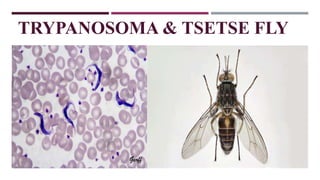
- 1. TRYPANOSOMA & TSETSE FLY
- 2. PRESENTATION OUTLINE INTRODUTION CLASSIFICATION MORPHOLOGY LIFECYCLE METHOD OF DIAGNOSIS TREATMENT AND PREVENTION
- 3. INTRODUCTION CONT.… Trypanosomiasis is a parasitic disease caused by the protozoan parasite Trypanosoma, which is transmitted to humans and animals by tsetse flies. The disease is commonly known as sleeping sickness in humans and nagana in animals. There are two types of trypanosomiasis: African trypanosomiasis (also called sleeping sickness): This type is found in sub- Saharan Africa, and it is caused by Trypanosoma brucei. The disease can be acute or chronic and it affects the central nervous system causing symptoms such as fever, headaches, joint pains, itching, and behavioral changes, including confusion, disruption of the sleep cycle, and even coma.
- 4. INTRODUCTION CONT.… American trypanosomiasis (also called Chagas disease): This type is found in Central and South America, and it is caused by Trypanosoma cruzi. This disease can also have acute and chronic phases, and it affects the heart, esophagus, and colon causing symptoms such as fever, muscle pain, enlarged liver or spleen, and digestive problems. There are two types of African trypanosomiasis (also called sleeping sickness); each is named for the region of Africa in which they were found historically. East African trypanosomiasis is caused by the parasite Trypanosoma brucei rhodesiense. West African trypanosomiasis is caused by the parasite Trypanosoma brucei gambiense. Both types of African trypanosomiasis are transmitted by the tsetse fly which is found only in rural Africa The two forms differ in their clinical presentation, with T. brucei gambiense causing a chronic, slowly progressive form of the disease, and T. brucei rhodesiense causing a more acute and rapidly progressive form
- 5. TRYPANOSOMA CLASSIFICATION Trypanosoma is a genus of unicellular parasitic protozoan that belongs to the family Trypanosomatidae, which is part of the order Kinetoplastida, phylum sarcomastogophora and kingdom protista. The Trypanosoma genus is further divided into several subgenera and species, based on their morphology, host range, and pathogenicity. There are over 30 known species of Trypanosoma, with the most important ones in terms of human and animal health being: Trypanosoma brucei, which causes African sleeping sickness in humans and nagana in animals. Trypanosoma cruzi, which causes Chagas disease in humans and animals
- 6. MORPHOLOGY
- 7. MORPHOLOGY Trypanosoma is a unicellular protozoan parasite that has a characteristic elongated shape, ranging from 15 to 40 micrometers in length, with a diameter of about 1-2 micrometers. The organism has a single flagellum that extends from its anterior end and is used for movement, and an undulating membrane that runs along its body and aids in locomotion. The cell body of Trypanosoma is covered by a flexible and protective surface membrane, which is composed of glycoproteins and lipids, and can vary in structure and composition among different species. Beneath the surface membrane, there is a layer of microtubules that forms the cytoskeleton of the organism, providing structural support and aiding in cell division.
- 8. MORPHOLOGY CONT.. Kinetoplast is a specialized mitochondrion that contains a large network of DNA fibers, called the kinetoplast DNA (kDNA), that encodes for several mitochondrial proteins. The kDNA is arranged in a unique circular or linear structure and is located close to the base of the flagellum. Trypanosoma can also change its surface proteins, a phenomenon called antigenic variation, which allows the organism to evade the host immune system and establish chronic infections. This process involves the switching of expression of genes encoding for surface proteins, resulting in the production of different protein variants that are recognized differently by the host immune system.
- 9. LIFECYCLE The life cycle of Trypanosoma brucei involves two hosts, an insect vector (tsetse fly) and a mammalian host, and has several stages, including the following: Epimastigote: The T. brucei parasite first develops in the midgut of the tsetse fly vector after ingestion of infected blood. In the midgut, the parasite transforms into the epimastigote stage, which multiplies by binary fission. Metacyclic trypomastigote: The epimastigotes then migrate to the salivary glands of the tsetse fly, where they differentiate into the infective metacyclic trypomastigote stage, which can be transmitted to the mammalian host during a blood meal. Bloodstream trypomastigote: Once the metacyclic trypomastigote enters the mammalian host bloodstream, it transforms into the bloodstream trypomastigote stage, which is the most commonly known form of the parasite.
- 10. LIFECYCLE CONT. The bloodstream trypomastigotes multiply by binary fission and can invade and replicate within various tissues, including the lymphatic system, spleen, and brain. Intracellular amastigote: Within the mammalian host, some of the bloodstream trypomastigotes can transform into the intracellular amastigote stage, which can invade and replicate within the cells of various organs and tissues, including the central nervous system. Tsetse fly transmission: If a tsetse fly bites an infected mammalian host, it ingests the bloodstream trypomastigotes. In the midgut of the tsetse fly, the parasites transform into the epimastigote stage, starting the cycle again.
- 11. LIFECYCLE CONT..
- 12. LABORATORY INVESTIGATION The laboratory diagnosis of trypanosomiasis, caused by Trypanosoma spp., involves the identification of the parasite in clinical specimens obtained from the patient or the insect vector. Some of the common methods used for laboratory diagnosis of trypanosomiasis include: Blood smear examination: Blood smears are prepared from the patient's blood and examined microscopically for the presence of trypanosomes. Thick and thin blood films are usually used, with the thin films being stained with Giemsa or Wright stains. The parasites can be identified based on their characteristic morphology, such as elongated shape, undulating membrane, and single flagellum.
- 13. Blood Smear Showing Trypomastigote Of Trypanosoma Brucei
- 14. LABORATORY DIAGNOSIS CONT.. Lymph node aspiration: In some cases, the trypanosomes can be detected by examining lymph node aspirate samples, which are collected using fine needles and examined microscopically for the presence of the parasites. Cerebrospinal fluid (CSF) examination: If the patient has neurological symptoms, a lumbar puncture can be performed to collect CSF for examination. The CSF sample is examined microscopically for the presence of trypanosomes. Serological tests: Serological tests can be used to detect antibodies produced by the host against Trypanosoma spp. The tests can include enzyme-linked immunosorbent assay (ELISA), immunofluorescence assay (IFA), and immunochromatographic tests. However, serological tests may not be useful in the early stages of the disease, as it may take several weeks for detectable antibodies to develop.
- 15. LABORATORY DIAGNOSIS CONT.… Molecular tests: Polymerase chain reaction (PCR) can be used to detect the DNA of Trypanosoma spp. in clinical specimens, including blood, CSF, or lymph node aspirates. PCR is highly sensitive and specific and can be useful in detecting low-level parasitemia in early stages of the disease The laboratory diagnosis of trypanosomiasis requires careful specimen collection, processing, and examination by trained personnel to ensure accurate results
- 16. TREATMENT AND PREVENTION The treatment and prevention of trypanosomiasis, caused by Trypanosoma spp., depend on the species of the parasite, the stage of the disease, and the severity of the symptoms. Some of the common methods used for the treatment and prevention of trypanosomiasis include: Chemotherapy: Chemotherapy is the mainstay of treatment for trypanosomiasis. The drugs used for treatment depend on the species of the parasite and the stage of the disease. For example, suramin and pentamidine are used for the treatment of early- stage African trypanosomiasis, while melarsoprol and eflornithine are used for the treatment of late-stage African trypanosomiasis. Benznidazole and nifurtimox are used for the treatment of Chagas disease.
- 17. TREATMENT AND PREVENTION CONT.… Animal reservoir control: In endemic areas, trypanosomiasis can be transmitted from infected animals to humans. Therefore, controlling the infection in animal reservoirs, such as cattle, can help reduce the risk of human infection. Vector control: Since tsetse flies are the vectors for African trypanosomiasis, vector control measures, such as insecticide-treated screens, traps, and insecticide spraying, can be used to reduce the population of tsetse flies in endemic areas. Screening and surveillance: Early detection and treatment of infected individuals can help prevent the spread of the disease.. Education and awareness: Education and awareness programs can help raise awareness about the risk factors and symptoms of trypanosomiasis and promote the use of preventive measures, such as insecticide-treated bed nets and protective clothing.
- 18. TSETSE FLY
- 19. INTRODUCTION Is a type of bloodsucking insect that is found only in Africa. It is known for being a carrier of African trypanosomiasis, also known as sleeping sickness, a parasitic disease that affects humans and animals. It also known as tik-tik flies. Having size of 6–15mmin length. They occur only in tropical Africa and are important as vectors of African trypanosomiasis(protozoa) in both humans and animals. The tsetse are obligate parasites that live by feeding on the blood of vertebrate animals. Tsetse can be distinguished from other large flies by observed features. Tsetse fold their wings completely when they are resting so that one wing rests directly on top of the other over their abdomens.
- 20. CLASSIFICATION The tsetse fly belongs to the family Glossinidae, which is a family of bloodsucking flies found only in Africa. The Glossina subgenus includes most of the tsetse fly species and is further divided into several groups based on morphological and genetic characteristics. Kingdom: Animalia (animals) Phylum: Arthropoda (arthropods) Class: Insecta (insects) Order: Diptera (true flies) Suborder: Brachycera (short-horned flies) Family: Glossinidae (tsetse flies) Genus: Glossina
- 21. DISTINCTIVE MORPHOLOGICAL FEATURES Tsetse flies are robust sparsely bristled insects that usually range from 6 to 16 mm (0.2 to 0.6 inch) in length The tsetse fly (Glossina spp.) has several distinct morphological features that distinguish it from other types of flies. Some of these features include: Long proboscis: Tsetse flies have a long, pointed proboscis that they use to pierce the skin of their host and feed on blood. Large eyes: Tsetse flies have large compound eyes that help them locate their host and avoid predators. Wings: Tsetse flies have two wings that are held upright when the fly is at rest. Mouthparts: The mouthparts of tsetse flies are modified for feeding on blood, with sharp, needle-like structures that pierce the skin of their host.
- 22. MORPHOLOGICAL FEATURES CONT.… Brownish coloration: Tsetse flies are typically brown or gray in color, with distinctive patterns on their wings. Size: Tsetse flies are relatively large, with a body length of up to 15mm. Antennae: Tsetse flies have short, bristly antennae that are used for sensing their environment and locating their host.
- 23. LIFECYCLE Tsetse fly have an unusual lifecycle which may be due to the richness of their food source. A female fertilizes only one egg at a time and retains each egg within her uterus to have the offspring develop internally during the first three larval stages (adenotrophic viviparity). During this time, the female feeds the developing offspring with a milky substance secreted by a modified gland in the uterus.
- 24. LIFECYLE CONT.… In the third larval stage, the tsetse larva leaves the uterus and begins its independent life. The newly independent tsetse larva crawls into the ground, and develops a hard outer shell called the puparial case, in which it completes its morphological transformation into an adult fly. Typically producing 4 generations per year
- 25. HOST AND DISEASE The tsetse-vectored trypanosomiases affect various vertebrate species including humans, antelopes, bovine cattle, camels, horses, sheep, goats, and pigs. Sleeping sickness: Two different types of human sleeping sickness are caused by different subspecies of trypanosome parasites Gambiense sleeping sickness (Trypanosoma brucei gambiense) is generally considered to be a chronic disease and is found mostly in West and Central Africa Rhodesiense sleeping sickness (Trypanosoma brucei rhodesiense) is an acute disease that occurs mainly in East Africa.
- 26. In domestic animals: Animal trypanosomiasis, it occurs in cattle or horses. These diseases reduce the growth rate, milk productivity, and strength of farm animals, generally leading to the eventual death of the infected animals
- 27. SYMPTOMS A bite by the tsetse fly is often painful and can develop into a red sore, also called a chancre. Fever, severe headaches, irritability, extreme fatigue, swollen lymph nodes, and aching muscles and joints are common symptoms of sleeping sickness. Some people develop a skin rash. Progressive confusion, personalitychanges, andother neurologic problems occur after infection has invaded the central nervous system. If left untreated,infection becomes worse and death will occur within months.
- 28. PREVENTION AND CONTROL Land clearing Pesticide campaigns Sterile insect technique
- 29. THANK YOU
Editor's Notes
- The life cycle of T. brucei is complex and involves various transformations, with different morphological and biochemical characteristics at each stage, which allows the parasite to adapt to different environments within its hosts. The cycle can take several weeks to months, and is crucial for the transmission and survival of the parasite.
- It takes about 3 wks from the time of blood meal for fly to become infective. Then it remains infective for life (6Months
- .
- It is important to note that the prevention and control of trypanosomiasis require a multidisciplinary approach involving healthcare professionals, researchers, policymakers, and the affected communities.