Intro to ra lausanne 2018
•
12 likes•771 views

This is a lecture that Dr Amit Pawa gave in Lausanne, Switzerland in October 2018 as part of the 4th Romandie Day of Regional Anaesthesia. In it he covers some tips and tricks as part of an introduction to Ultrasound guided regional anaesthesia - note - versions of this course have been delivered at courses in the UK in the past

Recommended
Recommended
More Related Content
What's hot
What's hot (20)
Similar to Intro to ra lausanne 2018
Similar to Intro to ra lausanne 2018 (20)
More from Amit Pawa
More from Amit Pawa (9)
Recently uploaded
Recently uploaded (20)
Intro to ra lausanne 2018
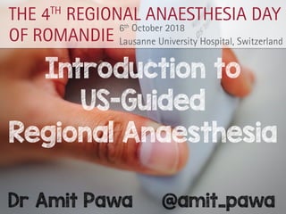
- 1. Introduction to US-Guided Regional Anaesthesia Dr Amit Pawa @amit_pawa THE 4TH REGIONAL ANAESTHESIA DAY OF ROMANDIE 6th October 2018 Lausanne University Hospital, Switzerland Sempach | Switzerland AK2357_04.2018 OF ROMANDIE 6th October 2018 Lausanne University Hospital, Switzerland
- 3. Declaration I love Ultrasound Guided RA!… RA-UK President Consult Honoraria from ! Sponsored by:
- 5. Purpose of this lecture Gain Tips On How to… 1. Generate best image 2. Interpret best image 3. Needle best image 4.Achieve best results @amit_pawa
- 6. Why Ultrasound for RA?
- 8. The Future #POCUS Airway Breathing Circulation @amit_pawa
- 9. Why Ultrasound for RA? US Machines everywhere! Familiarity (CVC) We know some anatomy Reliable Non-opiate analgesia @amit_pawa
- 10. Accuracy Reduce Less Intraneural Injections Rapid Block Onset Quick & Painless block insertion Longer Block Durations Why Ultrasound for RA? Reduce LA Dose Identify Aberrant anatomy Avoid “Tiger Territory” L.A.S.T
- 11. The Anatomy Lesson of Dr Nicolaes Tulp - 1632 - Rembrandt Learning Anatomy
- 12. The Anatomy Lesson of Dr Nicolaes Tulp - 1632 - Rembrandt Learning Anatomy
- 13. Machines are getting smaller! @amit_pawa
- 14. When did it all begin?… 1994 @amit_pawa
- 15. Prince - “The Most Beautiful Scan in the world” “Could u be, the most beautiful nerve in the world, Its plain to see, the body anatomy all told. When the probe, makes the images of you come to life, I know I, can remove all the pain of the knife…” " # # " 1994
- 16. Back to 2018…
- 17. 1. Generate Best Image Probe Handling Probe Manipulation - P.A.R.T Machine Manipulation/Optimisation Short / Long Axis imaging Ergonomics @amit_pawa
- 18. Probe Handling My Hand My 6yr old’s Hand Not like this… @amit_pawa
- 19. Stable & precise Probe Handling Treat it like a paintbrush My Hand My 6yr old’s Hand @amit_pawa
- 25. Probe Selection Superficial High Frequency “Resolves” Deeper Low Frequency “Penetrates” >10 MHz 2-5 MHz
- 26. Select the Correct Mode/ Preset @amit_pawa
- 30. Add some colour… • Veins: compressible, non pulsatile • Arteries: non- compressible, pulsatile • Detects flow • Red flow towards probe • Blue away from probe @amit_pawa
- 31. Short/Long Axis Imaging Short Axis = Cross Section Long Axis = Longitudinal Scan @amit_pawa
- 34. Purpose of this lecture Gain Tips On How to… 1. Generate best image 2.Interpret best image 3.Needle best image 4.Achieve best results @amit_pawa
- 35. 2. Interpret Best Image Anatomy Sonoanatomy - Template Dynamic - traceback to known anatomical relations “Only See what you Look for, Only Look for what you Know” Know common anomalies @amit_pawa
- 36. Anatomy This is the best time to revise your old anatomy notes Attend a cadaveric course Dig out your old text books Buy an App!! @amit_pawa
- 37. @amit_pawa
- 38. Axilla Coracobrachialis MCN Humerus Biceps Brachii Long Head ofTriceps/ ConjointTendon (TM/LD)AA av AV M U R L A T E R A L M E D I A L London Society of Regional Anaesthesia !"#$%$&'()#*+,(-*. “Sono”Anatomy = Ultrasound Anatomy @amit_pawa
- 39. Nerve Proximal - Interscalene Distal - Musculocutaneous Hyperechoic rim Hypoechoic content (neural tissue) Hyperechoic rim Honeycomb content – fascicles & connective tissue
- 40. Nerve Proximal - Interscalene Distal - Musculocutaneous Hyperechoic rim Hypoechoic content (neural tissue) Hyperechoic rim Honeycomb content – fascicles & connective tissue
- 41. Sonoanatomy Cross sectional anatomy Schematic Pattern recognition Apply to real US Ultrasound is dynamic! @amit_pawa
- 42. Sternocleidomastoid Anterior Scalene CA IJV Middle Scalene phrenic n vagus n Medial Lateral Interscalene @amit_pawa
- 43. Sternocleidomastoid Anterior Scalene CA IJV Middle Scalene phrenic n vagus n Medial Lateral Interscalene @amit_pawa
- 46. Cephalad Caudad Pec Major Pec Minor AA AVL P M Clavicle Infraclavicular Block @amit_pawa
- 47. Cephalad Caudad Pec Major Pec Minor AA AVL P M Clavicle Infraclavicular Block @amit_pawa
- 49. Pec Major Pec Minor AA AVL P M Clavicle Cephalad Caudad @amit_pawa
- 50. © - Amit Pawa
- 51. Many more examples… Axilla C oracobrachialis MCN Humerus BicepsBrachii Long Head ofTriceps/ ConjointTendon (TM/LD) AA av AVM U R L A T E R A L M E D I A L London Society of Regional Anaesthesia !"#$%$&'()#*+,(-*. Friday, 21 June 2013 E.O.m I.O.m T.A.m Rectus m skin Q.L.m L.D.m Peritoneum ABDOMEN London Society of Regional Anaesthesia !"#$%$&'()#*+,(-*. Friday, 15 June 2012 Friday, 21 June 2013 Subgluteal Sciatic Gluteus Maximus Quadratus femorisSN Fat GT IT Lateral Medial London Society of Regional Anaesthesia !"#$%$&'()#*+,(-*. SFA Lateral sartorius vastus medialis adductor longus adductor magnus FV Adductor Canal London Society of Regional Anaesthesia !"#$%$&'()#*+,(-*. Obturator Block FV Pectineus Adductor Longus Adductor Brevis Adductor Magnus Lateral Medial Ant Post FA London Society o !"#$%$
- 52. Ultrasound is Dynamic Confirm what you think you know Trace to known anatomical relations - Forearm peripheral nerves Scan from antecubital fossa to wrist & back Nerves follow a characteristic path @amit_pawa
- 53. Ultrasound is Dynamic Confirm what you think you know Trace to known anatomical relations - Infraclavicular brachial plexus Scan from medial to lateral, the cords rotate around axillary artery @amit_pawa
- 54. @amit_pawa You only SEE what you LOOK for Caution
- 55. Caution @amit_pawa You only LOOK for what you KNOW Caution
- 57. 1st Rib Pleura Plexus SCA Supraclavicular Block @amit_pawa
- 58. Had a pre-procedure doppler been performed & attention paid to Pleura(!)... @amit_pawa
- 59. @amit_pawa
- 60. Purpose of this lecture Gain Tips On How to… 1. Generate best image 2.Interpret best image 3.Needle best image 4.Achieve best results @amit_pawa
- 61. 3. Needle Best Image In-Plane / Out-of-Plane needling S.T.A.R Ergonomics again @amit_pawa
- 62. Needling In-Plane Out - of - Plane @amit_pawa
- 64. Needling - OOP Needle tip deeper than initially seen Target Often use hydrolocation to assist here
- 66. Needling - IP Visualise shaft & tip @amit_pawa
- 67. From Above NOT in-plane From In-front “Looks” in-plane
- 68. S.T.A.R. What about when needling in-plane & not seeing needle entirely… Remember P.A.R.T.? Anything Better? A Randomized Controlled Trial Evaluating the See, Tilt, Align, and Rotate (STAR) Maneuver on Skill Acquisition for Simulated Ultrasound- Guided Interventional Procedures Nicholas C. K. Lam, MD, Steven J. Fishburn, MD, Angie R. Hammer, BS, Timothy R. Petersen, PhD, Neal S. Gerstein, MD, Edward R. Mariano, MD, MAS Received August 12, 2014, from the Department of Anesthesiology and Critical Care Medicine, University of New Mexico, Albuquerque, New Mexico USA (N.C.K.L., S.J.F., A.R.H., T.R.P., N.S.G.); Department of Anesthesiology, Perioper- ative and Pain Medicine, Stanford University School of Medicine, Stanford, California USA (E.R.M.); and Anesthesiology and Perioperative Care Service, VA Palo Alto Health Care System, Palo Alto, California USA (E.R.M.). Revision requested August 26, 2014. Revised manuscript accepted for publication September 1, 2014. This work was presented in part at the American Society of Anesthesiologists Annual Meeting;October2013;SanFrancisco,California. Dr Lam received support for this project from the Father Meldon Hickey Memorial Fund administered by the University of New Mexico Foundation.DrMarianohasreceivedunrestricted Objectives—Achieving the best view of the needle and target anatomy when perform- ingultrasound-guidedinterventionalproceduresrequirestechnicalskill,whichnovices may find difficult to learn. We hypothesized that teaching novice performers to use 4 sequentialsteps(see,tilt,align,androtate[STAR]method)toidentifytheneedleunder ultrasoundguidanceismoreefficientthantrainingwiththecommonlydescribedprobe movements of align, rotate, and tilt (ART). Methods—Thisstudycompared2instructionalmethodsfortransducermanipulation including alignment of a probe and needle by novices during a simulated ultrasound- guidednerveblock.Right-handedvolunteersbetweentheagesof18and55yearswho hadnopreviousultrasoundexperiencewererecruitedandrandomizedto1of2groups; one group was trained to troubleshoot misalignment with the ART method, and the otherwastrainedwiththenewSTARmaneuver.Participantsperformedthetask,con- sisting of directing a needle in plane to 3 targets in a standardized gelatin phantom 3 times. The performance assessor and data analyst were blinded to group assignment. Results—Thirty-five participants were recruited. The STAR group was able to com- pletethetaskmorequickly(P<.001)andvisualizedtheneedleinagreaterproportion of the procedure time (P = .004) compared to the ART group. All STAR participants were able to complete the task, whereas 41% of ART participants abandoned the task ctiveperformanceofallultrasound-guidedproceduresrequires wledge of relevant sonoanatomy and proficiency in real-time dle guidance. In ultrasound-guided regional anesthesia, the in- e technique (directing the needle within the same plane as the ne | J Ultrasound Med 2015; 34:1019–1026 | 0278-4297 | www.aium.org
- 69. wellas the proportion of attempt time in which the needle was not fully visible. If an attempt lasted 600 seconds, the participant was offered the option to terminate the attempt, and its duration was recorded as 600 seconds. attempt time in which the inserted portion of the needle was not fully visible on the ultrasound screen and the par- ticipants’self-reportedfatigueduringandaftertheir3task attempts. J Ultrasound Med 2015; 34:1019–10261022 Figure 2. The STAR technique. A, “See,” where the needle is in relation to the probe. B, “Tilt” the probe to optimize the reflected signal from the target. C, “Align” the probe in the direction of the needle. D, “Rotate” if the needle is partially visualized. S.T.A.R. well asthe proportionof attempt time in which the needle was not fully visible. If an attempt lasted 600 seconds, the participant was offered the option to terminate the attempt, and its duration was recorded as 600 seconds. attempt time in which the inserted portion of the needle was not fully visible on the ultrasound screen and the par- ticipants’self-reportedfatigueduringandaftertheir3task attempts. J Ultrasound Med 2015; 34:1019–10261022 Figure 2. The STAR technique. A, “See,” where the needle is in relation to the probe. B, “Tilt” the probe to optimize the reflected signal from the target. C, “Align” the probe in the direction of the needle. D, “Rotate” if the needle is partially visualized. wellas the proportionofattempt time in which the needle was not fully visible. If an attempt lasted 600 seconds, the participant was offered the option to terminate the attempt, and its duration was recorded as 600 seconds. attempt time in which the inserted portion of the needle was not fully visible on the ultrasound screen and the par- ticipants’self-reportedfatigueduringandaftertheir3task attempts. J Ultrasound Med 2015; 34:1019–10261022 Figure 2. The STAR technique. A, “See,” where the needle is in relation to the probe. B, “Tilt” the probe to optimize the reflected signal from the target. C, “Align” the probe in the direction of the needle. D, “Rotate” if the needle is partially visualized. wellastheproportionofattempt time in which the needle was not fully visible. If an attempt lasted 600 seconds, the participant was offered the option to terminate the attempt, and its duration was recorded as 600 seconds. attempt time in which the inserted portion of the needle was not fully visible on the ultrasound screen and the par- ticipants’self-reportedfatigueduringandaftertheir3task attempts. J Ultrasound Med 2015; 34:1019–10261022 Figure 2. The STAR technique. A, “See,” where the needle is in relation to the probe. B, “Tilt” the probe to optimize the reflected signal from the target. C, “Align” the probe in the direction of the needle. D, “Rotate” if the needle is partially visualized. itute of Ultrasound in Medicine | J Ultrasound Med 2015; 34:1019–1026 | 0278-4297 | www.aium.org See Tilt Align Rotate
- 70. Ergonomics again! Variations with In-Plane needling @amit_pawa
- 71. Ergonomics In-Plane Needling Along Visual Axis is better Novice Learner In-Plane Ultrasound Imaging Which Visualization Technique? Melanie Speer, MBChB, FANZCA* Neil McLennan, MBChB, FANZCA† and Chris Nixon, MBChB*† Background and Objectives: Needle guidance under ultrasound is an acquired skill requiring fine motor control. Maintaining the image of an advancing needle in the plane of an ultrasound beam may be per- formed with the probe and needle orientated along the visual axis (AL) or across the visual axis (AC). This study was undertaken to deter- mine if orientation affected task performance. Methods: Twenty-four relative novices were tasked to perform guided punctures to a target in a pork phantom using each technique 5 times. The technique first used was randomly chosen from a sealed envelope. The time taken to guide the needle to target and the accuracy of needle imaging were recorded. Results: The mean time to locate the target was significantly faster for the AL technique, compared with the AC technique (group AL, 35.7, vs group AC, 58.6 seconds; P < 0.0001, Wilcoxon matched-pairs signed rank test). The mean imaging quality score was also significantly better when needle advancement was along the visual axis (group AL, 1.37, vs group AC, 1.64; P = 0.05). Conclusions: Advancing the needle along the visual axis was associ- ated with improved task completion speed and quality of needle imag- ing. This ergonomic pattern, therefore, may be the more appropriate choice for novices learning ultrasound-guided in-plane needle imaging. (Reg Anesth Pain Med 2013;38: 350–352) Ultrasound-guided regional anesthesia is widely used for achieving reliable peripheral nerve blockade with or with- out nerve stimulation. A block needle may approach a target from a position perpendicular to the ultrasound beam or parallel to the ultrasound beam, referred to as “out of plane” and “in plane,” respectively. The in-plane approach has the advantage of enabling the user to see the needle shaft and tip as it is di- rected toward a target but requires skill and may result in a false The aim of this study was to assess any potential advan- tage in terms of accuracy or speed to novices performing an in-plane needle imaging task using these 2 techniques. METHODS Ethics approval for this study was obtained from the Northern X Regional Ethics Committee (NTX/10/EXP/145) in Auckland, New Zealand. Medical students and first-year residents in the Department of Anesthesia were invited to participate in the study. After ob- taining written informed consent, each subject completed a brief questionnaire detailing previous ultrasound experience. Those whose experience was greater than 10 nerve blocks were excluded from study. Experience of ultrasound-guided vascular access was not regarded as an exclusion criterion because this is performed using an out-of-plane technique in our hospital. Each participant watched an educational presentation (Power- Point; Microsoft Corp, Redmond, Washington) detailing the study method and objectives immediately after testing. This included a brief introduction to ultrasound, a video demonstrating the task re- quired, and specific instructions to advance the needle only when the tip was visualized. This standardized the instructions given to all subjects, reducing instruction bias. A simulated peripheral nerve block was created using a por- cine phantom.6 An 18-gauge Tuohy needle (Perican; B. Braun, Melsungen AG, Germany) embedded in the phantom acted as a target. It was positioned at a depth of 1 to 3 cm below the surface of the phantom and imaged in cross-section. The position within the phantom was changed between subjects to avoid needle tracks left after previous attempts. A short bevel nonechogenic spinal needle (Pencan 20 g; B.Braun) was used to approach the target. ULTRASOUND ARTICLE was contacted and the buzzer sounded. A 30-second retrospec- tive video clip of each attempt was recorded immediately after the buzzer sounded. The video clips were later analyzed for the quality of needle imaging using a 4-point scoring system (Table 1) modified from Sites et al.6 Each video clip was scored independently by 3 investigators, blinded to the technique used and the subject’s experience. B, Department of and City Hospital, Level ukland, New Zealand onal Anesthesia and Pain Regional Anesthesia and Pain Medicine • Volume 38, Number 4, 2013 ional Anesthesia and Pain Medicine. Unauthorized reproduction of this article is prohibited. matched-pairs signed rank test for time and quality score data The preferred technique was analyzed using Fisher exact test. RESULTS Twenty-four novice ultrasound users were recruited for the study. Thirteen were medical students, and 11 were first-yea residents. The 13 medical students had never used ultrasound previously. All residents had previous ultrasound experience 5 for vascular access only, and 6 had previously performed ultrasound-guided nerve blocks. Of these 6 participants, 2 had performed 1 to 5 ultrasound-guided nerve blocks and 4 had per formed 6 to 10 blocks. the remaining 234 were analyzed for needle imaging qu Each clip was given a score from 0 to 3 with a score of 0 r senting excellent needle imaging, whereas a score of 3 poor. The mean quality score was 1.64 for the AC techn compared with 1.37 for the AL technique (P = 0.05, Wilc matched-pairs signed rank). Each participant was asked which technique they ferred. Overall, 6 (25%) of the 24 participants preferred techn AC, and 18 (75%) preferred technique AL. The preferred nique was not statistically correlated with the technique formed first (Fisher exact test, P = 0.64). DISCUSSION This study demonstrates an improved ability of novic guide a needle to a target in a pork phantom when the sound probe and needle were orientated along the visual This complements a previous ergonomic study by Lan et al,7 which demonstrated that the position of the ultras monitor affected the speed and accuracy of ultrasound-gu needle placement. Poor ergonomics was identified by et al6 as a key error made by anesthesia residents perfor ultrasound-guided nerve blocks. This was defined as an arc torso, nondominant hand holding the needle, or head tu 45 degrees or greater (as estimated by the reviewers). Poor nomics occurred 31 times and resulted in unintentional p movement in 70% of cases. A total of 398 errors were in 520 ultrasound-guided nerve blocks, of which the most mon was advancing the needle while the tip was not visua (174 cases, 44%). In our study, of the 234 procedures anal only 69 (29%) received a score of 0, indicating ideal ima of the needle tip and shaft. In the remaining 165 (71%), movement of the needle occurred without the needle an being fully imaged. FIGURE 1. Orientation of monitor screen, ultrasound probe, needle and operator. A, the across technique, B, the along technique. @amit_pawa
- 72. Purpose of this lecture Gain Tips On How to… 1. Generate best image 2.Interpret best image 3.Needle best image 4.Achieve best results @amit_pawa
- 73. 4. Achieve Best Results Single/Double/Triple Monitoring ?Self Injection Volume of LA? Avoid Wrong Sided Blocks Document clearly @amit_pawa
- 74. Single/Double/Triple Single Monitoring - US Only Double monitoring US plus either Pressure or PNS Triple Monitoring - US plus PNS plus Pressure Well known to our American colleagues… @amit_pawa
- 76. Why Bother… Review A systematic review and meta-analysis of ultrasound versus electrical stimulation for peripheral nerve location and blockade* S. Munirama1 and G. McLeod2,3 1 Consultant Anaesthetist, Department of Anaesthetics, Manchester Royal Infirmary, Manchester, UK 2 Consultant Anaesthetist, Department of Anaesthetics, Ninewells Hospital, Dundee, UK 3 Honorary Reader, Institute of Academic Anaesthesia, University of Dundee Medical School, Dundee, UK Summary We systematically reviewed peripheral nerve blockade guided by ultrasound versus electrical stimulation. We included 26 comparisons in 23 randomised controlled trials of 2125 participants. Ultrasound reduced the rate of pain during the procedure, relative risk (95% CI) 0.60 (0.41–0.89), p = 0.01. Ultrasound with or without electrical stimula- tion reduced the rate of analgesic or anaesthetic rescue versus electrical stimulation alone, relative risk (95% CI) 0.40 (0.29–0.54) and 0.29 (0.16–0.52), respectively, p < 0.0001 for both. The rate of rescue was unaffected by the addition of electrical stimulation to ultrasound, relative risk (95% CI) 1.07 (0.54–2.10), p = 0.85. Ultrasound, with or without electrical stimulation, reduced the pooled rate of vascular puncture, relative risk (95% CI) 0.23 (0.15–0.37), p < 0.0001. There was no difference in the rate of postoperative neurological side-effects, relative risk (95% CI) 0.76 (0.53–1.09), p = 0.13. ................................................................................................................................................................. Correspondence to: S. Munirama Email: shilpa.m@hotmail.co.uk Accepted: 5 March 2015 *Presented in part at the European Society of Regional Anaesthesia and Pain Therapy’s Annual Congress, Glasgow, 2013. Anaesthesia 2015 doi:10.1111/anae.13098 Review A systematic review and meta-analysis of ultrasound versus electrical stimulation for peripheral nerve location and blockade* S. Munirama1 and G. McLeod2,3 1 Consultant Anaesthetist, Department of Anaesthetics, Manchester Royal Infirmary, Manchester, UK 2 Consultant Anaesthetist, Department of Anaesthetics, Ninewells Hospital, Dundee, UK 3 Honorary Reader, Institute of Academic Anaesthesia, University of Dundee Medical School, Dundee, UK Summary We systematically reviewed peripheral nerve blockade guided by ultrasound versus electrical stimulation. We included 26 comparisons in 23 randomised controlled trials of 2125 participants. Ultrasound reduced the rate of pain during the procedure, relative risk (95% CI) 0.60 (0.41–0.89), p = 0.01. Ultrasound with or without electrical stimula- tion reduced the rate of analgesic or anaesthetic rescue versus electrical stimulation alone, relative risk (95% CI) 0.40 (0.29–0.54) and 0.29 (0.16–0.52), respectively, p < 0.0001 for both. The rate of rescue was unaffected by the addition of electrical stimulation to ultrasound, relative risk (95% CI) 1.07 (0.54–2.10), p = 0.85. Ultrasound, with or without electrical stimulation, reduced the pooled rate of vascular puncture, relative risk (95% CI) 0.23 (0.15–0.37), p < 0.0001. There was no difference in the rate of postoperative neurological side-effects, relative risk (95% CI) 0.76 (0.53–1.09), p = 0.13. ................................................................................................................................................................. Correspondence to: S. Munirama Anaesthesia 2015 doi:10.1111/anae.13098 @amit_pawa
- 77. Use of PNS Use PNS as a safety measure - the “Brakes” Leave on in background at 0.5mA Not to identify nerve If get twitches at 0.5mA Make sure not intraneural - withdraw Assess injection pressure (manual? or with device?) @amit_pawa
- 79. Injection pressure monitoring Compressed Air Injection Technique (CAIT) 50% compression ensures pressure < 1 atm
- 80. The Injection Small Aliquots Low pressure injection If resistance to injection - STOP If Spread of LA not visible - STOP (?DON’T?) Aim for circumferential spread of target - Scan distal/prox to injection site You can... @amit_pawa
- 82. Do Self Injection! Pappin D, Christie I. The Jedi Grip: a novel technique for administering local anaesthetic in ultrasound- guided regional anaesthesia. Anaesthesia. 2011 Sep; 66(9):845. Bedforth N, Townsley P. Single-handed ultrasound-guided regional anaesthesia. Anaesthesia. 2011 Sep; 66(9):846.
- 83. Do Self Injection! Pappin D, Christie I. The Jedi Grip: a novel technique for administering local anaesthetic in ultrasound- guided regional anaesthesia. Anaesthesia. 2011 Sep; 66(9):845. Bedforth N, Townsley P. Single-handed ultrasound-guided regional anaesthesia. Anaesthesia. 2011 Sep; 66(9):846.
- 84. What Volume of Depends... 1. Indication 2. Location - peripheral nerve VS plexus 3. Adequacy of LA spread 4.Body Weight 5. Any other blocks? @amit_pawa
- 86. London Society of Regional Anaesthesia 6&':":,-%9'$27%#$;
- 87. Stop Before You Block Dr Craig Johnstone @c_johnstone1980
- 89. PLEASE DOCUMENT ANY FURTHER DETAILS OVERLEAF GUYS AND ST THOMAS’ HOSPITAL NHS FOUNDATION TRUST ANAESTHETIC RECORD SHEET FOR CENTRAL AND PERIPHERAL REGIONAL ANAESTHESIA 6928-tba_Layout 1 27/01/2014 11:39 Page 1 London Society of Regional Anaesthesia
- 90. How Will I Know When I’m Good?
- 92. Feasibility of eye-tracking technology to quantify expertise in ultrasound-guided regional anesthesia T. Kyle Harrison1,2 · T. Edward Kim1,2 · Alex Kou1,2 · Cynthia Shum1 · Edward R. Mariano1,2 · Steven K. Howard1,2 · The ADAPT (Anesthesiology-Directed Advanced Procedural Training) Research Group Received: 5 August 2015 / Accepted: 21 February 2016 © Japanese Society of Anesthesiologists (outside the USA) 2016 Short communication Ultrasound-guided regional anesthesia (UGR an advanced procedural skill set that incorp sonographic knowledge of relevant anatomy technical proficiency in needle manipulation achieve a successful outcome. Arguably, e accumulate a substantial fund of knowledg deploy that knowledge through iterative pra ing, and multiple successive trials until reli cal performance and successful patient outco achieved. It is important to determine the Abstract Ultrasound-guided regional anesthesia (UGRA) requires an advanced procedural skill set that incorporates both sonographic knowledge of relevant anatomy as well as technical proficiency in needle manipulation in order to achieve a successful outcome. Understanding how to differentiate a novice from an expert in UGRA using a quantifiable tool may be useful for comparing educational interventions that could improve the rate at which one develops expertise. Exploring the gaze pattern of individu- als performing a task has been used to evaluate expertise in many different disciplines, including medicine. However, the use of eye-tracking technology has not been previously
- 93. Purpose of this lecture Gain Tips On How to… 1. Generate best image 2.Interpret best image 3.Needle best image 4.Achieve best results
- 94. Summary Sonoanatomy Learn how to “Drive” US machine Ergonomics ‘See the Needle’ Double/Triple monitoring Stop Before You Block Document clearly @amit_pawa