1. A Rapid, Integrated Approach Applied to Breast Cancer
Diagnostics: An Emerging Solution to Current Inaccuracies
Amanda Chargin2 , Keith Shults2, and Bruce Patterson1
1 IncellDx, Inc. Menlo Park, CA 2 Penfold Patterson Research Institute Frankfort, MI
Background
The current practice in breast cancer diagnostics revolves
around IHC studies for ER/PR and HER2 protein levels that
may be reflexed to FISH in cases where the HER2 protein
level is equivocal. Genomic studies may also be performed
in an attempt to generate risk profiles for use in deciding an
appropriate treatment strategy. In review of the current
literature, it has been reported that IHC may be inaccurate
in 20% of all cases. Additionally, the cost of current genomic
studies is prohibitive to the general public costing on
average $4,000. To address these inaccuracies and provide
an alternative cost-effective approach, we built a cell based
diagnostic with the ability to simultaneously detect proteins,
mRNA expression, and complete cell cycle analysis. We
have termed this technology Cellular Multiplex™.
Materials and Methods
In a preliminary study to characterize differences between normal breast epithelium and breast
tumors utilizing our Cellular Multiplex™, we procured ex vivo fine needle aspirations (FNA) which
were placed in two sample fixatives: the current standard ThinPrep™, and IncellFP™ a proprietary
fixative optimized for multiparametric analysis. We then applied our assay to detect proteins (ER,
PR, HER2, E-cadherin), mRNA expression (HER2), and complete cell cycle analysis utilizing DAPI
DNA dye. Unlike current methods dedicated to the detection of single alterations, this approach
provides an all-encompassing view of the cellular environment at multiple regulatory levels.
Proof of concept was determined using a model composed of WBCS, MCF-7, and SK-BR-3 cells to
mimic a FNA and to establish analytic performance prior to sampling human tissue. Here we report
an interim analysis of 16 tumors and 5 normal from a study of 40 breast tumors and 10 normal
breast samples. Performance of the assay is compared to known receptor and pathology status
obtained by pathology slide review and IHC.
Results
Normal breast cells obtained by breast reduction procedures
were tested with the Cellular Multiplex™. This provided
insight into the normal expression levels of the hormone
receptors ER/PR and HER2 as well as gave a point of
reference for normal cell cycle distributions and DNA content.
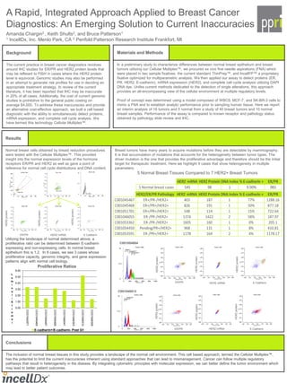
Utilizing the landscape of normal determined above, a
proliferative ratio can be determined between E-cadherin
expressing and non-expressing cells. In normal breast
epithelium this is 1.2. In 8 cases, we see 3 cases whose
proliferative capacity, genomic integrity, and gene expression
patterns align with normal cell biology.
Breast tumors have many years to acquire mutations before they are detectable by mammography.
It is that accumulation of mutations that accounts for the heterogeneity between tumor types. The
driver mutation is the one that provides the proliferative advantage and therefore should be the initial
target for therapeutic treatment. Here we highlight 9 cases that show heterogeneity in multiple
parameters:
5 Normal Breast Tissues Compared to 7 HER2+ Breast Tumors
Conclusions
The inclusion of normal breast tissues in this study provides a landscape of the normal cell environment. This cell based approach, termed the Cellular Multiplex™,
has the potential to limit the current inaccuracies inherent using standard approaches that can lead to mismanagement. Cancer can follow multiple regulatory
pathways that result in heterogeneity in the disease. By integrating cytometric principles with molecular expression, we can better define the tumor environment which
may lead to better patient outcomes.
ER/PR HER2 mRNA E-Cadherin
HER2protein
HER2protein
HER2protein
ER/PR HER2 mRNA E-Cadherin
HER2protein
HER2protein
HER2protein
ER/PR HER2 mRNA E-Cadherin
HER2protein
HER2protein
HER2protein
0.00
1.00
2.00
3.00
4.00
5.00
6.00
COO1042971
COO1045465
COO1045467
COO1045469
COO1046041
COO1049303
COO1046510
COO1049308
L
u
m
i
n
a
l
/
S
t
r
o
m
a
l
Proliferative Ratios
E-cadherin+/E-cadherin- Post G1
C001046513
C001054804
HER2 mRNA HER2 Protein DNA Index % E-cadherin + ER/PR
5 Normal breast cases 545 98 1 9.90% 983
HER2/ER/PR Pathology HER2 mRNA HER2 Protein DNA Index % E-cadherin + ER/PR
C001045467 ER+/PR-/HER2+ 403 187 1 77% 1288.16
C001045468 ER+/PR+/HER2+ 826 191 1 50% 877.18
C001051701 ER+/PR+/HER2+ 548 114 1 15% 722.64
C001046055 ER-/PR-/HER2+ 1374 1422 2 58% 187.97
C001053362 ER-/PR-/HER2+ 1605 160 1 0% 205.1
C001054450 Pending/PR+/HER2+ 968 131 1 8% 410.81
C001053591 ER-/PR+/HER2+ 1178 164 2 4% 1178.17