2. 620
Ravage et al.
in distant organs, as well. The systemic effects occur in cases with extensive
thermal trauma, but can be seen even in burn injuries covering only one quarter
of the total body surface area (TBSA). In a second-degree thermal injury model
in rats involving 25-30% TBSA, secondary lung injury has been observed. Till,
et al. (1983) described systemic complement activation, as detected by reductions in hemolytic activity of individual complement components (C3, C4, C6)
and crossed immunoelectrophoresis analysis of the conversion of serum C3
(2). Chemotaxis assays showed C5a-mediated activation of blood neutrophils
to occur, as well. Secondary accumulation of polymorphonuclear leukocytes
(PMN) has been shown to occur in lung tissues, followed by development of
acute pulmonary injury, related to production of neutrophil-derived toxic oxygen metabolites (2). Employing the same animal model of dermal burn injury,
PMN influx into lung tissues was reduced substantially (up to 77%) using antibodies to adhesion molecules (LFA-1, Mac-1, ICAM-1, E and L-selectin) (3).
The process of local edema formation in the burn wound appears to be more
complex, as it involves both the direct effect of heat and the consequences of
inflammatory mediators locally generated in response to the thermal insult. It
has been shown that under conditions of limited thermal, chemical or physical
trauma, two waves of increased vascular permeability occur in the skin (4). The
"early phase" of increased permeability—approximately one hour after thermally
injury—was shown to develop as a result of complement activation with anaphylatoxin release and mast cell secretion of histamine. This lead to an enhancement
of xanthine oxidase activity and increased production of oxygen radicals, damaging endothelial cells (5). This one hour injury has been shown to be neutrophil
independent (6).
The pathophysiology of "late phase" dermal microvascular injury (at
approximately four hours post-burn) is less well understood. The delay in onset
of this injury would indicate that a series of elaborate events must take place
before maximal tissue damage is achieved. Recent data from our group suggest
that, in contrast to the "early phase" edema formation, blood neutrophils are
involved in the pathogenesis of the "late phase" injury. Since antibodies to neutrophils, as well as, to E- and L-selectin and ICAM-1 were shown to effectively
reduce vascular leakage in the dermal burn wound at four hours post-burn (3),
we sought to clarify the mechanism for neutrophil recruitment and infiltration by
focusing on the upstream inflammatory mediators that may regulate these events.
The current studies were designed to elucidate the role of the cytokines IL-6,
IL-1 and TNFa, as well as complement, and to further clarify the role of neutrophils in the development of microvascular injury in the second-degree, "late
phase" dermal burn wound. Here, we present evidence which demonstrates that
the four hour burn injury is complement independent, involves the proinflammatory cytokines IL-6, IL-1 and TNFa and may result from the tissue-damaging
effects of neutrophil-derived reactive oxygen species.
3. Mediators of Injury in Dermal Burn Wounds
621
MATERIALS AND METHODS
Animal Model of Thermal Injury
The experimental burn model used in the present study has been described previously (2, 6,
7). Adult male, specific pathogen-free Long-Evans rats (300-350 g, Marian Sprague-Dawley, Indianapolis, Indiana) were used in all experiments. Ketamine hydrochloride (100 mg/kg body weight)
(Fort Dodge Laboratories, Fort Dodge, Iowa) and xylazine (13 mg/kg body weight) (Bayer Corporation, Shawnee Mission, Kansas) were administered intraperitoneally and intramuscularly, respectively, throughout the experiment. This ensured that the animals were properly anesthetized for the
entire procedure, from the induction of the burn injury to the time of sacrifice. The skin over the
lumbrosacral and dorsal flank areas was shaved and exposed to 70°C water for 30 s. This resulted
in a deep second-degree skin burn involving 25 to 30% of the total body surface area. Animals
were sacrificed at 4 h by cervical dislocation. Control animals were exposed to 22°C water. All
experiments were in accord with the standards in The Guide for the Care and Use of Laboratory
Animals, and were supervised by veterinarians from the Unit for Laboratory and Animal Care of
the University of Michigan Medical School.
Measurement of Microvascular Injury. Local microvascular injury was assessed by measurement of extravasation of radiolabeled bovine serum albumin ( 125 I-BSA) into the burned skin. Immediately prior to thermal injury, burn or sham-treated animals received an intravenous injection of 0.5
uCi of 125I-BSA in 0.5 ml sterile phosphate buffered saline (PBS). Using a template, four uniform
skin samples, each one square inch in size, were excised from the burned area on each animal. For
calculations of the permeability index, the amount of radioactivity ( I25 I-BSA) in skin biopsies was
compared to the amount of radioactivity present in 1.0 ml of blood obtained from the inferior vena
cava at the time of sacrifice (4 h).
Measurement of Skin Myeloperoxidase (MPO) Content. Local accumulation of neutrophils
was assessed by measurement of myeloperoxidase in skin biopsies. Animals received burn or sham
treatment as described above. At time of sacrifice (4 h), four 4 mm punch biopsies from standardized
areas of the wound were taken from each animal and instantly frozen in liquid nitrogen. The biopsies
were homogenized in 500 ul of PBS pH 7.4, containing 0.1% Tween 20, sonicated on ice and
insoluble material removed by centrifugation at 3000 rpm for 10 min. 5 ul of tissue extract (PBS
pH 7.4 and 0.1% Tween 20) were incubated with 100 ul of 2,2'-Azino-di-[3-ethylbenzthiozoline
sulfonate (6)] diammonium salt solution (ATBS substrate) (Boehringer Mannheim, BIOCHEMICA,
Germany) and the maximum velocity of the substrate/MPO chromogenic reaction (Vmax) measured
by monitoring the 96 well low-protein binding flat bottom plates (Corning Glass Works, Corning,
New York) at 405 nm over a two minute period (BioTek Elx808 microplate reader) (BIO-TEK
Instruments, INC., Winooski, Vermont). Kinetic calculations were performed using KC3 software
(BIO-TEK Instruments, Inc.). MPO concentrations in samples were determined using a standard
curve of purified MPO (CALBIOCHEM, San Diego, California). MPO values are reported as units
of activity/biopsy.
Interventional Studies
Cytokine Blockade. Irrelevant IgG antibody, anti-mouse IL-6 polyclonal antibody, and antirat IL-10 monoclonal antibody were obtained from R&D Systems, Minneapolis, Minnesota. Antirat TNFa polyclonal antibody were purchased from PeproTech, INC., Rocky Hill, New Jersey. In
4. 622
Ravage et al.
each case, antibodies were given in a total amount of 500 ug per animal, in 0.5 ml sterile PBS,
administered intravenously in two equal doses at 30 and 120 min post-burn.
Complement Depletion. Cobra venom factor (CVF) was purified from crude lyophilized
cobra venom (Naja naja kaouthia) (Sigma Chemical Company, St. Louis, Missouri) by ion exchange
chromatography and gel filtration (8). Complement depletion was achieved by serial intraperitoneal
injections of 4 X 20 units CVF in 12 h intervals, resulting in undetectable levels of serum hemolytic
complement activity (CH50 Assay). The experiments were performed 12 h after the final injection
of CVF.
C5a Blockade. Isolation of polyclonal antibody to C5a was performed as described by Mulligan, et al. (9). Briefly, animals were immunized with rat C5a. Obtained serum was IgG purified by
acid elution of Sepharose G beads (Pharmacia Biotech AB, Uppsala, Sweden), followed by extensive
dialysis against PBS. Characterization of the anti-rat C5a antibody was performed by immunoprecipitation and Western blot analysis showing a single band at 14 kDa. This antibody was given in
a total amount of 400 ug per animal, administered intravenously in two equal doses at 30 and 120
min post-burn.
Neutrophil Depletion. Neutrophil depletion was induced by the intraperitoneal injection of
1.0 ml of rabbit antiserum to rat PMN (Accurate, Westbury, New York) 16 h prior to the experiment.
This procedure reduced neutrophil counts in peripheral blood by >90 percent.
Hydroxyl Radical Scavenger Administration. Dimethyl thiourea (DMTU) (Sigma Chemical
Co.) (1000 mg/kg body weight) in 1.0 ml sterile PBS was injected intraperitoneally 10 min prior
to thermal injury. Dimethyl sulfoxide (DMSO) (Sigma Chemical Co.) (500 ug) in 1.0 ml sterile
PBS was injected intraperitoneally 10 min prior to thermal injury. The effectiveness of the chosen
concentrations of scavengers was demonstrated in earlier studies (7).
Statistical Analysis. Data sets were analyzed using one-way ANOVA. Individual group
means were compared with the Tukey multiple comparison test. All values were expressed as mean
± SEM. Significance was assigned where P < 0.05. For percentage change between groups, values obtained from negative controls were subtracted from each data point. Statistical analysis was
performed using SigmaStat 2.0 (Jandel Scientific Software, San Rafael, California).
RESULTS
Protective Effects of Neutrophil Depletion in Dermal Burn Injury. Neutropenia was achieved by intraperitoneal injection of antiserum to rat PMN.
Extravasation of 125I-bovine serum albumin into the skin 4 h after thermal trauma
was used to measure tissue injury. The results of neutropenia on the development
of increased vascular permeability in the skin are shown in Figure 1. Negative
controls had a permeability index of 0.049 ± 0.039. Neutrophil depletion was
associated with a 58% (P < 0.001) attenuation of the dermal vascular permeability four hours after thermal injury (permeability index of 0.405 ± 0.038 in
neutrophil depleted rats versus an index of 0.898 ± 0.039 in nontreated rats).
Thus, availability of PMNs seems to be required for the full development of
dermal microvascular injury four hours after thermal trauma.
Failure of Complement Depletion to Protect Against Dermal Microvascular Injury. Complement depletion was induced with CVF as described above.
Extravasation of 125I-bovine serum albumin into the skin four hours after ther-
5. Mediators of Injury in Dermal Burn Wounds
623
Fig. 1. Effects of complement depletion and neutrophil depletion on dermal vascular injury 4 h
after thermal trauma to skin as measured by leakage of 125I-labeled bovine serum albumin. For
each vertical bar, N = 4.
mal trauma was used to assess tissue injury. The results of complement depletion
on the development of increased vascular permeability in the skin are shown in
Figure 1. Complement depleted rats had a permeability index of 0.88 ± 0.065
versus an index of 0.898 ± 0.039 in non-treated rats (PN.S). Thus, complement
depletion was not associated with a significant reduction in dermal vascular permeability compared with positive controls.
Failure of C5a Blockade to Protect Against Dermal Microvascular Injury.
In the presence of antibody to C5a, vascular permeability index was calculated.
Anti-C5a-treated rats had an index of 0.89 ± 0.054, as compared with an index of
6. 624
Ravage et al.
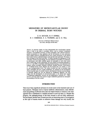
Fig. 2. Effects of cytokine blockade on dermal vascular injury 4 h after thermal trauma to skin as
measured by leakage of 125I-labeled bovine serum albumin. For each vertical bar, n = 5 or higher.
and 0.89 ± 0.047 in positive controls (PN.S.). C5a blockade was not associated
with a significant reduction in dermal vascular permeability compared with positive controls. Thus, C5a does not seem to be required for dermal microvascular
injury four h after thermal trauma, providing further evidence that development
of the "late phase" burn wound is complement independent.
Protective Effects of Cytokine Blockade Against Dermal Microvascular
Injury. Protection against increased vascular permeability in skin 4 h after thermal injury was evaluated through the use of blocking antibodies to cytokines.
Vascular permeability index was calculated as described above. The negative
control skin permeability index was 0.049 ± 0.002; this value increased to 0.885
± 0.048 in positive controls, as shown in Figure 2. Treatment with anti-IL-6 was
7. Mediators of Injury in Dermal Burn Wounds
625
Fig. 3. Effects of hydroxyl radical scavengers on dermal vascular injury 4 h after thermal trauma
to skin as measured by leakage of 125I-labeled bovine serum albumin. For each vertical bar, n = 4.
associated with a 53% (P < 0.001) reduction in dermal vascular permeability to
0.47 ± 0.031. Treatment with antibody to IL-1 resulted in a decrease of dermal
vascular permeability by 34% (P < 0.001) to a value of 0.63 ± 0.068. Similarly, animals treated with anti-TNFa had a mean dermal vascular permeability
index of 0.60 ± 0.068, which was 37% (P < 0.001) lower than the positive control group. Thus, dermal microvascular injury at four hours after thermal trauma
requires the cytokines IL-6, IL-1 and TNFa for full development.
Protective Effects of Hydroxyl Radical Scavengers in the Skin Burn Wound.
The effects of hydroxyl radical scavengers on vascular permeability in skin
4 h after thermal injury was assessed by determination of vascular permeability index. The data are shown in Figure 3. Treatment with dimethyl thiourea
(DMTU) was associated with a 56% (P < 0.001) reduction in vascular perme-
8. 626
Ravage et al.
Table 1. Protection Against Neutrophil Influx into Thermally Injured Skin
Treatment
No. of
animals
None
anti-IL-6
anti-IL-1
anti-TNFoi
anti-PMN
4
4
4
4
4
MPO Value
(x ± SEM)
23.81
8.70
9.34
7.41
6.94
± 0.20
+0.81
± 1.60
±0.11
± 0.33
Significance
(P values)
Change
(%)a
<0.001
<0.001
<0.001
<0.001
<0.001
-67
-64
-73
-75
a
For percentage change between groups, values obtained from negative controls were subtracted
from each data point.
ability as compared to the positive controls (0.426 + 0.070 for DMTU treated
rats, 0.898 ± 0.039 in non-treated rats). Treatment with dimethyl sulfoxide
(DMSO) resulted in a 46% (P < 0.001) reduction in dermal vascular permeability (0.508 ± 0.081 with DMSO treatment, 0.898 ± 0.039 without). Thus,
blockade of hydroxyl radicals results in a marked protective effect on dermal
microvascular injury four hours after thermal trauma, indicating an important
role for these radicals in the "late phase" local burn injury.
Role of PMN in the Dermal Burn Wound. Skin samples from injured animals treated with irrelevant antibodies or specific antibodies directed against the
cytokines IL-6, IL-1 and TNFa or against rat PMNs were collected and assessed
for MPO activity as a measure of tissue accumulation of neutrophils. Negative
control animals showed values of MPO content of 1.31 ± 0.019, increasing to
23.8 ± 0.20 in samples from positive controls. The results of interventional studies of vascular leakage (Figure 1) were noticeably similar to PMN accumulation
in these treatment groups (Table 1). Anti-IL-6 treated animals showed a MPO
value of 8.7 ± 1.6 which represents a 67% (P < 0.001) reduction in PMN accumulation. Anti-IL-1-treated animals showed a 64% (P < 0.001) decrease in MPO
value to 9.3 ± 3.2. Anti-TNFa-treated animals displayed an MPO value of 7.4
± 0.23, a reduction of 73% (P < 0.001) as compared to positive controls. As
would be expected, neutrophil depletion resulted in a 75% (P < 0.001) reduction in MPO value to 6.9 ± 0.66. Therefore, blockade of IL-6, IL-1 and TNFa
greatly reduced tissue MPO activity, demonstrating a clear requirement for these
cytokines in neutrophil accumulation during the "late phase" dermal burn injury.
DISCUSSION
Our data indicate that development of the late phase of microvascular leakage (four h post-burn) in thermally injured rat skin requires the pro-inflammatory
9. Mediators of Injury in Dermal Burn Wounds
627
cytokines IL-6, IL-1 and TNFa. The ability of blocking antibodies to each of the
aforementioned cytokines to attenuate both vascular injury and polymorphonuclear leukocyte (PMN) influx into the burn wound suggests that these cytokines
represent upstream mediators in this inflammatory process. These cytokines may
affect vascular endothelial cells (EC), which are known to actively participate
in the development of inflammatory reactions by controlling fluid leakage and
promoting adhesion and activation of leukocytes, or target PMN. With regard to
IL-1 and TNFa, there is a large body of evidence detailing the ability of these
cytokines to activate EC to synthesize and express adhesion molecules (10-14).
The adhesion molecules ICAM-1 and E- and L-selectin have previously been
shown to be required for full development of vascular injury in the "late phase"
edema formation of thermally injured rat skin (3). In addition, IL-1 and TNFa
have been shown to induce cellular production of IL-6 (15, 16).
There are several ways in which IL-6 may exert pro-inflammatory effects
on increased vascular permeability and PMN influx in acute local thermal injury.
There is in vitro evidence that IL-6 increases endothelial permeability by rearranging actin filaments and by changing the shape of endothelial cells (17). Biffl,
et al., have demonstrated that with platelet-activating factor, IL-6 potentiates
PMN priming and delays PMN apoptosis (18, 19), both effects which contribute
to PMN-mediated tissue damage. Furthermore, Mullen et al. demonstrated that
IL-6 is capable of interacting synergistically with TNFa to augment the effect
of TNFa on PMN phagocytosis and superoxide production in vitro (20). The
authors hypothesized IL-6 to be a more distal mediator of the cytokine cascade,
which may modulate an inflammatory response to trauma initiated by other, more
proximal cytokines. This supports our data which show IL-6 blockade to be the
most effective of the cytokine interventions in reducing dermal vascular injury
after thermal trauma. Lastly, expression of ICAM-1 on myocytes is induced by
IL-6 in a cardiac ischemia/reperfusion injury model (21). Given the established
role of ICAM-1 and E- and L-selectin in the development of "late phase" edema
in thermally injured skin, it is possible that IL-6 may exert a similar effect on
EC in the pathogenesis of burns, resulting in increased PMN recruitment and
infiltration into injured tissues.
PMN depletion resulted in a significant decrease in both vascular leakage
and PMN accumulation in skin at 4 h. This is in contrast to the 1 h burn wound
for which PMN involvement was not required (7). The delay in burn wound
edema formation may be explained by the time necessary for the cytokine cascade to cause expression of adhesion molecules in the injured skin and activate
PMNs. The notion that increased tissue damage then results from PMN-derived
reactive oxygen species is supported by our data showing the ability of hydroxyl
radical scavengers in these experiments to reduce vascular injury. The fact that
PMN depletion, or any of the other interventions, was only able to result in a
maximum reduction of approximately 60% in vascular leakage is most likely
10. 628
Ravage et al.
due to the possibility that a large percentage of injury and cell death (approximately 40%) is directly heat-related. A similar phenomenon was observed in the
assay of skin MPO content.
It is known that the burn model utilized in these experiments produces systemic complement activation that initiates a series of events leading to the "early
phase" edema formation in the burned skin. Interestingly, neither complement
depletion nor C5a blockade was able to attenuate local vascular permeability
at four hours. In this case, the effects of cytokines and PMNs may be able to
produce maximal injury even in the absence of complement. Another possibility
is that the role of complement is fulfilled in the early phase, whereas the late
phase is more dependent on the ensuing inflammatory reaction. Despite the fact
that our data does not support a role for complement in the "late phase" burn
wound, a complete lack of involvement of complement components cannot be
stated. Recently, studies have revealed local complement production in numerous
and varied tissues in an ischemia/reperfusion injury in rabbit (22). Complement
proteins may be present in thermally injured tissue and contributing to the development of the 4 h injury, but were not effectively blocked by our antibody to
C5a or were able to be produced in significant amounts in skin, though serum
complement levels were undetectable following treatment with CVF. Administered in one dose immediately prior to induction or thermal trauma, 400 ug of
anti-C5a was able to slightly, though not significantly, reduce vascular leakage
at four hours (data not shown).
We therefore conclude that the development of the "late phase" dermal vascular injury following thermal trauma to the skin is largely mediated by the
pro-inflammatory actions of cytokines, in particular IL-6. IL-6 may modulate
the effects of TNFa and IL-1, resulting in EC expression of E- and L-selectin
and ICAM-1. IL-6 also may act directly on EC to alter cell structure and/or
promote upregulation of adhesion molecules. PMN-generated reactive oxygen
species appear to be responsible for the local tissue damage. Complement does
not appear to play a significant role, if any, in the pathogenesis of the "late phase"
second-degree burn wound.
REFERENCES
1. MANN, R. and D. HEIMBACH. 1996. Prognosis and treatment of burns. West. J. Med.
165:215-220.
2. TILL, G. O., C. BEAUCHAMP, D. MENPACE, W. TOURTELLOTTE Jr., R. KUNKEL, K. J. JOHNSON,
and P. A. WARD. 1983. Oxygen radical dependent lung damage following thermal injury of rat
skin. J. Trauma. 23:269-273.
3. MULLIGAN, M. S., G. O. TILL, C. W. SMITH, D. C. ANDERSON, M. MIYASAKA, T. TAMATANI,
R. F. TODD, III, T. B. ISSEKUTZ, and P. A. WARD. 1994. Role of leukocyte adhesion molecules
in lung and dermal vascular injury after thermal trauma of skin. Am. J. Pathol. 144:1008-1015.
11. Mediators of Injury in Dermal Burn Wounds
629
4. DEMLING, R. H. 1985. Burns. N. Engl. J. Med. 313: 1389-1398.
5. FREIDL, H. P., G. O. TILL, O. TRENTZ, and P. A. WARD. 1989. Roles of histamine, complement
and xanthine oxidase in thermal injury of skin. Am. J. Pathol. 135:203-217.
6. TILL, G. O., L. S. GUILDS, M. MAHROUGUI, H. P. FREIDL, O. TRENTZ, and P. A. WARD. 1989.
Role of xanthine oxidase in thermal injury of skin. Am. J. Pathol. 135:195-202.
7. TILL, G. O., J. R. HATHERILL, W. w. TOURTELLOTTE, M. J. LUTZ, and P. A. WARD. 1985. Lipid
peroxidation and acute lung injury after thermal trauma to skin: Evidence of a role for hydroxyl
radical. Am. J. Pathol. 135:195-202.
8. BALLOW, M. and C. G. COCHRANE. 1969. Two anti-complementary factors in cobra venom.
Hemolysis of guinea pig erythrocytes by one of them. J. Immunol. 103:944-952.
9. MULLIGAN, M. S., E. SCHMID, B. BECK-SCHIMMER, G. O. TILL, H. P. FRIEDL, R. B. BRAUER,
T. E. HUGLI, M. MIYASAKA, R. L. WARNER, K. J. JOHNSON, and P. A. WARD. 1996. Requirement and role of C5a in Acute Lung Inflammatory Injury in Rats. J. Clin. Invest. 98:503-512.
10. PROBER, J. S. and R. S. COTRAN. 1990. The role of endothelial cells in inflammation. Transplantation. 50:537-544.
11. BEVILACQUA, M. P., J. S. PROBER, D. L. MENDRICK, R. S. COTRAN, and M. A. GIMBRONE, JR.
1987. Identification of an inducible endothelial-leukocyte adhesion molecule. Proc. Natl. Acad.
Sci. U.S.A. 84:9238-9242.
12. BEVILACQUA, M. P., S. STENGELIN, M. A. GIMBRONE, and B. SEED. 1989. Endothelial leukocyte
adhesion molecule 1: An inducible receptor for neutrophils related to complement regulatory
proteins and lectins. Science 243:1160-1165.
13. OSBORN, L., C. HESSION, R. TIZARD, C. VASSALLO, S. LUHOWSKYJ, G. Cm-Rosso, and R.
LOBB. 1989. Direct expression cloning of vascular cell adhesion molecule 1, a cytokine-induced
endothelial protein that binds to lymphocytes. Cell 60:577-584.
14. DUSTIN, M. A. and T. A. SPRINGER. 1988. Lymphocyte function-associated antigen-1 (LFA-l)
interaction with intercellular adhesion molecule-1 (ICAM-1) is one of at least three mechanisms
for lymphocyte adhesion to cultures endothelial cells. J. Cell Biol. 107:321-331.
15. CONTENT, J., L. DE WIT, P. POUPART, G. OPDENAKKER, J. VAN DAMME, and A. BILLIAU. 1985.
Induction of a 26-kDa-protein mRNA in human cells treated with and interleukin-1-related,
leukocyte-derived factor. Eur. J. Biochem. 152:253-257.
16. KASID, A., E. P. DIRECTOR, and S. A. ROSENBERG. 1989. Regulation of interleukin-6 (IL-6)
by JL-2 and TNF-a in human peripheral blood mononuclear cells. Ann. N.Y. Acad. Sci.
557:564-566.
17. MARUO, N., I. MORITA, M. SHIRAO, and S. MUROTA. 1992. IL-6 increases endothelial permeability in vitro. Endocrinology 131:710-714.
18. BIFFL, W. L., E. E. MOORE, F. A. MOORE, V. S. CARL, F. J. KIM, and R. J. FRANCIOSE.
1994. Interleukin-6 potentiates neutrophil priming with platelet-activating factor. Arch. Surg.
129:1131-1136.
19. BIFFL, W. L., E. E. MOORE, F. A. MOORE, and C. C. BARNETT. 1996. Interleukin-6 delays neutrophil apoptosis via a mechanism involving platelet-activating factor. J. Trauma 40:575-579.
20. MULLEN, P. G., A. C. J. WINDSOR, C. J. WALSH, A. A. FOWLER, III, and H. J. SUGARMAN.
1995. Tumor necrosis factor-a and IL-6 selectively regulate neutrophil function in vitro. J. Surg.
Res. 58:124-130.
21. YOUKER, K., C. W. SMITH, D. C. ANDERSON, D. MILLER, L. H. MICHAEL, R. D. ROSSEN, and
M. L. ENTMAN. 1992. Neutrophil adherence to isolated adult cardiac myocytes: Induction by
cardiac lymph collected during ischemia and reperfusion. J. Clin. Invest. 89:602-609.
22. YASOJIMA, K., K. S. KILGORE, R. A. WASHINGTON, B. R. LUCCHESI, and P. L. MCGEER. 1998.
Complement gene expression by rabbit heart: Upregulation by ischemia and reperfusion. Circ.
Res. 82:1224-1230.