Tulathromycin, a macrolide antibiotic, has anti-inflammatory and immunomodulatory effects in addition to its antimicrobial properties. This study investigated these effects of tulathromycin both in vitro using pig leukocytes and in vivo in pigs challenged with Actinobacillus pleuropneumoniae or zymosan. In vitro, tulathromycin induced neutrophil apoptosis in a time- and concentration-dependent manner and enhanced clearance of apoptotic neutrophils by macrophages. In vivo, tulathromycin promoted leukocyte apoptosis and clearance, reduced the proinflammatory mediator leukotriene B4, and attenuated lung damage in A. pleuropneumoniae-infected
1. AJVR •Vol 76 • No. 6 • June 2015 507
Self-perpetuating inflammation contributes to the
pathogenesis of Actinobacillus pleuropneumoni-
ae infection in swine.1 Excessive extravasation of
Immunomodulatory effects of tulathromycin
on apoptosis, efferocytosis, and proinflammatory
leukotriene B4 production in leukocytes
from Actinobacillus pleuropneumoniae–
or zymosan-challenged pigs
Stephanie C. Duquette MSC
Carrie D. Fischer PhD
Alison C.Williams
Saman Sajedy
Troy D. Feener MSC
Amol Bhargava MSC
Kristen L. Reti MSC
Gregory P. Muench DVM
Douglas W. Morck DVM,PhD
Jim Allison BVSC
Merlyn J. Lucas DVM,MS
Andre G. Buret PhD
Received June 16, 2014.
Accepted November 14, 2014.
From the Department of Biological Sciences, In-
flammation Research Network (Duquette, Fischer,
Williams, Sajedy, Feener, Bhargava, Reti, Morck, Buret),
the Animal Health Unit (Muench, Morck), the Depart-
ment of Comparative Biology and Experimental Medi-
cine (Morck), Faculty of Veterinary Medicine, University
of Calgary, Calgary, AB T2N 4N1, Canada; Zoetis, 100
Campus Dr, Florham Park, NJ 07932 (Allison); andVeter-
inary Research and Development,Zoetis,333 Portage St,
Kalamazoo, MI 49001 (Lucas).
Address correspondence to Dr. Buret (aburet@
ucalgary.ca)
OBJECTIVE
To investigate the anti-inflammatory and immunomodulatory properties of
tulathromycin in vitro and in experimental models of Actinobacillus pleuropneu-
moniae–induced pleuropneumonia and zymosan-induced pulmonary inflam-
mation in pigs.
ANIMALS
Blood samples from six 8- to 30-week-old healthy male pigs for the in vitro
experiment and sixty-five 3-week-old specific pathogen–free pigs.
PROCEDURES
Neutrophils and monocyte-derived macrophages were isolated from blood
samples. Isolated cells were exposed to tulathromycin (0.02 to 2.0 mg/mL)
for various durations and assessed for markers of apoptosis and efferocy-
tosis. For in vivo experiments, pigs were inoculated intratracheally with
A pleuropneumoniae,zymosan,or PBS solution (control group) with or without
tulathromycin pretreatment (2.5 mg/kg, IM). Bronchoalveolar lavage fluid was
collected 3 and 24 hours after inoculation and analyzed for proinflammatory
mediators, leukocyte apoptosis, and efferocytosis.
RESULTS
In vitro, tulathromycin induced time- and concentration-dependent apoptosis
in neutrophils, which enhanced their subsequent clearance by macrophages.
In the lungs of both A pleuropneumoniae– and zymosan-challenged pigs, tu-
lathromycin promoted leukocyte apoptosis and efferocytosis and inhibited
proinflammatory leukotriene B4 production, with a concurrent reduction in
leukocyte necrosis relative to that of control pigs.Tulathromycin also attenu-
ated the degree of lung damage and lesion progression in A pleuropneumoniae–
inoculated pigs.
CONCLUSIONS AND CLINICAL RELEVANCE
Tulathromycin had immunomodulatory effects in leukocytes in vitro and anti-
inflammatory effects in pigs in experimental models of A pleuropneumoniae
infection and nonmicrobial-induced pulmonary inflammation. These data
suggested that in addition to its antimicrobial properties, tulathromycin may
dampen severe proinflammatory responses and drive resolution of inflamma-
tion in pigs with microbial pulmonary infections. (Am J Vet Res 2015;76:507–
519)
neutrophils and their subsequent uncontrolled death
at the site of inflammation can lead to local release
of proteolytic enzymes, reactive oxygen species, and
potent proinflammatory mediators that exacerbate
the inflammatory response and amplify the degree
of tissue injury.2 Leukotriene B4, a proinflammatory
lipid mediator derived from arachidonic acid, acts as
a potent neutrophil chemoattractant and is an impor-
tant marker of inflammation.3,4 Similarly, the release
of LDH is a prominent marker of cellular necrosis
and hallmark of pulmonary tissue damage5 caused by
A pleuropneumoniae in swine.
In homeostatic conditions, infiltrating immune
cells undergo programmed cell death (apoptosis) and
ABBREVIATIONS
BALF Bronchoalveolar lavage fluid
HBSS Hanks balanced salt solution
HI-FBS Heat-inactivated fetal bovine serum
IL Interleukin
LDH Lactate dehydrogenase
LTB4 Leukotriene B4
NAD Nicotinamide adenine dinucleotide
NF-κB Nuclear factor κB
TUNEL Terminal deoxynucleotidyl transferase–mediated
dUTP nick-end labeling
14-06-0160r.indd 507 5/18/2015 11:07:38 AM
2. 508 AJVR •Vol 76 • No. 6 • June 2015
are subsequently cleared from the tissues by resident
macrophages.6 During apoptosis, senescent cells be-
come desensitized to inflammatory stimuli and cellu-
lar organelles are disassembled and degraded within
apoptotic bodies while cellular membranes remain
intact; this prevents release of injurious cellular con-
tents into the extracellular environment.7,8 Apop-
totic bodies then signal for their own nonphlogistic
clearance by resident macrophages.9–11 In situations
of severe inflammation, excessive recruitment of
neutrophils can overwhelm homeostatic clearance
mechanisms.The neutrophils then undergo necrosis,
which is associated with a loss of cellular membrane
integrity and the release of cytotoxic cellular con-
tents, consequently damaging surrounding cells and
perpetuating the inflammatory response.12 This exag-
gerated and self-amplifying inflammatory response is
common in bacterial-induced pneumonia, including
infection with A pleuropneumoniae. Medications
capable of breaking this amplification cycle and re-
storing homeostatic balance through resolution of
inflammation would be useful in the treatment of in-
flammatory disease.
Actinobacillus pleuropneumoniae is a highly
contagious, gram-negative coccobacillus13,14 that is
transmitted rapidly through pig herds in which ad-
equate biosecurity is lacking. Infection with NAD-
dependent strains of A pleuropneumoniae can result
in signs of severe disease within 12 hours after expo-
sure and in death within 24 to 36 hours thereafter.15
Gross examination of lungs from infected pigs reveals
demarcated lesions in the middle, cranial, and caudal
lobes that are characterized by pulmonary consolida-
tion, fibrosis, and necrosis.1,16 Histologic examination
of the lungs 3 hours after intratracheal inoculation
with A pleuropneumoniae reveals alveolar edema, fi-
brin,and severe neutrophil infiltration.1 Large coalesc-
ing foci of necrosis, hemorrhage, and edema can be
seen 24 hours after inoculation.1
The vast degree of infiltration and activation of
neutrophils within the lungs contributes considerably
to disease progression. Indeed, treatment of pigs with
indomethacin, a cyclooxygenase inhibitor capable of
inhibiting neutrophil activation, prior to intratracheal
inoculation with A pleuropneumoniae delayed devel-
opment of pulmonary lesions in 1 study.1 Those find-
ings suggested a primary role for neutrophil activation
in tissue damage associated with A pleuropneumoni-
ae infection. Moreover, treatment with tilmicosin, a
macrolide with proapoptotic effects in neutrophils,
led to a significant decrease in the number of pulmo-
nary lesions following experimental A pleuropneu-
moniae infection in piglets,17 providing further evi-
dence of a role for neutrophils in mediating necrotic
tissue damage associated with infection.It follows that
medications that promote apoptosis of neutrophils
and their subsequent nonphlogistic removal by resi-
dent phagocytes may be particularly efficacious in the
treatment of inflammatory diseases such as A pleuro-
pneumoniae pleuropneumonia in swine.
Macrolide antimicrobials can modulate the host
immune response18 and thus may be useful in the
treatment of infectious diseases involving a patho-
genic inflammatory response. Indeed, macrolides ex-
ert several anti-inflammatory effects by inhibiting re-
cruitment of neutrophils19; reducing the production
of proinflammatory cytokines and chemokines such
as tumor necrosis factor-β, IL-1β,20,21 and IL-822–24; pre-
venting the activation of proinflammatory NF-κB.25,26
Some macrolides can also promote cellular death by
apoptosis,27–31 which may provide additional benefit
by contributing to the resolution of inflammation.
However,the precise mechanisms underlying the anti-
inflammatory and proapoptotic properties of macro-
lides remain incompletely understood.
Tulathromycin is a macrolide antimicrobial de-
rived from erythromycin that is approved for use in
the treatment and prevention of respiratory disease
in swine and cattle. Success of tulathromycin for use
in treating respiratory diseases may be attributed, at
least in part, to its preferential accumulation within
pulmonary tissues.18,32 This antimicrobial has a high
affinity for absorption within neutrophils and macro-
phages,32,33 which aids in its rapid delivery to the site
of inflammation.The anti-inflammatory and proapop-
totic properties of tulathromycin have been demon-
strated in bovine neutrophils31 and macrophages,34
and these properties may enhance the antimicrobial’s
efficacy in the treatment of bovine respiratory disease.
Moreover, our research group has shown that tulath-
romycin also exerts proresolution effects (promoting
the resolution of inflammation) in bovine neutrophils
by influencing the production and release of lipid
mediators.35
The purpose of the study reported here was to
investigate whether tulathromycin would confer
anti-inflammatory and proresolution benefits through
mechanisms similar to those in cattle when used for
the treatment of respiratory disease in swine. Specifi-
cally, we wanted to investigate immunomodulatory
effects of tulathromycin on porcine neutrophils and
macrophages in 3 complementary experiments: ex-
perimentally induced A pleuropneumoniae infection
in young pigs, zymosan-triggered pulmonary inflam-
mation in the absence of bacterial stimuli in young
pigs, and in vitro assessment of the effects of tulath-
romycin on leukocytes obtained from young healthy
pigs.We hypothesized that tulathromycin would exert
proapoptotic effects on neutrophils and facilitate non-
phlogistic removal of the dying cells by macrophage
efferocytosis. Moreover, we hypothesized that tulath-
romycin would have additional anti-inflammatory ef-
fects in vivo by inhibiting the production of potent
proinflammatory mediators such as LTB4, as has been
demonstrated in a model of bovine respiratory dis-
ease.31 Taken together, the findings would further elu-
cidate mechanisms through which tulathromycin may
contribute to the resolution of severe inflammation
that is characteristic of bacterial pleuropneumonia in
swine.
14-06-0160r.indd 508 5/18/2015 11:07:38 AM
3. AJVR •Vol 76 • No. 6 • June 2015 509
Materials and Methods
Animals
Six healthy 8- to 30-week old castrated male pigs
initially weighing 15 to 20 kg were obtained from a
herd free of Mycoplasma hyopneumoniae infection
and retained as blood donors to assess the effects of
tulathromycin on leukocytes in vitro.Sixty-five healthy
3- to 4-week-old weaned male pigs weighing 4.5 to
11 kg were obtained from the same herd and used for
in vivo experiments involving infection with live A pleu-
ropneumoniae or inoculation with abiotic zymosan.
Protocol for the in vitro experiment
To obtain leukocytes for the in vitro experiment,
blood samples were collected from the cranial vena
cava of the healthy male pigs via 16-gauge, 2-inch
needles into evacuated tubes containing 1.5 mL of
citrate-dextrose solution.a Neutrophils were extracted
and purified by means of differential centrifugation
and hypotonic lysis. Briefly, blood was centrifuged
at 1,200 X g for 20 minutes at 4°C. Plasma and buffy
coat fractions were removed, and the remaining cells
were washed with an equal volume of HBSSb with-
out calcium chloride and recentrifuged at 1,200 X g
for 10 minutes at 4°C. The resulting cellular pellet
was resuspended in HBSS, and contaminating eryth-
rocytes were eliminated by the addition of 20 mL of
cold filter-sterilized hypotonic lysis solution (10.6mM
Na2HPO4 and 2.7mM NaH2PO4).Isotonicity was subse-
quently restored with 10 mL of cold 3X hypertonic re-
storing solution (10.6mM Na2HPO4, 2.7mM NaH2PO4,
and 462mM NaCl).The lysis procedure was repeated
twice, and the cell pellet was resuspended in HBSS
containing 10% HI-FBSc to optimize the cell environ-
ment.With a hemocytometer,d neutrophil viability was
assessed on the basis of the percentage of cells that
excluded 0.1% trypan blue solution.e Differential cell
counts were performed on stainedf cytocentrifugeg
preparations to assess neutrophil purity. All neutro-
phil preparations used for in vitro experiments were
assessed to be > 90% pure and > 90% viable.
For monocyte isolation and macrophage differ-
entiation, whole blood was pooled and centrifuged
at 1,200 X g for 20 minutes at 4°C to isolate the
buffy coat.The buffy coat was diluted 1:1 with filter-
sterilized saline (0.9% NaCl) solution, layered onto a
polysucrose and sodium diatrizoate gradient, and cen-
trifuged at 1,500 X g for 40 minutes at 4°C.The cell
suspension was then collected, washed with HBSS,
and centrifuged at 500 X g for 10 minutes at 4°C.Con-
taminating erythrocytes were removed by hypotonic
lysis in 10 mL of cold sterile double-distilled water
for 30 seconds, followed by the addition of 20 mL
of cold 2X HBSS to restore isotonicity. Mononuclear
cells were then resuspended in Iscove modified Dul-
becco medium containing 10% HI-FBSc and cultured
for macrophage differentiation. Cells were counted
with a hemocytometerd and viability was assessed on
the basis of the percentage of cells that excluded 0.1%
trypan blue solution.e Differential cell counts were
performed on stainedf, cytocentrifugedg preparations
to assess monocyte purity.For macrophage differentia-
tion,mononuclear cells (2 X 10⁵ cells/well) were plat-
ed onto 48-well plates.h Cells were incubated in Iscove
modified Dulbecco medium containing 10% HI-FBS
for 60 minutes in a humidified incubator at 37°C with
5% CO2 and 95% air.Nonadherent cells were removed
by washing 3 times with warm (37°C) HBSS. Remain-
ing adherent monocytes were incubated at 37°C and
5% CO2 in Iscove modified Dulbecco medium contain-
ing 10% HI-FBS, penicillin (100 U/mL), streptomycin
(100 U/mL),and tylosin (80 µg/mL) for 7 days to allow
for macrophage differentiation. Culture medium was
replenished every 2 to 3 days.Macrophage differentia-
tion was > 95% pure throughout the study and > 95%
mature at 7 days.
Assessment of the effects of tulathromycin
on leukocytes—To assess in vitro proapoptotic ef-
fects of various concentrations of tulathromycin, pu-
rified porcine neutrophils (1 X 10⁶ cells) were incu-
bated with tulathromycini (0.02 mg/mL to 2.0 mg/
mL), staurosporine (1.0µM), or 10% HI-FBS in HBSS
(control treatment) at 37°C and 5% CO2 for 0.25 to
2 hours. Apoptosis was measured with an ELISA kitj
in accordance with the manufacturer’s instructions.
The ELISA specifically measured the histone region
(H1, H2A, H2B, H3, and H4) of mono- and oligonu-
cleosomes released during apoptosis.Data were calcu-
lated from light absorbances acquired at 405 nm.k The
TUNEL technique was used to detect apoptotic DNA
fragmentation in treated neutrophils. Briefly, cytocen-
trifuged preparations were fixed in 4% paraformalde-
hyde and made permeable with 0.1% Triton X-100l in
0.1% sodium citrate solution. Afterward, slides were
washed with PBS solution and incubated with TUNEL
reaction mixturem at 37°C for 1 hour in the dark, in
accordance with the manufacturer’s instructions.The
percentage of TUNEL-positive neutrophils within the
cell population was determined by use of fluorescent
microscopy; investigators did not have knowledge of
each sample’s identity. At least 100 cells/group were
counted within a randomly selected field of view.
Degree of caspase fragmentation was measured in
isolated neutrophils by means of western blot detec-
tion and densitometric analysis of active cleaved cas-
pase-3 fragments normalized to β-actin. Briefly, cells
obtained from each group were washed with HBSS
and lysed with a lysis buffer (1% octylphenoxypolye-
thoxyethanol, 0.1% SDS, and 0.5% sodium deoxycho-
late diluted in PBS solution) containing a protease
inhibitor.n Total protein concentrations were deter-
mined by means of a Bradford protein assayo in ac-
cordance with the manufacturer’s instructions and
standardized at 1 to 5 mg/mL.Whole cell lysates were
diluted 1:1 in 2X electrophoresis buffer (17% [vol/vol]
glycerol, 8% [vol/vol] β-mercaptoethanol, 5% [wt/vol]
SDS, 22% [vol/vol] 1M Tris-HCl [pH, 7.0], and 0.04%
[wt/vol] bromophenol blue) and boiled at 90°C for 3
minutes. Proteins were resolved on a 10% SDS-poly-
14-06-0160r.indd 509 5/18/2015 11:07:38 AM
4. 510 AJVR •Vol 76 • No. 6 • June 2015
acrylamide gel by means of electrophoresis and were
electrotransferred to nitrocellulose membranes. Mem-
branes were blocked in 5% (wt/vol) bovine serum
albumin in Tris-buffered saline solution with 0.05%
Tween 20p for 1 hour at room temperature (approx
22°C). Membranes were then probed with purified
rabbit polyclonal anticleaved caspase-3q (Asp175) or
purified rabbit polyclonal anti–β-actin.q After expo-
sure to each primary antibody, blots were incubated
with secondary antibodies (anti-rabbit IgG) conju-
gated with horseradish peroxidase at a final dilution
of 1:1,000 for 1 hour at room temperature and bands
were made visible with a chemiluminescence detec-
tion system,r in accordance with the manufacturer’s
instructions. Densitometric analysis was performed
with image processing and analysis software.s
To investigate whether tulathromycin-treated
neutrophils would be readily cleared by macrophages,
myeloperoxidase activity was assessed in neutrophil-
macrophage cocultures. For this portion of the in vi-
tro experiment, neutrophils were treated by incuba-
tion with tulathromycin (0.2 mg/mL) or HBSS (control
treatment) for 0.5 hours, washed with HBSS, and then
cocultured with monocyte-derived porcine macro-
phages in a humidified chamber at 37°C and 5% CO2
for 2 hours.Myeloperoxidase activity was measured in
coculture supernatants and monolayers with a kinetic
assay, as described elsewhere.36 Photometric develop-
ment of this assay was measured at 460 nmk at mul-
tiple intervals to yield enzyme activity data defined
as the change in optical density with time (mU/min).
Results are reported as fold change relative to activity
in control cocultures;activity for control cultures was
set to a value of 1.0.
Protocol for in vivo experiments
Following 7 days of acclimation, 3- to 4-week-old
pigs were randomly assigned via weight ranking and
drawing of group numbers to treatment groups in 2
in vivo experiments. In the first experiment involving
experimentally induced A pleuropneumoniae infec-
tion,3 treatment groups were used:one inoculated in-
tratracheally with 2 mL of endotoxin-free PBS solution
(vehicle [control treatment]; n = 12 pigs), one inocu-
lated intratracheally with 1.5 X 10⁷ CFUs of A pleuro-
pneumoniae (13), and another injected IM with tu-
lathromycinl (13) at the recommended dose (2.5 mg/
kg) 0.25 hours prior to intratracheal inoculation with
1.5 X 10⁷ CFUs of A pleuropneumoniae,as described
elsewhere.17
In the second in vivo experiment designed to
investigate zymosan-triggered pulmonary inflam-
mation in the absence of bacterial stimuli, 3 groups
were also used: one inoculated intratracheally with
2 mL of endotoxin-free PBS solution (vehicle [con-
trol group]; n = 7 pigs), one inoculated intratrache-
ally with zymosant (1 mg/kg; 7), and another inject-
ed IM with tulathromycin (2.5 mg/kg; 9) 0.25 hours
prior to intratracheal inoculation with zymosan (1
mg/kg). Tulathromycin treatment in both in vivo
experiments was intended to reflect metaphylactic
administration, and the timing of this treatment has
been validated.29–31,34,35,37
All pigs were given food and water ad libitum and
housed at the animal care facilities of the University
of Calgary.The environment was maintained at 21° to
23°C with 40% humidity, with the light cycle alternat-
ing between 12 hours of light and 12 hours of dark-
ness. To offer additional microenvironment tempera-
ture regulation,pigs were provided with areas of solid
flooring with black rubber mats,and 175-W red creep
heat lamps were hung over a portion of the matted
area to allow each pig to select an area within the pen
where it felt comfortable. The solid flooring was of
adequate area that all pigs could lie comfortably. Pigs
were observed at least twice daily for assessment of
sleeping and resting patterns to ensure they appeared
comfortable with no signs of heat or cold stress dur-
ing the study period. Pen washing was performed
sparingly as needed to decrease the possibility that
pigs might become chilled as a result of being wet.
Care and experimental practices were conducted in
accordance with standards of the Canadian Council
on Animal Care38 and approved by the University of
Calgary Life and Environmental Science Animal Care
Committee.
Preparation of bacterial isolates for inocula-
tion—Actinobacillus pleuropneumoniae serotype
1,u which had been isolated from a pig that died of
pleuropneumonia and preserved by freezing at –20°C,
was used in this study as described elsewhere.16 Bacte-
ria were cultured from the frozen stock by streaking of
thawed isolate on Brucella agar plates supplemented
with 0.2% β-NAD and 5% horse serum and incubating
overnight (10 to 14 hours) at 37°C in an atmosphere of
5% CO2.17Thirty colonies were isolated and resuspended
in 30 mL of PBS solution.Inocula were prepared by the
addition of 1 mL of the bacterial suspension to 24 mL
of prewarmed Brucella broth supplemented with 0.2%
β-NAD and 5% horse serum. Inoculated broth was incu-
bated for 3 to 4 hours in an orbital incubator at 150 revo-
lutions/min, 37°C, and 5% CO2 to generate mid to late
logarithmic-phase bacterial suspensions. Experimental
inocula were prepared by diluting A pleuropneumoniae
suspension in endotoxin-free PBS solution to achieve a
final concentration of 7.5 X 10⁶ CFUs/mL
Intratracheal inoculation—Pigs were anesthe-
tized by IM injection of azaperone (4 mg/kg) followed
by administration of 5% isoflurane in O2. Following
induction of inhalation anesthesia, while pigs were
still anesthetized, lidocaine spray was applied locally
to the caudal aspect of the pharynx.A sterile catheter
was inserted by use of an intubation tube, with the
catheter tip positioned at the tracheal bifurcation.Pigs
were challenged intratracheally with endotoxin-free
PBS solution containing 1.5 X 10⁷ CFUs of A pleu-
ropneumoniae or zymosan (1 mg/kg), as described
elsewhere.17 Inoculum bacterial load was verified by
spot plating on Brucella agar plates (data not shown).
14-06-0160r.indd 510 5/18/2015 11:07:38 AM
5. AJVR •Vol 76 • No. 6 • June 2015 511
Rectal temperatures were monitored prior to and
throughout the experimental period to assess the
health of all pigs involved.
BALF collection and analysis
Bronchoalveolar lavage was performed on pigs 3
or 24 hours after intratracheal challenge with A pleu-
ropneumoniae. Following sedation and induction of
anesthesia as described for the experimental inocula-
tion procedure, pigs were endotracheally intubated
and a sterile catheter was advanced through the tube
to the tracheal bifurcation.Lavage was performed with
3 sequential washes of 10 mL of endotoxin-free HBSS,
as described elsewhere.17 Collected BALF samples
were maintained on ice and processed immediately.
Following BALF collection at 3 or 24 hours,pigs were
euthanized while still anesthetized by intracardiac in-
jection with sodium pentobarbital in accordance with
the standards of the Canadian Council onAnimal Care.
Necropsies were immediately performed for gross
morphological analysis and acquisition of lung tissue
samples for histologic examination.
Cytocentrifuge samples were prepared by centrifu-
gationv of BALF (100 µL/slide) at 113 X g for 10 minutes
and stainingw or fixation with freshly prepared 4% para-
formaldehyde in PBS solution for cell identification and
apoptosis detection, respectively.The degree of neutro-
phil infiltration in each sample was calculated as the per-
centage of neutrophils within the total leukocyte popu-
lation detected within 3 microscope fields.Assessment
of efferocytosis and phagocytosis was based on enu-
meration of macrophages containing apoptotic bodies
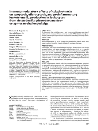
Figure 1—Effects of various concentrations of tulathromycin (TUL) on neutrophils isolated from the peripheral blood of 6 healthy
male 8- to 30-week-old pigs.A—Results of an ELISA to identify apoptotic mono- and oligonucleosomes in neutrophils.Values were
calculated as the ratio of light absorbance values for tulathromycin-treated cells versus values for control-treated cells (10% HI-FBS
in HBSS; n = 4 repetitions/group) at 0.5 (gray bars) and 2 (black bars) hours of incubation. B—Results ofTUNEL staining to identify
apoptotic neutrophils following neutrophil incubation withTUL, staurosporine (STA), or 10% HI-FBS in HBSS (control).Values were
calculated as the percentage of TUNEL-positive neutrophils within the microscopic field of view and are reported as a fold change
relative to the degree of staining in control-treated cells (n = 3 repetitions/group). C—Results of densitometric analysis of western
blot data for fragmentation of caspase-3 relative to β-actin in neutrophils.Values were calculated as the band density of cleaved
caspase-3 (17 kDa) relative to that of β-actin and are reported as a ratio relative to the control value (n = 3 repetitions/group).
D—Representative results of western blot analysis to detect caspase-3 fragmentation in neutrophils incubated withTUL at various
concentrations.Value in panels A, B, C are reported as mean ± SEM. *Value is significantly (P < 0.05) different from the control value.
14-06-0160r.indd 511 5/18/2015 11:07:39 AM
6. 512 AJVR •Vol 76 • No. 6 • June 2015
or particulate matter (zymosan).An aliquot of BALF from
each sample was serially diluted and plated onto Bru-
cella agar supplemented with 0.2% β-NAD and 5% horse
serum and incubated overnight at 37°C. Colonies of
A pleuropneumoniae on each plate were subsequently
counted.The remaining BALF was centrifuged at 1,500 X
g for 10 minutes.Aliquots of the BALF supernatants were
snap frozen in liquid nitrogen and stored at –70°C for fur-
ther analysis.Cell pellets were resuspended in HBSS,and
cells counts were performed with a hemacytometer.d Re-
maining leukocytes were divided into aliquots of equal
volumes for western blot analysis.Cell pellets were snap
frozen in liquid nitrogen and stored at –70°C for further
analysis.
Immunohistochemical analysis of lung
tissue—Tissue samples obtained from cranial lung
lobes during necropsy were fixed in formalin and em-
bedded in paraffin prior to sectioning. Five-microme-
ter segments were prepared,mounted onto slides,and
deparaffinized in preparation for H&E staining accord-
ing to described methods.39,40 Briefly, slides of tissue
sections were stained in hematoxylin,rehydrated,and
counterstained with eosin. Slides were examined by
investigators who were not aware of their origin, and
images were obtained at 400X magnification.x
Detection of LTB4—To assess immunomodula-
tory effects of tulathromycin in vivo, LTB4 concentra-
tions in the neutrophil supernatants extracted from
BALF samples were quantified by use of an ELISA kity
in accordance with the manufacturer’s instruction.
Concentrations of LTB4 were determined at 405 nm
by use of a microplate reader.k Reported specificity of
the ELISA was 100% for LTB4,0.03% for 5(S)-hydroxye-
icosatetraenoic acid, and < 0.01% for leukotrienes C4,
E4,and D4;it had a detection limit of 7 pg/mL
Detection of necrosis—Lactate dehydrogenase
activity was quantified in BALF supernatants with a cy-
totoxicity detection kit,z in which colorimetric develop-
ment was based on the reduction of tetrazolium to forma-
zan,in accordance with the manufacturer’s instructions.
Degree of caspase fragmentation was measured in BALF
neutrophils as described for the in vitro experiment.
Detection of efferocytosis—Efferocytosis was
identified by direct microscopic examination of cy-
tocentrifuged BALF preparations for macrophages
containing 1 or more apoptotic neutrophils within
phagocytic vacuoles.Findings are reported as the per-
centage of macrophages with efferocytosis among all
macrophages.
Statistical analysis
All data were analyzed by means of 1-way ANOVA.
Post hoc multiple comparisons were performed with
theTukey test.Values of P < 0.05 were considered sig-
nificant. Summary data are reported as mean ± SEM
(in some experiments, from multiple replicates of a
minimum of 3 independent experiments).
Results
Effects of tulathromycin on leukocytes in
vitro
After neutrophils from healthy 8- to 30-week-old
pigs were incubated with tulathromycin (0.02 mg/mL)
for 0.5 hours, the amount of apoptosis was significantly
Figure 2—Mean ± SEM myeloperoxidase activity (MPO) in
macrophage lysates (gray bars) and supernatants (black bars)
after coculture of neutrophils pretreated with tulathromycin
or HBSS (control) with monocyte-derived macrophages from
the same pigs as in Figure 1 (n = 3 repetitions/group). *Value is
significantly (P < 0.05) different from the corresponding control
value. See Figure 1 for remainder of key.
A pleuropneumoniae
Control group A pleuropneumoniae and tulathromycin
Variable 3 h 24 h 3 h 24 h 3 h 24 h
Bacteria (CFUs/mL) 1.58 X 10² 7.75 X 10² 8.65 X 105* 3.24 X 107† 7.82 X 10⁵a 6.69 X 10⁴‡
Neutrophils (%) 60.3 ± 6.6 43.4 ± 6.8 74.5 ± 5.2* 39.9 ± 10.2 74.8 ± 4.0* 46.4 ± 7.4
Rectal temperature (°C) 37.2 ± 0.3 38.2 ± 0.1 38.6 ± 0.6 39.4 ± 0.2 38.0 ± 0.3 38.1 ± 0.2
*Value is significantly (P < 0.05) different from the corresponding 3-hour control value. †Value is significantly
(P < 0.05) different from the corresponding 24-hour control value. ‡Value is significantly (P < 0.05) different from
the 24-hour value for untreated A pleuropneumoniae–inoculated pigs.
Table 1—Mean ± SEM rectal temperatures, numbers of Actinobacillus pleuropneumoniae CFUs, and
percentages of neutrophils recovered from BALF samples obtained from 3- to 4-week-old pigs 3 and
24 hours after intratracheal inoculation without (control) or with A pleuropneumoniae and pretreat-
ment with or without tulathromycin (2.5 mg/kg, IM; n = 6 to 7 pigs/group).
14-06-0160r.indd 512 5/18/2015 11:07:39 AM
7. AJVR •Vol 76 • No. 6 • June 2015 513
greater than that in untreated control neutrophils (Fig-
ure 1).After incubation for 2 hours, no effect of tulath-
romycin concentration (0.02 mg/mL to 2.0 mg/mL) on
the degree of apoptosis was detected.The lowest con-
centration (0.02 mg/mL) was therefore selected for all
subsequent testing.To corroborate these findings,the de-
gree of DNA fragmentation within the neutrophils was
examined,and results ofTUNEL staining confirmed that
incubation with tulathromycin significantly increased
the number ofTUNEL-positive neutrophils relative to the
degree of staining in control neutrophils. Neutrophils
incubated with staurosporine (1µM) as a positive con-
trol treatment also underwent apoptosis. Western blot
analysis revealed that incubation with tulathromycin
significantly increased fragmentation of caspase-3 in a
concentration-dependent manner in neutrophils in vitro.
After incubation of neutrophils with HBSS or tu-
lathromycin (0.02 mg/mL) for 2 hours and subsequent
coculturing with macrophages from the same pig,my-
eloperoxidase activity was assessed in macrophage
lysates and supernatant. In tulathromycin-treated co-
cultures, a significant increase was identified in intra-
cellular myeloperoxidase activity relative to that in the
control cocultures (Figure 2).
Effects of tulathromycin in vivo
In 3- to 4-week-old pigs inoculated intratrache-
ally with live A pleuropneumoniae (inoculated pigs),
treatment with tulathromycin 0.25 hours before inoc-
ulation induced leukocyte apoptosis, inhibited the in-
crease of LDH activity and LTB4 concentration in BALF,
and improved the histologic and macroscopic appear-
ance of lung tissue following inoculation. Inoculated
pigs had high numbers of bacteria and neutrophils
in BALF samples collected 3 hours after inoculation
(Table 1). Inoculated pigs treated with tulathromy-
cin had significantly fewer bacteria in their lungs at
24 hours after inoculation relative to the number of
bacteria in the lungs of untreated inoculated pigs, in
which bacterial loads remained significantly higher
than those in untreated sham-inoculated control pigs.
Neutrophil numbers in treated and untreated inocu-
lated pigs did not differ significantly from those for
control pigs at 24 hours after inoculation. Rectal tem-
peratures did not differ significantly among treatment
groups throughout the experimental infection period.
Densitometric analysis of BALF samples collected
3 hours after inoculation revealed no difference among
the 3 treatment groups (data not shown). However, at
Figure 3—Results of quantification of apoptosis in leukocytes obtained from BALF of 3- to 4-week old pigs that were untreated
and sham-inoculated (control; n = 12), untreated and intratracheally inoculated with Actinobacillus pleuropneumoniae (APP;13), or
treated with tulathromycin (2.5 mg/kg, IM) 0.25 hours before intratracheal inoculation with A pleuropneumoniae (APP+TUL;13).
A—Photomicrograph illustrating different degrees of TUNEL staining (green regions) in leukocytes. Blue regions indicate Hoechst
nuclear counterstain. Notice the apoptotic leukocytes (arrowheads). Bars = 10 µm. B—Results of densitometric analysis of western
blots of caspase-3 fragmentation in BALF samples obtained 24 hours after intratracheal inoculation.Values were calculated as the
band density of cleaved caspase-3 (17 kDa) relative to that of β-actin and are reported as a ratio relative to the control value (n
= 5 to 6 samples/group). C—Results of LDH quantification in BALF samples to assess the degree of cellular necrosis.Values were
calculated as the ratio of light absorbance values relative to control values. Data in panels B and C are reported as mean ± SEM.
*Value is significantly (P < 0.05) different from the control value. See Figure 1 for remainder of key.
14-06-0160r.indd 513 5/18/2015 11:07:39 AM
8. 514 AJVR •Vol 76 • No. 6 • June 2015
24 hours after inoculation, an apparent increase in
the numbers of TUNEL-positive leukocytes was de-
tected within BALF samples (Figure 3), and many of
these apoptotic cells appeared to be contained within
phagocytic vacuoles of macrophages.A significant in-
crease in caspase-3 fragmentation was identified in
samples collected 24 hours after inoculation. Consis-
tent with these results, treatment with tulathromycin
inhibited the increase in LDH and necrosis caused by
A pleuropneumonia infection.
As early as 3 hours after inoculation, the BALF
concentration of LTB4 was significantly higher in
untreated inoculated pigs than in control pigs.Treat-
ment with tulathromycin inhibited this increase
(Figure 4). Notably, these differences were identi-
fied in the absence of a significant difference in bac-
terial loads between treated and untreated inoculat-
ed pigs (Table 1). By 24 hours after inoculation, the
BALF LTB4 concentration was likewise significantly
greater in A pleuropneumoniae–inoculated pigs
versus control pigs, and treatment with tulathromy-
cin prevented this increase.
Necropsy and histologic evaluation of tissue sam-
ples obtained from the cranial lung lobes of pigs euth-
anized 3 hours after inoculation revealed that untreat-
ed inoculated pigs had evidence of edema and severe
inflammatory infiltration and accumulation within the
alveoli. On the other hand, control pigs evaluated at
the same point had a distinct alveolar structure and
clear alveolar spaces (Figure 5). Conversely, treated
A pleuropneumoniae–inoculated pigs had markedly
less edema and inflammatory infiltrate within the alve-
olar spaces and an apparent reduction in inflammation
relative to results for control pigs.Macroscopic exami-
nation of lung samples from untreated inoculated pigs
euthanized 24 hours after inoculation revealed areas
of apparent edema, tissue consolidation, necrotic foci,
and fibrotic lesions that were absent in samples from
control pigs at the same assessment point. In treated
inoculated pigs at 24 hours after inoculation, these le-
sions were dramatically reduced in severity.
Effects of zymosan in vivo
The degree of pulmonary neutrophil infiltration
in zymosan-inoculated pigs that had been euthanized
and evaluated at 24 hours after inoculation was sig-
nificantly greater than that in control pigs at the same
evaluation point, and this increase was not altered by
treatment with tulathromycin (Table 2). Rectal tem-
peratures did not differ significantly among treatment
groups throughout the experimental infection period.
Fragmentation of caspase-3 in BALF leukocytes
was evident as early as 3 hours after inoculation with
zymosan, and treatment with tulathromycin signifi-
cantly increased the degree of fragmentation relative
to that in the control pigs (Figure 6).However,this ef-
fect was no longer evident in pigs evaluated 24 hours
after inoculation (data not shown).
Differential cell counts performed on BALF sam-
ples from pigs euthanized 3 hours after inoculation
revealed that the degree of efferocytosis among mac-
rophages was significantly greater in tulathromycin-
treated inoculated pigs versus in control pigs, but not
significantly different from the degree in untreated
inoculated pigs (Figure 7). At the same evaluation
point, the percentage of phagocytosis-positive macro-
phages was similarly greater in treated and untreated,
inoculated pigs than in control pigs.By 24 hours after
inoculation, the degree of efferocytosis was similarly
enhanced in treated and untreated, inoculated pigs
(data not shown).
Analyses involving BALF samples obtained 3 hours
after zymosan inoculation revealed a slight increase in
LTB4 concentrations in all pigs relative to expected
values17 (Figure 8). By 24 hours after inoculation,
LTB4 concentrations were significantly higher in un-
treated zymosan-inoculated pigs than in control pigs,
and treatment of inoculated pigs with tulathromycin
significantly inhibited this increase.
Discussion
Medications that possess dual antimicrobial and
Figure 4—Mean ± SEM LTB4 concentrations in BALF samples
collected from the pigs in Figure 3 at 3 hours (n = 6 to 7/
group; A) and 24 hours (6 to 7/group; B) after intratracheal
inoculation with A pleuropneumonia. *Value is significantly (P <
0.05) different from the control value. †Value is significantly (P
< 0.05) different from the value for untreated pigs intratrache-
ally inoculated with A pleuropneumoniae.See Figures 1 and 3 for
remainder of key.
14-06-0160r.indd 514 5/18/2015 11:07:40 AM
9. AJVR •Vol 76 • No. 6 • June 2015 515
anti-inflammatory properties may be more efficacious
than other medications for the treatment of infectious
diseases in which the pathogenesis includes an inflam-
matory component.17,41 Previous research has estab-
lished that tulathromycin31,34 and other macrolides30,41
have immunomodulatory and anti-inflammatory char-
acteristics. Results from the present study suggested
that tulathromycin had similar cellular effects in 3- to
4-week-old pigs with experimentally induced A pleu-
ropneumoniae infection or zymosan-induced pulmo-
nary inflammation. Findings from the in vitro experi-
ment involving leukocytes from 8- to 30-week old pigs
indicated that tulathromycin promoted apoptosis in
porcine neutrophils in a dose- and time-dependent
manner,presumably as determined by the presence of
mono- and oligonucleosomes. Supporting these find-
ings were results of TUNEL staining and detection of
caspase-3 fragmentation (the main caspase involved in
Figure 5—Representative photomicrographs (A–C) and photographs (D–F) of pulmonary tissue from untreated sham-inoculated
pigs (A and D), untreated pigs intratracheally inoculated with A pleuropneumoniae (B and E), and pigs treated with tulathromycin
(2.5 mg/kg, IM) 0.25 hours before intratracheal inoculation with A pleuropneumoniae (C and F). Photomicrographs show samples of
cranial lobe lung tissue collected from pigs euthanized at 3 hours after inoculation. Notice the extensive neutrophil recruitment
and a loss of apparent alveolar structure (arrowhead). H&E stain; bars = 100 µm. Photographs show evidence of severe pulmonary
edema (arrows) and fibrotic lesions (arrowhead). See Figure 1 for remainder of key.
Zymosan
Control Zymosan and tulathromycin
Variable 3 hours 24 hours 3 hours 24 hours 3 hours 24 hours
Neutrophils (%) 61.0 ± 27.1 23.3 ± 3.1 95.5 ± 0.9 68.7 ± 5.6* 72.8 ± 13.6 68.2 ± 3.4*
Rectal temperature (°C) 37.5 ± 0.4 37.6 ± 0.5 37.3 ± 0.4 36.9 ± 0.2 37.4 ± 0.2 37.3 ± 0.2
*Value is significantly (P < 0.05) different from the corresponding 24-hour control value.
Table 2—Mean ± SEM rectal temperatures and percentages of neutrophils recovered from BALF
samples from 3- to 4-week-old pigs at 3 and 24 hours after intratracheal inoculation without (control)
or with zymosan (n = 3 to 5/group) and with or without pretreatment with tulathromycin (2.5 mg/
kg, IM; 6 to 7 pigs/group).
14-06-0160r.indd 515 5/18/2015 11:07:41 AM
10. 516 AJVR •Vol 76 • No. 6 • June 2015
the execution phase of apoptosis7) in BALF samples
obtained from the 3- to 4-week-old pigs.We also found
that induction of apoptosis by tulathromycin facili-
tated cellular clearance by porcine macrophages, as
indicated by intracellular myeloperoxidase activity in
macrophage-neutrophil cocultures.
The aforementioned in vitro findings prompted
us to investigate the effects of tulathromycin in an
experimental model of A pleuropneumoniae infec-
tion. By 3 hours after intratracheal inoculation of pigs
with A pleuropneumoniae,we identified a significant
increase in the degree of fragmentation of caspase-3
within BALF leukocytes, which was accompanied by
an apparent increase in the number ofTUNEL-positive
BALF leukocytes. Moreover, treatment with tulathro-
mycin prevented the increase in tissue necrosis caused
by infection. These effects were observed without a
decrease in the total number of infiltrating neutrophils
within the lungs throughout infection,suggesting that
tulathromycin did not impact neutrophil recruitment,
as reported elsewhere.31 Furthermore, the findings
indicated that tulathromycin inhibited the accumula-
tion of proinflammatory LTB4 within bronchoalveo-
lar spaces of A pleuropneumoniae–inoculated pigs.
Together, these findings suggested that tulathromycin
had anti-inflammatory and proresolution benefits in
A pleuropneumoniae–challenged lungs.This hypoth-
Figure 6—Mean ± SEM band density values from densitomet-
ric analysis of caspase-3 fragmentation in leukocytes isolated
from BALF samples obtained from 3- to 4-week old pigs that
were untreated and sham-inoculated (control; n = 7), untreat-
ed and intratracheally inoculated with zymosan (ZYM; 7), or
treated with tulathromycin (2.5 mg/kg, IM) 0.25 hours before
intratracheal inoculation with zymosan (ZYM+TUL;9).Samples
were obtained 3 hours after inoculation. See Figures 1 and 3
for remainder of key.
Figure 7—Mean ± SEM percentage of macrophages with
evidence of efferocytosis and phagocytosis in BALF samples
obtained from the same pigs as in Figure 6 at 3 hours after
intratracheal inoculation with ZYM (n = 3 to 5 samples/group).
A minimum of 3 microscopic fields were used to calculate the
values. *Value is significantly (P < 0.05) different from the con-
trol value. See Figures 1 and 6 for remainder of key.
Figure 8—Mean ± SEM LTB4 concentrations in BALF samples
obtained from the same pigs as in Figure 6 at 3 hours (A) and
24 hours (B) after intratracheal inoculation with ZYM (n = 3 to
5 samples/group).*Value is significantly (P < 0.05) different from
the control value.†Value is significantly (P < 0.05) different from
the value for untreated ZYM-inoculated pigs. See Figures 1 and
6 for remainder of key.
14-06-0160r.indd 516 5/18/2015 11:07:41 AM
11. AJVR •Vol 76 • No. 6 • June 2015 517
esis was also supported by the pulmonary histopatho-
logic findings and gross pathological lesions, consid-
ering that treatment with tulathromycin reduced the
severity of inflammatory infiltrate, edema, and lung le-
sions relative to that in untreated but similarly inocu-
lated pigs, thereby suggesting an improved prognosis
after inoculation.The slight increase evident in BALF
LTB4 concentrations of all pigs relative to expected
values17 was likely a result of the lavage procedure.
Results of the A pleuropneumonia infection ex-
periment prompted us to evaluate anti-inflammatory
and proresolution properties of tulathromycin inde-
pendent of its antimicrobial effects with an in vivo
model of pulmonary inflammation (caused by zymo-
san) in the absence of live bacteria. Similar to results
obtained for the A pleuropneumonia infection ex-
periment, tulathromycin had proapoptotic properties
early in the nonbacterial, zymosan-induced inflam-
mation experiment, without impacting neutrophil
recruitment to the bronchoalveolar spaces. Tulathro-
mycin also appeared to promote the preferential ef-
ferocytosis of apoptotic leukocytes at the same time
point, suggesting enhanced nonphlogistic clearance
of the dying cells. Perhaps most striking, however,
was the observation that tulathromycin inhibited LTB4
production in zymosan-challenged lungs, a distinct
anti-inflammatory effect in the absence of bacterial
stimuli. These findings suggested that tulathromycin
had immunomodulatory properties independent of
its antimicrobial effects and that these properties may
be particularly advantageous in the treatment of infec-
tious inflammatory diseases.
Severe tissue damage and rapid pathogenesis of
A pleuropneumoniae–induced pneumonia is in part
a result of local cellular necrosis and self-perpetuating
recruitment of neutrophils to the site of infection.
Investigators in a previous study1 determined the ef-
ficacy of drugs that target neutrophil activation in
delaying lesion development associated with A pleu-
ropneumoniae infection. Our findings provided evi-
dence of the anti-inflammatory and immunomodulato-
ry properties of tulathromycin in a clinically relevant
model of bacterial pneumonia within a target species.
Within the lungs of pigs, A pleuropneumoniae exo-
toxins (I, II, and III) cause lysis of infiltrating neutro-
phils and macrophages as well as alveolar epithelial
and endothelial cells.42,43 Cell lysis causes the release
of cytotoxic cellular contents into the extracellular mi-
lieu, which damages surrounding cells and promotes
the secretion of proinflammatory mediators. These
mediators in turn amplify leukocyte recruitment and
exacerbate inflammatory injury.12 Findings of the
present study suggested that tulathromycin was able
to interrupt the inflammatory cascade by promoting
controlled apoptosis of leukocytes within the lungs
and that this proapoptotic effect was associated with
an improvement in pulmonary histologic findings and
a decrease in lesion development in infected pigs.Cor-
respondingly, tulathromycin inhibited the increase in
tissue necrosis caused by infection. In vitro evidence
further supported the in vivo findings and was con-
sistent with previous reports31,34 of the mechanisms
through which tulathromycin exerts proapoptotic
effects. Similarly, other macrolides including azithro-
mycin and tilmicosin appear to have a propensity
for the induction of neutrophil apoptosis.28–30,38,44,45
Moreover, effects in the present study occurred with-
out impacting the degree of neutrophil recruitment to
the lungs,similar to findings for models of pneumonia
in cattle31 and consistent with reports17,29,43 for other
macrolides.
Precise mechanisms through which tulathromy-
cin exerts its anti-inflammatory and immunomodulato-
ry effects are incompletely understood, but it appears
to signal through caspase-3.31,34 Controlled apoptosis
of infiltrating leukocytes is an essential component in
the resolution of inflammation following infection.7
Additional investigation is warranted to determine the
apoptotic pathway through which tulathromycin acts
and whether it acts through similar mechanisms in
porcine cells as it does in bovine cells.31,34
During severe inflammation, infiltrating neutro-
phils release LTB4, a potent neutrophil chemoattrac-
tant derived from arachidonic acid that contributes
considerably to the self-amplifying recruitment of
neutrophils to the site of inflammation3,4,17 and the
development of pulmonary lesions in A pleuropneu-
moniae infection.1 For the resolution of inflamma-
tion to occur following infection, proinflammatory
signals must first be dampened to curtail leukocyte
recruitment. Other macrolides can inhibit the pro-
duction of proinflammatory cytokines41,46 and sup-
press NF-κB activation.47–49 Results of the present
study indicated that treatment with tulathromycin
inhibited LTB4 production in infected lungs as well
as in inflamed zymosan-challenged lungs in pigs.
These findings are consistent with those for an ex-
perimental model of Mannheimia haemolytica–
induced pneumonia in cattle,31 and similar obser-
vations have been reported for tilmicosin, another
macrolide that also promotes apoptosis.39 Notably,
our findings were observed in the presence of a
bacterial challenge as well as in a nonbacterial in-
flammatory challenge, suggesting that the immu-
nomodulatory and anti-inflammatory properties of
tulathromycin may be independent of its antimicro-
bial properties.Additional research is warranted to
investigate whether these anti-inflammatory effects
of tulathromycin were independent of its proapop-
totic properties. Nevertheless, the inhibition of pro-
inflammatory mediator production may contribute
considerably to the promotion of inflammation res-
olution following infection.
Cellular death by apoptosis is a critical step in
the resolution of inflammation following infection be-
cause it facilitates nonphlogistic removal of infiltrat-
ing cells from the site of injury.7,50,51 The process of
apoptosis is characterized by several morphological
changes including cellular shrinkage, chromatin con-
densation, and DNA fragmentation.52,53 Cells under
14-06-0160r.indd 517 5/18/2015 11:07:42 AM
12. 518 AJVR •Vol 76 • No. 6 • June 2015
going apoptosis also lose some of their functional
characteristics and become hyporesponsive to exter-
nal stimuli, which helps facilitate their nonphlogistic
removal from the site of inflammation.8,54 In addition,
apoptotic cells express signals necessary to trigger
their phagocytic uptake by macrophages in a process
known as efferocytosis.11,55 For example, apoptotic
cells express phosphatidylserine on the outer surface
of the cell membrane to signal their death to adjacent
phagocytes.56 Receptors associated with efferocy-
tosis of apoptotic cells include a phosphatidylserine
receptor, CD36, CD14, and CD68.57,58 Recognition of
apoptotic cells by macrophages facilitates clearance
of these cells from the tissues, which prevents dam-
age from postapoptotic necrosis59 and can also trigger
an anti-inflammatory phenotype in efferocytic macro-
phages that promotes the release of anti-inflammatory
mediators such as transforming growth factor-β and
IL-10.10,50 Together, these processes help resolve in-
flammation and restore tissue homeostasis.
Findings in the present study suggested that tu-
lathromycin promoted apoptosis within infected and
inflamed lungs in pigs and that tulathromycin also
enhanced efferocytosis of apoptotic cells.The results
were consistent with reported results involving bo-
vine cells35 and were corroborated by our in vitro
findings,which suggested that neutrophils that under-
went tulathromycin-induced apoptosis were phagocy-
tosed by macrophages with greater avidity than were
control cells. Collectively, the findings indicated that
tulathromycin had immunomodulatory properties in
leukocytes that conferred anti-inflammatory and pro-
resolution benefits in the context of bacterial pneu-
monia as well as pulmonary inflammation in pigs.
These benefits may explain at least in part the efficacy
of tulathromycin in the treatment of respiratory dis-
ease in pigs caused by A pleuropneumoniae.60 Addi-
tional research is warranted to investigate the effects,
if any,that tulathromycin may have on the signaling of
proresolution lipid mediators.
Acknowledgments
Supported in part by Zoetis.
Presented in abstract form at Experimental Biology 2013, Boston,April
2013, and Interscience Conference on Antimicrobial Agents and Che-
motherapy 2013, Denver, September 2013.
The authors thank Barbara Smith, James Cotton, Jennifer Beatty, Joey
Lockhart, Lynne Buret, Marie Halliez, and Sarah Akierman for technical
assistance.
Footnotes
a. Vacutainer,Becton-Dickinson,Franklin Lakes,NJ.
b. Sigma-Aldrich,Oakville,ON,Canada.
c. PAA Laboratories Inc,Dartmouth,Mass.
d. VWR Scientific,Edmonton,AB,Canada.
e. Gibco,Grand Island,NY.
f. Diff-Quik stain set,Siemens Healthcare Diagnostics,Tarrytown,
NY.
g. CytoSpin 4 cytocentrifuge,Thermo Scientific, Burlington, ON,
Canada.
h. Costar,Cambridge,Mass.
i. Draxxin,Zoetis,Kalamazoo,Mich.
j. Cell Death ELISA,Roche Diagnostics,Laval,QC,Canada.
k. SpectraMAX M2e microplate reader, Molecular Devices, Men-
lo Park,Calif.
l. Sigma-Aldrich,Oakville,ON,Canada.
m. Cell DeathTUNEL kit,Roche Diagnostics,Laval,QC,Canada.
n. Cell lysis buffer solution,Roche Diagnostics,Laval,QC,Canada.
o. Bio-Rad Laboratories,Mississauga,ON,Canada.
p. TWEEN 20,Roche Diagnostics,Laval,QC,Canada.
q. Cell Signaling,Beverly,Mass.
r. Amersham ECL Western Blotting Detection Reagent, GE
Healthcare Life Sciences,Pittsburgh,Pa.
s. ImageJ, version 1.48, National Institutes of Health, Bethesda,
Md. Available at: rsbweb.nih.gov/ij/index.html. Accessed Jun
16,2014.
t. Sigma-Aldrich,Oakville,ON,Canada.
u. Provided by Dr.M.Gottschalk,University of Montreal,St-Hyan-
cinth,QC,Canada.
v. CytoSpin 4 cytocentrifuge,Thermo Scientific, Burlington, ON,
Canada.
w. Diff-Quik stain set,Siemens Healthcare Diagnostics,Tarrytown,
NY.
x. Retiga 2000X with Q Capture Suite software, Q Imaging, Sur-
rey,BC,Canada
y. LTB4 ELISA Cayman Chemical Co,Ann Arbor,Mich.
z. Cytotoxicity detection kit (LDH),Roche Diagnostics,Laval,QC,
Canada.
References
1. BertramTA.Pathobiology of acute pulmonary lesions in swine
infected with Haemophilus (Actinobacillus) pleuropneu-
moniae. Can Vet J 1988;29:574–577.
2. Gompertz S, Stockley RA. Inflammation—role of the neutro-
phil and eosinophil.Semin Respir Infect 2000;15:14–23.
3. Canetti C, Silva JS, Ferreira SH, et al.Tumour necrosis factor-α
and leukotriene B4 mediate neutrophil migration in immune
inflammation.Br J Pharmacol 2001;134:1619–1628.
4. Palmblad J, Malmsten CL, Uden AM, et al. Leukotriene B4 is a
potent and stereospecific stimulator of neutrophil chemotaxis
and adherence.Blood 1981;58:658–661.
5. Drent M,Cobben NA,Henderson RF,et al.Usefulness of lactate
dehydrogenase and its isoenzymes as indicators of lung dam-
age or inflammation.Eur Respir J 1996;9:1736–1742.
6. Savill J,Dransfield I,Gregory C,et al.A blast from the past:clear-
ance of apoptotic cells regulates immune responses. Nat Rev
Immunol 2002;2:965–975.
7. Haslett C. Granulocyte apoptosis and its role in the resolution
and control of lung inflammation. Am J Respir Crit Care Med
1999;160:S5–S11.
8. Whyte MK, Meagher LC, MacDermot J, et al. Impairment of
function in aging neutrophils is associated with apoptosis. J
Immunol 1993;150:5124–5134.
9. Savill J, Fadok V, Henson P, et al. Phagocyte recognition of cells
undergoing apoptosis.Immunol Today 1993;14:131–136.
10. Fadok VA,Bratton DL,Konowal A,et al.Macrophages that have
ingested apoptotic cells in vitro inhibit proinflammatory cyto-
kine production through autocrine/paracrine mechanisms in-
volvingTGF-β,PGE2,and PAF.J Clin Invest 1998;101:890–898.
11. Savill JS,Wyllie AH, Henson JE, et al. Macrophage phagocytosis
of aging neutrophils in inflammation. Programmed cell death
in the neutrophil leads to its recognition by macrophages. J
Clin Invest 1989;83:865–875.
12. Kono H,Rock KL.How dying cells alert the immune system to
danger.Nat Rev Immunol 2008;8:279–289.
13. Rycroft AN, Garside LH. Actinobacillus species and their role
in animal disease.Vet J 2000;159:18–36.
14. SebunyaTN,Saunders JR.Haemophilus pleuropneumoniae in-
fection in swine:a review.J AmVet Med Assoc 1983;182:1331–
1337.
15. Dom P, Haesebrouck F. Comparative virulence of NAD-depen-
dent and NAD-independent Actinobacillus pleuropneumoni-
ae strains.Zentralbl Veterinarmed B 1992;39:303–306.</jrn>
16. Marsteller TA, Fenwick B. Actinobacillus pleuropneumoniae
disease and serology.Swine Health Prod 1999;7:161–165.
17. Nerland EM, LeBlanc JM, Fedwick JP, et al. Effects of oral ad-
14-06-0160r.indd 518 5/18/2015 11:07:42 AM
13. AJVR •Vol 76 • No. 6 • June 2015 519
ministration of tilmicosin on pulmonary inflammation in pig-
lets experimentally infected with Actinobacillus pleuropneu-
moniae. Am J Vet Res 2005;66:100–107.
18. Villarino N, Brown SA, Martin-Jimenez T. Understanding the
pharmacokinetics of tulathromycin: a pulmonary perspective.
J Vet Pharmacol Ther 2014;37:211–221.
19. Miyajima M,Suga M,Nakagawa K,et al.Effects of erythromycin
on experimental extrinsic allergic alveolitis. Clin Exp Allergy
1999;29:253–261.
20. Konno S, Asano K, Kurokawa M, et al. Antiasthmatic activity
of macrolide antibiotic, roxithromycin: analysis of possible
mechanisms in vitro and in vivo. Int Arch Allergy Immunol
1994;105:308–316.
21. Kadota J, Matsubara Y, Ishimatsu Y, et al. Significance of IL-1β
and IL-1 receptor antagonist (IL-1Ra) in bronchoalveolar la-
vage fluid (BALF) in patients with diffuse panbrochiolitis
(DPB).Clin Exp Immunol 1996;103:461–466.
22. Khair OA, Devalia JL,Abdelaziz MM, et al. Effect of erythromy-
cin on Haemophilus influenzae endotoxin-induced release of
IL-6, IL-8 and sICAM-1 by cultured human bronchial epithelial
cells.Eur Respir J 1995;8:1451–1457.
23. Oishi K,Sonoda F,Kobayashi S,et al.Role of interleukin-8 (IL-8)
and an inhibitory effect of erythromycin on IL-8 release in the
airways of patients in chronic airway diseases. Infect Immun
1994;62:4145–4152.
24. Fujii T, Kadota J, Morikawa T, et al. Inhibitory effect of erythro-
mycin on interleukin 8 production by 1 alpha,25-dihydroxyvi-
tamin D3-stimulatedTHP-1 cells.Antimicrob Agents Chemoth-
er 1996;40:1548–1551.
25. Miyachi Y,Yoshioka A, Imamura S, et al. Effect of antibiotics on
the generation of reactive oxygen species. J Invest Dermatol
1986;86:449–453.
26. Hand WL, Hand DL, King-Thompson NL.Antibiotic inhibition of
the respiratory burst response in human polymorphonuclear leu-
kocytes.Antimicrob Agents Chemother 1990;34:863–870.
27. Aoshiba K, Nagai A, Konno K. Erythromycin shortens neutro-
phil survival by accelerating apoptosis. Antimicrob Agents
Chemother 1995;39:872–877.
28. Buret AG. Immuno-modulation and anti-inflammatory ben-
efits of antibiotics: the example of tilmicosin. Can J Vet Res
2010;74:1–10.
29. ChinAC,Morck DW,Merrill JK,et al.Anti-inflammatory benefits
of tilmicosin in calves with Pasteurella haemolytica–infected
lungs.Am J Vet Res 1998;59:765–771.
30. Chin AC, Lee WD, Murrin KA, et al.Tilmicosin induces apop-
tosis in bovine neutrophils in the presence of in the absence
of Pasteurella haemolytica and promotes neutrophil phago-
cytosis by macrophages. Antimicrob Agents Chemother
2000;44:2465–2470.
31. Fischer CD, Beatty JK, Zvaigzne CG, et al. Anti-inflammato-
ry benefits of antibiotic-induced neutrophil apoptosis: tu-
lathromycin induces caspase-3-dependent neutrophil pro-
grammed cell death and inhibits NF-κB signaling and CXCL8
transcription. Antimicrob Agents Chemother 2011;55:338–
348.
32. Evans NA.Tulathromycin:an overview of a new tiamilide antibi-
otic for livestock respiratory disease.Vet Ther 2005;6:83–95.
33. Siegel T. Cellular uptake of the triamilide tulathromycin by
bovine and porcine phagocytic cells in vitro. J Anim Sci
2004;82:186.
34. Fischer CD, Beatty JK, Duquette SC, et al. Direct and indirect
anti-inflammatory effects of tulathromycin in bovine macro-
phages:inhibition of CXCL-8 secretion,induction of apoptosis,
and promotion of efferocytosis. Antimicrob Agents Chemoth-
er 2013;57:1385–1393.
35. Fischer CD, Duquette SC, Renaux BS, et al.Tulathromycin ex-
erts pro-resolving effects in bovine neutrophils by inhibiting
phospholipases and altering leukotriene B4, prostaglandin E2,
and lipoxin A4 production. Antimicrob Agents Chemother
2014;58:4298–4307.
36. Krueger AJ,Yang JJ, Roy TA, et al.An automated myeloperoxi-
dase assay.Clin Chem 1990;36:158.
37. Lee WD,Flynn AN,LeBlanc JM,et al.Tilmicosin-induced bovine
neutrophil apoptosis is cell-specific and downregulates spon-
taneous LTB4 synthesis without increasing Fas expression. Vet
Res 2004;35:213–224.
38. Canadian Council onAnimal Care.Standards and policies.Avail-
able at: www.ccac.ca/en_/standards/guidelines.Accessed Mar
20,2015.
39. Llewellyn BD. Nuclear staining with alum hematoxylin. Bio-
tech Histochem 2009;84:159–177.
40. Luna LG. American Registry of Pathology manual of his-
tologic staining methods of the Armed Forces Institute of
Pathology. 3rd ed. New York: McGraw Hill Publishers,
1960;122–132.
41. Tamaoki J, Kadota J, Takizawa H. Clinical implications of
the immunomodulatory effects of macrolides. Am J Med
2004;117:5S–11S.
42. Chiers K,De WaeleT,Pasmans F,et al.Virulence factors of Acti-
nobacillus pleuropneumoniae involved in colonization, per-
sistence and induction of lesions in its porcine host. Vet Res
2010;41:65.
43. Kamp EM, Stockhofe-Zurwieden N, Van Leengoed LA, et al.
Endobronchial inoculation with Apx toxins of Actinobacillus
pleuropneumoniae leads to pleuropneumonia in pigs. Infect
Immun 1997;65:4350–4354.
44. Koch CC, Esteban DJ, Chin AC, et al.Apoptosis, oxidative me-
tabolism and interleukin-8 production in human neutrophils
exposed to azithromycin: effects of Streptococcus pneumoni-
ae. J Antimicrob Chemother 2000;46:19–26.
45. Ishimatsu Y, Kadota J, Iwashita T, et al. Macrolide antibiotics in-
duce apoptosis of human peripheral lymphocytes in vitro. Int
J Antimicrob Agents 2004;24:247–253.
46. Zalewska-Kaszubska J,Gorska D.Anti-inflammatory capabilities
of macrolides.Pharmacol Res 2001;44:451–454.
47. Aoki Y, Kao PN. Erythromycin inhibits transcriptional activa-
tion of NF-κB, but not NFAT, through calcineurin-indepen-
dent signaling in T cells. Antimicrob Agents Chemother
1999;43:2678–2684.
48. Leiva M, Ruiz-Bravo A, Moreno E, et al.Telithromycin inhibits
the production of proinflammatory mediators and the activa-
tion of NF-κB in in vitro-stimulated murine cells.FEMS Immu-
nol Med Microbiol 2008;53:343–350.
49. Ou XM, Feng YL, Wen FQ, et al. Macrolides attenuate mucus
hypersecretion in rat airways through inactivation of NF-κB.
Respirology 2008;13:63–72.
50. Serhan CN, Savill J. Resolution of inflammation: the beginning
programs the end.Nat Immunol 2005;6:1191–1197.
51. Gilroy DW, Lawrence T, Perretti M, et al. Inflammatory resolu-
tion:new opportunities for drug discovery.Nat Rev Drug Dis-
cov 2004;3:401–416.
52. Maianski NA,Maianski AN,KuijpersTW,et al.Apoptosis of neu-
trophils.Acta Haematol 2004;111:56–66.
53. Scheel-Toellner D, Wang KQ, Webb PR, et al. Early events
in spontaneous neutrophil apoptosis. Biochem Soc Trans
2004;32:461–464.
54. Van Oostveldt K,Paape MJ,Dosogne H,et al.Effect of apoptosis
of phagocytosis, respiratory burst and CD18 adhesion recep-
tor expression of bovine neutrophils. Domest Anim Endocri-
nol 2002;22:37–50.
55. Savill J. Recognition and phagocytosis of cells undergoing
apoptosis.Br Med Bull 1997;53:491–508.
56. Naito M,Nagashima K,MashimaT,et al.Phosphatidylserine ex-
ternalization is a downstream event of interleukin-1-β-convert-
ing enzyme family protease activation during apoptosis.Blood
1997;89:2060–2066.
57. Ravichandran KS. Beginnings of a good apoptotic meal:
the find-me and eat-me signaling pathways. Immunity
2011;35:445–455.
58. Hart SP, Dransfield I, Rosi AG. Phagocytosis of apoptotic cells.
Methods 2008;44:280–285.
59. Silva MT. Secondary necrosis: the natural outcome of the com-
plete apoptotic program.FEBS Lett 2010;584:4491–4499.
60. Hart FJ,Kilgore RW,MeinertTR,et al.Efficacy of tulathromycin
in the treatment of respiratory disease in pigs caused by Acti-
nobacillus pleuropneumoniae. Vet Rec 2006;158:433–436.
14-06-0160r.indd 519 5/18/2015 11:07:42 AM