
Recommended
Recommended
More Related Content
What's hot
What's hot (19)
Viewers also liked
Viewers also liked (12)
Similar to Integr Cancer Ther-2015-Quinn-1534735415617014
Similar to Integr Cancer Ther-2015-Quinn-1534735415617014 (20)
Integr Cancer Ther-2015-Quinn-1534735415617014
- 1. Integrative Cancer Therapies 1–7 © The Author(s) 2015 Reprints and permissions: sagepub.com/journalsPermissions.nav DOI: 10.1177/1534735415617014 ict.sagepub.com Research Articles Introduction The anthracycline doxorubicin (DOX; trade name: Adriamycin) is a frequently prescribed chemotherapy agent used in the treatment of a number of cancers. Unfortunately, negative side effects in healthy tissues commonly occur and limit dosage. Beyond toxicities seen in cardiac and hepatic tissues, DOX has been shown to induce thymic involution with concurrent decreases in local thymocyte (T-cell) number.1-3 When lymphocytes are exposed to DOX in vitro, elevation in markers of cellular apoptosis is observed.4 In addition to prevention of DNA synthesis, the antitumor effects of DOX may be attributed to the generation of reac- tive oxygen species (ROS).5 ROS manifest in the form of superoxide, hydrogen peroxide and hydroxyls, which can damage membranes and initiate cell death. Acute, exhaustive exercise elicits glucocorticoid produc- tion and decreases in circulating lymphocytes, with subse- quent reduction in thymus gland size.6 Chronic, submaximal exercise, however, does not provoke these adverse impacts but stimulates favorable elevations in levels of free-radical- scavenging enzymes.7,8 Likewise, prolonged aerobic train- ing prior to DOX exposure has shown to increase antioxidant expression and activity in cardiac tissue.9-14 It is possible that similar adaptations to chronic endurance training may also mitigate DOX effects through increased handling of ROS in the thymus. Cancer patients receiving chemotherapy exhibit decreased immune responses following treatment and may be more susceptible to disease.15 Because DOX use is widely prescribed in cancer patients, chronic endurance training prior to treatment may be useful in enhancing immune systems of this population. The effect of submaxi- mal, chronic exercise prior to DOX administration on the thymus, however, remains unknown. Therefore, the pur- pose of this study was to examine the effects of chronic, submaximal endurance exercise prior to acute DOX admin- istration on thymus size, viable T-cell number, and thymic levels of lipid peroxidation. It was postulated that chronic endurance exercise would attenuate thymic deficiencies associated with DOX administration. 617014ICTXXX10.1177/1534735415617014Integrative Cancer TherapiesQuinn et al research-article2015 1 University of Northern Colorado, Greeley, CO, USA Corresponding Author: David Hydock, School of Sport and Exercise Science, University of Northern Colorado, 501 20th Street, Greeley, CO 80639, USA. Email: David.Hydock@unco.edu Effects of Chronic Endurance Exercise on Doxorubicin-Induced Thymic Damage Colin J. Quinn, PhD1 , Patrick D. Burns, PhD1 , Noah M. Gibson, MS1 , Alex Bashore, BS1 , Reid Hayward, PhD1 , and David S. Hydock, PhD1 Abstract The use of prior exercise training has shown promise in minimizing doxorubicin (DOX)-induced physical impairments. The purpose of this study was to compare changes in thymus mass, thymocyte (T-cell) number, and tissue peroxidation following chronic endurance exercise and DOX treatment in the rat. The thymus mass, number of viable T-cells, and levels of malondialdehyde and 4-hydroxyalkenals (MDA+4-HAE) were compared 3 days post-injection between rats assigned to the following treatment conditions: (a) 10 weeks of endurance training, followed by a saline injection 24 hours after the last training session (TM+SAL); (b) treadmill training as above, followed by a single, bolus 10-mg/kg injection of DOX (TM+10); (c) treadmill training with 12.5 mg/kg of DOX (TM+12.5); (d) sedentary (without exercise) and a saline injection (SED+SAL); (e) sedentary with 10 mg/kg of DOX (SED+10); and (f) sedentary with 12.5 mg/kg (SED+12.5). Thymic mass and T-cell numbers significantly decreased following DOX injections. TM rats exhibited significantly less lipid peroxidation compared with paired-dose SED groups. TM+10 did not significantly differ from SED+SAL in thymic levels of lipid peroxidation. We conclude that chronic endurance exercise decreases levels of lipid peroxidation in the thymus seen with acute DOX treatment. Keywords Adriamycin, anthracycline, lipid peroxidation, physical activity, thymocyte by guest on June 2, 2016ict.sagepub.comDownloaded from
- 2. 2 Integrative Cancer Therapies Materials and Methods Animals A total of 48 male, 10-week-old Sprague-Dawley rats (Harlan, Indianapolis, IN) were housed in the University of Northern Colorado Animal Research Facility in 12-hour light/dark conditions at thermoneutrality (21.0°C ± 1.0°C) and fed rodent lab chow (Harlan Teklad 2016) and distilled water ad libitum. The animal use protocol was approved by the University of Northern Colorado Animal Care and Use Committee and is in accordance with the Animal Welfare Act. Initially, rats were randomly assigned to treadmill exercise (TM, n = 23) or sedentary (SED, n = 25) conditions for 10 weeks. Following the activity period, rats were ran- domly assigned to receive either DOX or saline injections. Experimental Design Rats assigned to TM trained for 10 weeks using a progres- sive training protocol on a motorized treadmill (Table 1). An initial acclimation to the treadmill was performed at week 0. Exercise duration and intensity (initial setting: 25 m/min, 0% slope, 20 min/session, 5 d/wk) gradually increased until week 8, when it reached final conditions (30 m/min, 18% slope, 60 min/session, 5 d/wk).Acontrol group (SED+SAL/10/12.5) remained sedentary (normal cage activity) for 10 weeks. TM received bolus DOX (Sagent: Schaumburg, IL) or saline injections 24 hours after the last training session. Injections were administered intraperito- neally according to milligram per kilogram body weight. TM+10 and SED+10 animals received 10 mg/kg of DOX. TM+12.5 and SED+12.5 animals received 12.5 mg/kg of DOX. TM+SAL and SED+SAL animals received equiva- lent volumes of saline. Animals were anesthetized with heparinized sodium pentobarbital 72 hours after injections. After a tail-pinch reflex was absent, animals were killed humanely by aortic exsanguination, and the thymus was excised. Tissue Preparation Rat thymus glands were isolated and maintained in ice-cold phosphate buffered solution (PBS 1×) supplemented with glucose. After cleansing and wet weight recording, the thy- mus was briefly exposed (<10 s) to hypotonic PBS (10×) to lyse any remaining red blood cells on the tissue and quickly restored to 1× PBS. Lymphocytes were expressed from the thymus by gently teasing organs between frosted-edge glass slides into a cell culture dish containing 5 mL PBS. Slides were rinsed with PBS over the collecting dish to ensure a complete thymocyte delivery. After gentle mixing, 10 µL of thymocytes in PBS were added to a polypropylene tube with an equal volume of trypan blue (Sigma-Aldrich, St Louis, MO). The blend of thymocyte solution/trypan blue was thoroughly mixed via pipette and centrifuged before being added to a hemocytometer slide for manual viable cell count. An upright 10× microscope and CellSens soft- ware (Olympus, Tokyo, Japan) were used to examine the hemocytometer slide. Exclusion of trypan blue indicated viable status, whereas inclusion staining denoted perfora- tion of cell membranes in nonviable thymocytes. Immediately after T-cell collection, thymus tissue was flash frozen in liquid nitrogen and stored at −80°C until biochem- ical analysis. Lipid Peroxide Determination Lipid peroxidation was assessed in thymic tissue as an indi- cator of DOX-induced cellular oxidative damage. Malondialdehyde and 4-hydroxyalkenals (MDA+4-HAE) were determined using a commercially available kit (Oxis International, Inc, Portland, OR). Thymic tissues were homogenized in RIPA buffer and centrifuged at 3000g for 10 minutes. Supernatant was removed, and protein concen- tration was analyzed using the Bradford protocol.16 Total protein concentrations were standardized, and a 200-µL ali- quot of each sample was added to 650 µL of N-methyl-2- phenylinodole and briefly vortexed. Next, 150 µL of methanesulfonic acid was added, vortexed, and incubated (45°C) for 60 minutes. Following centrifugation at 15,000g for 10 minutes, supernatant was transferred to a cuvette, and absorbency was measured at 586 nm. A standard curve of provided reagents and different concentrations of MDA provided a linear regression analysis for MDA+4-HAE at r² = 0.99990. All samples were run in duplicate, and if absor- bency differed by >5%, samples were reassayed. Statistical Analysis All data are presented as mean ± standard error of the mean. A 2-factor (Activity × Drug) ANOVA was performed to determine differences in physical characteristics (body and tissue masses), viable T-cell numbers, and MDA+4HAE concentrations. When a significant difference was detected between groups, a Bonferroni post hoc analysis determined which groups differed. Significance was set at a P < .05 level. Statistical analyses were performed using the Prism software package (GraphPad, LaJolla, CA). Results Animal Characteristics All TM rats completed exercise training. Animal character- istics are presented in Table 2. Body mass (BM) from the SED+12.5 group was significantly less than that in SAL groups. Rats receiving DOX displayed significant reduction in BM, except TM+12.5. This group did not significantly by guest on June 2, 2016ict.sagepub.comDownloaded from
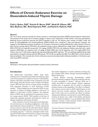
- 3. Quinn et al 3 differ from the TM+SAL group. All rats receiving DOX possessed significantly lower thymus masses than SAL ani- mals, as seen in Figure 1. Thymus mass relative to BM was also significantly lower in rats receiving DOX. Thymocyte Viability Figure 2 illustrates viable T-cell numbers between groups. There was a significant drug effect observed (P = .0107) with DOX administration decreasingT-cell numbers. No significant exercise effect or interaction was observed, and post hoc test- ing did not reveal any between-group differences (P > .05). Lipid Peroxidation As shown in Figure 3, a significant drug effect was observed in MDA+4HAE levels (P < .0001), indicating that DOX treatment elevated oxidative stress levels. A significant activity effect (P < .0001) was seen, with chronic exercise suppressing levels of lipid peroxidation. Additionally, a sig- nificant interaction (P = .0385) was observed. TM+10 exhibited significantly less MDA+4HAE than SED+10 and was not significantly different from SED+SAL. TM+12.5 thymus had significantly less MDA+4HAE than the SED+12.5 as well. Discussion To our knowledge, this is the first study to examine the effects of chronic exercise prior to DOX treatment on the thymus. We examined changes in tissue mass, local T-cell count, and MDA+HAE. Our main finding was that chronic endurance exercise significantly reduced levels of oxidative stress following DOX injections. This finding is in agree- ment with previous studies suggesting that chronic exercise elevates antioxidant enzyme levels and activity. However, exercise did not prevent thymic involution and thymocyte loss. Although T-cell number was not significantly different between TM and SED animals receiving 10 mg/kg (P = .1701), a trend of higher viable T-cell count with endur- ance exercise was noted. DOX is an antineoplastic agent used in a wide variety of cancers. In spite of its efficacy combating cancerous cells, accompanying side effects limit dosing. The most recog- nized side effect accompanying DOX administration is car- diotoxicity because DOX accumulates in cardiac cells and undergoes redox cycling resulting in the formation of ROS.5,17 ROS-associated damage occurs as peroxidizing of Table 2. Physical Characteristics of Subjects.a SED+SAL (n = 8) SED+10 mg/kg (n = 8) SED+12.5mg/kg (n = 7) TM+SAL (n = 9) TM+10 mg/kg (n = 9) TM+12.5 mg/kg (n = 7) Body mass (g) 432.8 ± 6.3 401.0 ± 10.8 375.9 ± 7.6b,c 441.9 ± 9.2 394.9 ± 16.3 422.6 ± 16.1 Δ Body mass (g) 2.8 ± 4.9 −38.7 ± 3.8b,c −32.1 ± 8.5b,c 0.8 ± 2.9 −44.0 ± 6.4b,c −26.9 ± 7.1b Thymus mass (g) 265.7 ± 13.6 123.0 ± 10.3b,c 122.8 ± 15.7b,c 258.8 ± 17.6 113.1 ± 13.6b,c 116.4 ± 9.9b,c Thymus/Body (mg/kg) 614.8 ± 35.3 282.7 ± 43.4b,c 319.1 ± 136.4b,c 556.5 ± 46.4 288.8 ± 29.9b,c 275.0 ± 20.3b,c Abbreviations: SED, sedentary; SAL, saline treated; TM, treadmill trained; 10 mg/kg, 10 mg/kg doxorubicin treated; 12.5 mg/kg, 12.5 mg/kg doxorubicin treated; Δ Body mass, change in body mass before and after injections. a Data are mean ± standard error of the mean. b Significantly different compared with sedentary/saline group (P < .05). c Significantly different compared with exercise/saline group (P < .05). Figure 1. Thymus size comparison: A. SED+DOX; B. SED+SAL. Abbreviations: SED, sedentary; DOX, doxorubicin; SAL, saline treated. Table 1. Progressive Treadmill Training Protocol. Week 1 2 3 4 5 6 7 8 9 10 Speed (m/min) 25 25 25 30 30 30 30 30 30 30 Incline (%) 0 0 0 3 6 9 12 15 18 18 Duration (minutes) 20 30 30 60 60 60 60 60 60 60 by guest on June 2, 2016ict.sagepub.comDownloaded from
- 4. 4 Integrative Cancer Therapies the lipid constituting membranes, at the cellular, mitochon- drial, and nuclear levels.18 Additionally, DOX intercalates DNA and stabilizes topoisomerases, preventing synthesis and replication. DOX-affected cells may undergo apoptotic events with eventual cell death. Although cardiac tissue has received a majority of the attention regarding associated toxicity, DOX has also been shown to induce thymic invo- lution and T-cell senescence.1 Additionally, isolated rat thy- mocytes exposed to DOX undergo DNA fragmentation, with subsequent increases in ROS scavenging enzyme activities of superoxide dismutase (SOD) and catalase activities.4 Although elevated antioxidant enzymes could not prevent apoptosis, the stimulation of antioxidant enzymes suggests that apoptosis may be partly a result of free radical formations. In vitro exposure of murine lym- phocytes to DOX induces rapid DNA degradation in mostly noncycling cells, with greater cell death dependent on DOX concentrations.19 The immune system functions to identify and eliminate foreign antigens that enter the body.20 Leukocytes, or white blood cells, originate in bone marrow, but mature in organs, including the thymus, spleen, lymph nodes, and peripheral lymphoid organs. Leukocytes that act directly on antigens are B- and T-cells, with B-cells creating antibodies specific to antigens and T-cells destroying targeted cells. B-cells mature in bone marrow, whereas T-cells mature in the thy- mus. T-cells, or thymocytes, are cytotoxic (CD8+ ), helper (response-enhancing, CD4+ ), or regulatory (Treg) cells. Various conditions of stress have been shown to decrease T-cell number and lead to thymic involution, including anx- iety, physical stress, and heat/cold exposure.21-24 Physical stress, such as exercise, may alter immunologi- cal capacity. Nieman’s25 J-shaped model of exercise and immunity suggests that exercise can enhance or reduce immune function depending on frequency, duration, and intensity of exercise performed. Evidence suggests that strenuous acute bouts of exercise decrease circulating lym- phocyte number while increasing apoptosis, as evidenced by DNA fragmentation.26,27 Conversely, submaximal exer- cise in mice demonstrates fewer apoptotic and higher num- bers of viable T-cells compared with sedentary controls.28 Although peroxidation of lipids of cellular and mitochon- drial membranes have been primarily implicated in the induction of apoptosis, isolated rat nuclei exposed to DOX also exhibit membrane peroxidation.29 In addition to ele- vated ROS following strenuous exercise, increased gluco- corticoids, intracellular Ca2+ , and inflammatory signals may induce apoptosis during times of suppressed immune func- tion.30 A rise in glucocorticoid release and intracellular Ca2+ levels may further induce apoptosis in lymphocytes.31 Glucocorticoids released in response to exercise, such as cortisol, have been shown to induce pyknosis and DNA fragmentation in isolated thymocytes.32 In accordance with the J-shaped model, studies examin- ing response to chronic and acute endurance exercise have revealed opposing lymphocytic effects. Chronic exercise improved thymic structural quality and circulating lympho- cyte populations, whereas acute, exhaustive bouts had Figure 2. Graphical representation of viable T-cell number: data are mean ± standard error of the mean. Significant drug effect (P = .0107); no activity or interaction effect (P > .05). Abbreviations: SED, sedentary; TM, treadmill trained; SAL, saline treated; 10 mg/kg, 10 mg/kg doxorubicin treated; 12.5 mg/kg, 12.5 mg/kg doxorubicin treated. Figure 3. Graphical representation of lipid peroxidation in the thymus: data are mean ± standard error of the mean. Abbreviations: SED, sedentary; TM, treadmill trained; SAL, saline treated; 10 mg/kg, 10 mg/kg doxorubicin treated; 12.5 mg/kg, 12.5 mg/kg doxorubicin treated; DOX, doxorubicin. a Significantly different as compared with sedentary/saline group (P < .05). b Significantly different as compared with exercise/saline group (P < .05). c Significantly different as compared with sedentary/DOX 10-mg/kg group (P < .001). d Significantly different as compared with sedentary/DOX 12.5-mg/kg group (P < .01). by guest on June 2, 2016ict.sagepub.comDownloaded from
- 5. Quinn et al 5 negative effects on the thymus, lymphocyte populations, and other immune organs.33 Specifically, thymic tissues express significantly elevated membrane lipid peroxidation after mice were acutely run to exhaustion.27 Furthermore, T-cells exhibited higher levels of intracellular Ca2+ and lipid peroxidation after exercise.34 Navalta et al35 demonstrated that exercise intensity affects circulating lymphocyte apop- tosis, with significance occurring above 60% of VO2max in untrained human subjects performing incremental treadmill tests to exhaustion. To determine training effects on lymphocyte apoptosis in response to chronic training, Mooren et al36 compared trained runners against poorly trained individuals and found cell death induced by exercise in the poorly trained indi- viduals. Enhanced serum antioxidant enzyme production with chronic wheel running (10 months) in mice demon- strated reduced ROS-induced apoptosis in immune cells.37 An increase in antioxidant enzymes through chronic exer- cise training may attenuate ROS-induced damage seen in lymphocytes.7 Additionally, 8 weeks of treadmill- and swim-training induced significantly higher levels of CD4+ and CD8+ T-cells in the thymus of Sprague-Dawley rats.38 Although acute, exhaustive exercise may elevate glucocor- ticoids, which initiate lymphocytic cell death, chronic sub- maximal training may not trigger such levels. It has been long known that exhaustive exercise in rats induces significant thymic atrophy.21 Additionally, studies demonstrate that lymphocyte apoptosis occurs immediately following high-intensity exercise.26 Using a submaximal, chronic training program, however, may enhance antioxi- dant enzyme concentrations and alleviate ROS-related lym- phocyte apoptosis without the negative side effects seen with exhaustive bouts. Sugiura et al39 determined that 12 weeks of forced, submaximal running in mice (15 m/min) significantly increased spleen and liver mass while slightly enlarging thymic tissue compared with sedentary animals. Using a swim-training model for 8 weeks, Pereira et al7 demonstrated that lipid peroxidation levels were signifi- cantly decreased in thymus tissues versus sedentary con- trols. Additionally, citrate synthase and GPX levels were significantly elevated. Short-term exercise (3 weeks) in hemodialysis patients does not restore T-cell numbers com- parable to normal healthy individuals, but T-cell activity is elevated when compared with sedentary hemodialysis patients.40 It is suggested that chronic, moderate exercise may increase total T-cell number with amplified activity.41 This study did not examine the effects of exercise intensity with DOX treatment on thymic changes. The thymus naturally reduces the size and total number of T-cells in an age-dependent fashion, leading to decreased immune resistance, referred to as immunosenescence.1,42 As humans age, the number of cytotoxic CD8+ T-cells tend to decrease with senescence. Moderate exercising older adults exhibit greater proportions of CD8+ T-cells when compared with sedentary individuals.43 Patients receiving chemother- apy also have a suppressed immune system. As demon- strated in this study, acute DOX treatment significantly reduces size of thymic tissue and reduces viable T-cell num- bers. There was an exercise effect, with endurance-trained animals presenting significantly less thymic lipid peroxida- tion when compared with sedentary paired groups. Furthermore, the level of MDA+4HAE in endurance- trained rats receiving 10 mg/kg DOX (TM+10) was not sig- nificantly different from that in sedentary/saline animals (SED+SAL). A significant drug effect was noted with DOX treatment in total T-cell number, and prior exercise did not significantly attenuate these losses. The addition of DOX- induced thymus dysfunction may further compromise the ability of patients to stave off further infections experienced during treatment and shortly thereafter. A prior endurance exercise program may reduce the thymic-related losses in size and T-cell number. Clinical implications of this study suggest that chroni- cally trained individuals may demonstrate greater immune response following chemotherapy treatment than sedentary counterparts. Clearly, this study demon- strates that DOX treatment results in significant thymic atrophy. Production of naïve T-cells is governed by thy- mus output and not replication of the circulating cell pool.44 Thymic involution, or atrophy, results in decreased T-cell development and thymopoiesis.45 Conversely, increased size of thymus may complement T-cell produc- tion and new antigen resistance in the time following treatment. DeNardo et al46 suggest that, in addition to increased macrophage number, an abundance of T-cells indicate greater survival rate among breast cancer patients. Additionally, Feng et al47 showed that mice in an oxidative stress model exhibit significantly less T-cell proliferation than controls. Exercise preconditioning lowered markers of oxidative stress and may attenuate decreased T-cell production following DOX treatment. The optimal cumulative dose of DOX for induction of congestive heart failure (CHF) has been established at 12.45 mg/kg in rats.48 The doses used (10 and 12.5 mg/kg) in this study may elicit outcomes similar to those experienced in clinical settings. However, clinical admin- istration of DOX is typically delivered in small doses over the course of treatment. The single bolus injection strategy is a limitation of the present study. Viable T-cell numbers in this study may have been overestimated in samples because of cells undergoing early stages of apoptosis. During early apoptosis, cell membranes remain intact and do not allow trypan blue entry into cells. The early-apoptotic T-cells were not distinguished in this study. Although this study did not examine circu- lating thymocytes, animals had not exercised for 3 days after injection, and lymphocyte circulation typically ele- vates following stressful conditions. by guest on June 2, 2016ict.sagepub.comDownloaded from
- 6. 6 Integrative Cancer Therapies Conclusion Data from the current study suggest significant decreases in thymic lipid peroxidation with chronic endurance exercise prior to DOX administration. Additionally, exercise precon- ditioning may have contributed to an increasing trend in T-cell count. Individuals who aerobically exercise at sub- maximal intensities prior to chemotherapy may offset thy- mocytic depressions typical with DOX treatment. The observed depression in levels of MDA+4HAE may be attributed to elevated antioxidant enzyme levels and activ- ity associated with chronic endurance exercise training. Acknowledgments The authors wish to thank Gregory DeKrey for his technical con- tribution to this investigation. Declaration of Conflicting Interests The author(s) declared no potential conflicts of interest with respect to the research, authorship, and/or publication of this article. Funding The author(s) received no financial support for the research, authorship, and/or publication of this article. References 1. Sultana R, Di Domenico F, Tseng M, et al. Doxorubicin-induced thymus senescence. J Proteome Res. 2010;9:6232-6241. 2. Bagchi D, Bagchi M, Hassoun EA, Kelly J, Stohs SJ. Adriamycin-induced hepatic and myocardial lipid peroxida- tion and DNA damage, and enhanced excretion of urinary lipid metabolites in rats. Toxicology. 1995;95(1-3):1-9. 3. Carvalho FS, Burgeiro A, Garcia R, Moreno AJ, Carvalho RA, Oliveira PJ. Doxorubicin-induced cardiotoxicity: from bioenergetic failure and cell death to cardiomyopathy. Med Res Rev. 2014;34:106-135. 4. Azmi S, Bhatia L, Khanna N, Dhawan D, Singh N. Adriamycin induces apoptosis in rat thymocytes. Cancer Lett. 1997;111(1-2):225-231. 5. Davies KJ, Doroshow JH. Redox cycling of anthracyclines by cardiac mitochondria: I. Anthracycline radical formation by NADH dehydrogenase. J Biol Chem. 1986;261:3060-3067. 6. Reyes MP, Lerner AM, Ho KL. Diminution in the size of the thy- musinmiceduringforcedswimming.JInfectDis.1981;143:292. 7. PereiraB,CostaRosaLF,SafiDA,MedeirosMH,CuriR,Bechara EJ. Superoxide dismutase, catalase, and glutathione peroxidase activities in muscle and lymphoid organs of sedentary and exer- cise-trained rats. Physiol Behav.1994;56:1095-1099. 8. Hoffman-Goetz L, Thorne R, Simpson J, Arumugam Y. Exercise stress alters murine lymphocyte subset distribu- tion in spleen, lymph nodes and thymus. Clin Exp Immunol. 1989;76:307-310. 9. Hydock DS, Lien CY, Jensen BT, Schneider CM, Hayward R. Exercise preconditioning provides long-term protec- tion against early chronic doxorubicin cardiotoxicity. Integr Cancer Ther. 2011;10:47-57. 10. Hydock DS, Lien C-Y, Schneider CM, Hayward R. Exercise preconditioning protects against doxorubicin-induced cardiac dysfunction. Med Sci Sports Exerc. 2008;40:808-817. 11. Chicco AJ, Hydock DS, Schneider CM, Hayward R. Low- intensity exercise training during doxorubicin treatment protects against cardiotoxicity. J Appl Physiol (1985). 2006;100:519-527. 12. Parry TL, Hydock DS, Jensen BT, Lien CY, Schneider CM, Hayward R. Endurance exercise attenuates cardiotoxicity induced by androgen deprivation and doxorubicin. Can J Physiol Pharmacol. 2014;92:356-362. 13. Kanter MM, Hamlin RL, Unverferth DV, Davis HW, Merola AJ. Effect of exercise training on antioxidant enzymes and cardiotoxicity of doxorubicin. J Appl Physiol (1985). 1985;59:1298-1303. 14. Chicco AJ, Schneider CM, Hayward R. Voluntary exercise protects against acute doxorubicin cardiotoxicity in the iso- lated perfused rat heart. Am J Physiol Regul Integr Comp Physiol. 2005;289:R424-R431. 15. Zhang H-G, Grizzle WE. Exosomes and cancer: a newly described pathway of immune suppression. Clin Cancer Res. 2011;17:959-964. 16. Bradford MM. A rapid and sensitive method for the quantita- tion of microgram quantities of protein utilizing the principle of protein-dye binding. Anal Biochem. 1976;72:248-254. 17. Doroshow JH, Davies KJ. Redox cycling of anthracyclines by cardiac mitochondria: II. Formation of superoxide anion, hydrogen peroxide, and hydroxyl radical. J Biol Chem. 1986;261:3068-3074. 18. Mimnaugh EG, Kennedy KA, Trush MA, Sinha BK. Adriamycin-enhanced membrane lipid peroxidation in iso- lated rat nuclei. Cancer Res. 1985;45:3296-3304. 19. Zaleskis G, Berleth E, Verstovsek S, Ehrke M, Mihich E. Doxorubicin-induced DNA degradation in murine thymo- cytes. Mol Pharmacol. 1994;46:901-908. 20. O’Leary A. Stress, emotion, and human immune function. Psychol Bull. 1990;108:363-382. 21. Andersen DH. The effect of food and of exhaustion on the pituitary, thyroid, adrenal and thymus glands of the rat. J Physiol. 1935;85:162-167. 22. Selye H. Thymus and adrenals in the response of the organism to injuries and intoxications. Br J Exp Pathol. 1936;17:234-248. 23. Concordet JP, Ferry A. Physiological programmed cell death in thymocytes is induced by physical stress (exercise). Am J Physiol. 1993;265:C626-C629. 24. Mars M, Govender S, Weston A, Naicker V, Chuturgoon A. High intensity exercise: a cause of lymphocyte apoptosis? Biochem Biophys Res Commun. 1998;249:366-370. 25. Nieman DC. Exercise, infection, and immunity. Int J Sports Med. 1994;15:S131-S141. 26. Niess A, Veihelmann S, Passek F, et al. Exercise induced oxida- tive stress: DNA damage and expression of stress proteins in leucocytes—an overview. Dtsch Z Sportmed. 1997;48:330-341. 27. Azenabor A, Hoffman-Goetz L. Intrathymic and intras- plenic oxidative stress mediates thymocyte and splenocyte damage in acutely exercised mice. J Appl Physiol (1985). 1999;86:1823-1827. 28. Hoffman-Goetz L, Zajchowski S, Aldred A. Impact of tread- mill exercise on early apoptotic cells in mouse thymus and spleen. Life Sci. 1998;64:191-200. by guest on June 2, 2016ict.sagepub.comDownloaded from
- 7. Quinn et al 7 29. Mimnaugh EG, Kennedy KA, Trush MA, Sinha BK. Adriamycin-enhanced membrane lipid peroxidation in iso- lated rat nuclei. Cancer Res. 1985;45:3296-3304. 30. Nieman DC. Immune response to heavy exertion. J Appl Physiol (1985). 1997;82:1385-1394. 31. Davies KJ, Quintanilha AT, Brooks GA, Packer L. Free radicals and tissue damage produced by exercise. Biochem Biophys Res Commun. 1982;107:1198-1205. 32. Thomas N, Edwards JL, Bell PA. Studies of the mechanism of glucocorticoid-induced pyknosis in isolated rat thymocytes. J Steroid Biochem. 1983;18:519-524. 33. Hoffman-Goetz L, Thorne R, Simpson JA, Arumugam Y. Exercise stress alters murine lymphocyte subset distribu- tion in spleen, lymph nodes and thymus. Clin Exp Immunol. 1989;76:307-310. 34. Azenabor AA, Hoffman-Goetz L. Effect of exhaustive exer- cise on membrane estradiol concentration, intracellular cal- cium, and oxidative damage in mouse thymic lymphocytes. Free Radic Biol Med. 2000;28:84-90. 35. Navalta JW, Sedlock DA, Park KS. Effect of exercise inten- sity on exercise-induced lymphocyte apoptosis. Int J Sports Med. 2007;28:539-542. 36. Mooren FC, Lechtermann A, Volker K. Exercise-induced apoptosis of lymphocytes depends on training status. Med Sci Sports Exerc. 2004;36:1476-1483. 37. Avula CP, Muthukumar AR, Zaman K, McCarter R, Fernandes G. Inhibitory effects of voluntary wheel exercise on apoptosis in splenic lymphocyte subsets of C57BL/6 mice. J Appl Physiol (1985). 2001;91:2546-2552. 38. Kwak Y-S, Um S-Y, Kim D-E, Hwang H-J. The effect of dif- ferent type of exercise on SOD, neutrophils and T lympho- cytes. Immune Netw. 2005;5:232-236. 39. Sugiura H, Nishida H, Washino K, Inaba R, Iwata H, Maeno H. Effects of exercise on immune functions in mice. Jpn J Hyg. 1993;48:845-851. 40. Endo F, Usuda S, Nakamura M, Kurashige S. The effects of rehabilitation on the immune responses in patients: I. The effects of exercise training on the T-cell activity in chronic hemodialysis patients. J Phys Ther Sci. 1995;7:15-20. 41. Ferry A, Rieu P, Laziri F, El Habazi A, Le Page C, Rieu M. Effect of moderate exercise on rat T-cells. Eur J Appl Physiol Occup Physiol. 1992;65:464-468. 42. Plecas-Solarovic B, Pesic V, Radojevic K, Leposavic G. Morphometrical characteristics of age-associated changes in the thymus of old male wistar rats. Anat Histol Embryol. 2006;35:380-386. 43. Yan H, Kuroiwa A, Tanaka H, Shindo M, Kiyonaga A, Nagayama A. Effect of moderate exercise on immune senes- cence in men. Eur J Appl Physiol. 2001;86:105-111. 44. Aspinall R, Andrew D. Thymic involution in aging. J Clin Immunol. 2000;20:250-256. 45. Lynch HE, Goldberg GL, Chidgey A, Van den Brink MR, Boyd R, Sempowski GD. Thymic involution and immune reconstitution. Trends Immunol. 2009;30:366-373. 46. DeNardo DG, Brennan DJ, Rexhepaj E, et al. Leukocyte com- plexity predicts breast cancer survival and functionally regu- lates response to chemotherapy. Cancer Discov. 2011;1:54-67. 47. Feng R, He W, Ochi H. A new murine oxidative stress model associated with senescence. Mech Ageing Dev. 2001;122:547-559. 48. Spivak M, Bubnov R, Yemets I, et al. Doxorubicin dose for congestive heart failure modeling and the use of general ultra- sound equipment for evaluation in rats. longitudinal in vivo study. Med Ultrason. 2013;15:23-28. by guest on June 2, 2016ict.sagepub.comDownloaded from