
Recommended
More Related Content
What's hot
What's hot (20)
Similar to Goniometer (range of motion )
Similar to Goniometer (range of motion ) (20)
Recently uploaded
Recently uploaded (20)
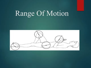
Goniometer (range of motion )
- 2. Why Is It Performed ? • Determining the presence of joint impairment • Developing treatment goals. • Evaluating progress or lack of progress. • Modifying treatment. • Motivating the subject. • Research
- 3. PLANES AND AXIS • Osteo-kinematic motions are described to be taking place in 3 cardinal planes and axis
- 5. A frontal or coronal axis lies parallel to the transverse suture of the skull. It is also horizontal and at right angle to the sagittal axis. Movement about frontal axis occurs in a sagittal plane. Flexion and extension (except of the thumb) occurs about a frontal axis and in a sagittal plane. A sagittal or antero-posterior axis lies parallel to the sagittal suture of the skull, i.e., in an antero-posterior direction. Movement about this axis occurs in a frontal plane. Abduction and adduction (except pf the thumb) and side flexion movements are said to take about a sagittal axis and in a frontal plane. A vertical axis lies parallel to the line of gravity and movement about it occurs in a horizontal plane. Rotation occurs about a vertical axis and in a horizontal plane
- 6. Joint Ranges Active ROM Passive ROM • Active motion is the unassisted voluntary movement of a joint. (Quality of ROM) • Passive motion is attained by the examiner without the patient’s assistance.(Quantity of ROM) • Normally, PROM is slightly greater than AROM because joints have a small amount of motion at the end range that is not under voluntary control.
- 7. MEASURING JOINT RANGE OF MOTION • Range Of Motion (ROM) is the arc of motion that occurs at a joint or a series of joints. • Three notation systems have been used to define ROM : 1. The 0 to 180 degree system 2. The 180 to 0 degree system 3. The 360 degree system Most commonly used is the 0 to 180 degree notation system
- 8. Prerequisite Knowledge For Measuring ROM a) Normal ROM’s (Range) b) Joint Structure And Function c) Recommended positioning for self and patient d) Bony landmarks related to each joint e) Alignment of Goniometer f) Normal end-feel g) Factors that can alter normal ROM
- 9. FACTORS DETERMINING AMOUNT OF ROM Integrity Of Joint SurfaceRELIABILI TY Amount Of Scarring Present AG E GEND ER Shape Of Articulati ng Surface Healt h Of Joint Various diseases/ pathologic al conditions Health Of Surroundi ng Tissues Mobilty & Pliabilty Of Soft Tissue
- 10. Common pathological causes of ROM Restriction • Skin/soft tissue contracture • Arthritis • Fracture • Burns • Muscle weakness/paralysis • Pain • Edema • Spasticity • Presence of foreign body in the joint
- 11. Prerequisite Skills For Measuring ROM • The therapist should be skilled in Correct positioning Stabilization for measurement Palpation Alignment Recording measurements accurately Documentation
- 12. Testing Procedure PLACE THE SUBJECT IN TESTING POSITION STABILIZE THE PROXIMAL JOINT SEGMENT MOVE THE DISTAL JOINT SEGMENT TO ZERO STARTING POSITION. SLOWLY MOVE THE DISTAL JOINT SEGMENT TO THE END OF PASSIVE ROM AND DETERMINE END FEEL MAKE VISUAL ESTIMATE OF THE ROM RETURN THE DISTAL JOINT SEGMENT TO THE STARTING POSITION PALPATE THE BONY ANATOMICAL LANDMARKS ALIGN THE GONIOMETER
- 13. RECORD THE STARTING POSITION. REMOVE THE GONIOMETER STABILIZE THE PROXIMAL JOINT SEGMENT MOVE THE DISTAL SEGMENT THROUGH FULL ROM REALIGN THE GONIOMETER. PALPATE THE ANATOMICAL LAND MARKS AGAIN IF NECESSARY RECORD THE ROM
- 14. Documentation • Hypo Mobility : A motion that does not start with 0 degree or ends prematurely indicates joint hypomobility Example : if knee joint has 30 degree of hypomobility in flexion, it would be recorded as 30 – 135 deg • Hyper Mobility : Joint hypermobility at the beginning of the range is noted by inclusion of a zero between the starting & ending measurements Example : if the elbow joint has 5 degree of hypermobility in extension and 140 degree of flexion , it would be recorded as 5 – 0 – 140 deg
- 15. What is Goniometry? • The term goniometry is derived from two Greek words : Gonia-metron • Therefore, goniometry refers to the measurement of angles, in particular the measurement of angles created at human joints by the bones. ANGL E MEASU RE
- 16. Types of Goniometer • Full Circle Manual Universal Goniometer (360) • Half circle manual Goniometer (180) • Gravity Goniometer :- • a) Double Inclinometer (used for spine goniometry) • b) Pendulum Inclinometer • c) BubbleGoniometer • Electrogoniometer • Digital Goniometer • Tape Measurements • Smartphone Devices • Use of malleable wires/sheets (in cases of deformities)
- 20. UNIVERSAL GONIOMETER • A universal Goniometer may be constructed of metal or plastic and it has 3 parts :- 1. Body of Goniometer2. Stationary arm 3. Movable arm (placed over the Joint being measured) (aligned parallel with the longitudinal axis of the fixed part) (aligned parallel with the longitudinal axis of the movable part)
- 22. Precautions !!! 1. Joint irritability status 2. Presence of Pain 3. Instability 4. Recent trauma 5. Is it really important to assess accurate ROM ??
- 23. END-FEEL • The end of each motion at each joint is limited from further movement by particular anatomical structures. • The type of structure that limits a joint motion has a characteristic feel, which may be detected by the therapist performing the passive ROM. • This feeling, which is experienced by the therapist as resistance or a barrier to further motion, is called the end-feel.
- 24. NORMAL END-FEEL DESCRIPTION EXAMPLE Soft Soft Tissue Approximation Knee flexion (contact between soft tissue of posterior leg and posterior thigh) Firm Muscular stretch Hip flexion with knee straight (passive elastic tension of hamstring muscles) Capsular stretch Extension of metacarpophalangeal joints of fingers Ligamentous stretch Forearm supination (tension in the palmar radioulnar ligament of the inferior radioulnar joint) Hard Bone contacting bone Elbow extension (olecranon process of the ulna and olecranon fossa
- 25. ABNORMAL END-FEEL DESCRIPTION EXAMPLES Soft Occurs sooner or later in the Soft tissue edema ROM than is usual or in a joint Synovitis that normally has a firm or hard end-feel . Feels boggy. Firm Occurs sooner or later in the Increased muscular tonus ROM than is usual or in a joint Capsular , muscular , that normally has a soft or ligamentous, and fascial hard end-feel. shortening Hard Occurs sooner or later in the Chondromalacia ROM than is usual or in a joint Osteoarthritis that normally has a soft or Loose bodies in joint firm end-feel. A bony grating Myositis ossificans or bony block is felt. Fracture Empty No real end-feel because pain Acute joint inflammation prevents reaching end of Bursitis ROM. No resistance is felt Abscess except for patient’s protective Fracture
- 26. Capsular & Non-capsular Pattern Of Movement Restriction • Cyriax proposed that pathological conditions involving the entire joint capsule cause a particular pattern of restriction involving most of the passive motions of the joint. This pattern is called as capsular pattern • Restriction caused by condition involving structures other than the entire joint capsule is called as non-capsular pattern • Example – Adhesive Capsulitis Shoulder
- 27. Shoulder ROM FLEXION: Motion: 0-180º Position: Subject supine with knees flexed or sitting. elbow extended with the palm facing the body Goniometer: Axis at the acromion process, laterally through the head of the humerus. Stationary arm is placed along the mid-axillary line of the trunk Moving arm place along the lateral mid-line of the humerus in line with the lateral epicondyle.
- 28. EXTENSION: Motion: 0-45º~60º from neutral position Position: Subject prone or sitting , elbow in slight flexion with the palm facing the body. Goniometer: Axis at the acromion process, laterally through the head of the humerus Stationary Arm aligned with mid- axillary line of the trunk Moving arm along the lateral mid-line of humerus in line with lateral epicondyle
- 30. ABDUCTION: Motion:0-180º Position: Supine, prone or sitting with the limb in anatomic position Goniometer: Axis at anterior portion of acromion process. Stationary arm at lateral aspect of anterior surface of chest parallel to midline of sternum. Moving arm on anterior aspect of arm parallel to midline of humerus and in line with medial epicondyle. OR Goniometer: Axis at the posterior portion of the acromion process; Stationary arm aligned parallel to spinous process of the vertebral colomn Moving arm aligned with the midline of the humerus in line with lateral epicondyle ADDUCTION: Motion: 0-30º Aligment of goniometer is same as abduction.
- 32. EXTERNAL ROTATION: Motion: 0-90º Position: Supine. Shoulder is abducted to 90º. Elbow flexed with forearm in neutral and perpendicular to table top such that the palm is facing the feet. Elbow not supported. Humerus is fully supported on the table. Stabilize the distal humerus, thorax, and scapula. Goniometer: Axis at olecranon process of the ulna. Stationary arm placed parallel to the table top or perpendicular to the floor. Moving arm along the ulnar shaft aligned with the styloid process of the ulna. INTERNAL ROTATION: Motion: 0-65~90º Positioning and goniometer alignment is same as in external rotation
- 34. Radio-ulnar ROM Supination: Motion: 0- 80º~ 90º Position: Subject sitting or supine, with the elbow flexed to 90º. Shoulder in zero degrees of its’ ROM. Position starts midway between Supination and Pronation. Goniometer: Axis is medial to the ulnar styloid process. Stationary arm is aligned parallel to the anterior midline of the humerus. Moving arm across the ventral aspect of the wrist on a line between and proximal to the styloid process of the radius and the ulna. Pronation: Motion: 0- 80º~ 90º Position: same for supination. Goniometer: Axis is lateral to the ulnar styloid process. Stationary arm is aligned parallel to the anterior midline of the humerus. Moving arm across the dorsum of the wrist on a line between and proximal to the styloid process of the radius and the ulna.
- 36. JOINT MOTION TESTING POSITION STABILIZATION MEASUREMENT S CERVICAL • FLEXION Sitting Shoulder & chest 1 cm– 4.3 cm • EXTENSION Shoulder & chest to prevent extension of thoracic & lumbar spine 18.5 cm–22.4cm • SIDE FLEXION To prevent side flexion of thoracic & lumbar spine 10.7cm-12.9cm • ROTATION To prevent rotation of thoracic & lumbar 11cm-13.2cm TAPE MEASUREMENTS OF THE SPINE
- 37. JOINT MOTION TESTING POSITION STABILIZATION MEASUREMEN TS THORACIC • FLEXION STANDING PELVIS To prevent anterior tilting 10 cms (4 inches) • EXTENSION •If the subject has balance problems or muscle weakness in the LE, measurement can be taken in prone/side lying To prevent posterior tilting • LATERAL FLEXION To prevent lateral tilting 15.9cm for rt LF 16.9cm for lt LF • ROTATION SITTING To prevent rotation 45 degree (universal goniometer )
- 38. JOINT MOTION TESTIN G POSITIO N STABILIZATIO N MEASUREMEN TS LUMBAR • FLEXION STANDING PELVIS To prevent anterior tilting 6.7cm in males 5.8cm in females Average 6.3cm- 6.9cm (Modified Schober test) •EXTENSION To prevent posterior tilting 1.6cm (Modified Schober Test) •LATER AL FLEXIO N To prevent lateral tilting 25 – 30 degree by AMA (double inclinometer)
- 39. Demonstration Schober’s Test For Lumbar Spine Flexion
- 49. HFD Thomas Test
- 50. KF D
- 51. Equinu s
- 52. TF Malalignment
- 53. Genu Recurvatum