
Recommended
More Related Content
What's hot
What's hot (20)
Similar to Uterine inversion
Similar to Uterine inversion (20)
Recently uploaded
Recently uploaded (20)
Uterine inversion
- 1. Edna Dahir
- 2. Introduction This is Rare.But Potentially Life Threatening Complication of the Third Stage Of Lobour. It Occurs in Approximately 1 in 20,000 Deliveries The Obstetric Inversion is almost always an Acute One & Usually Complete.
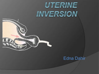
- 3. DEFINITION ‘‘Inversion of Uterus means Uterus is Turned Inside Out Partially OR Completely. Uterine inversion is the folding of the fundus into the uterine cavity in varying degrees.
- 4. CLASSIFICATION Inversion Of Uterus is Classified in Mainly 3 Types : A. According Types B. According Degrees C. According the Timing of Event
- 5. A. Types 1) Incomplete Inversion : When fundus of uterus has turned inside out, like toe of socks, but inverted fundus has not descended through Cx… 2) Complete Inversion : When the inverted fundus has passed completely through Cx to lie within the vagina or lie often outside the Vaginal Wall.
- 6. B. Degrees First degree: The uterus is partially turned out Second degree: The fundus has passed through the cervix but not outside the vagina Third degree: The fundus is prolapsed outside the vagina Fourth degree: The uterus, cervix and vagina are completely turned inside out and are visible
- 7. Universally…. First Degree : Incomplete Inversion Second Degree : Complete inversion in the vagina Third Degree : Complete inversion outside the Vagina
- 8. C. According to Timing of Event Acute : It occurs within 24 hrs of delivery. Sub-acute : It presents between 24 hrs & 4 wks of delivery. Chronic : It presents beyond 4 wks of delivery or in non pregnant stage.
- 9. CAUSES Excessive cord traction (esp. with an unseparated placenta) Excessive fundal pressure (esp. when uterus is poorly contracted Atonic) Placenta accreta Congenital predisposition
- 10. Conti… Spontaneous (40%) : Abnormal short umbilical cord or functionally shortened by being wrapped around the fetal body. Sudden rise in intra abdominal pressure due to maternal coughing or vomiting. Connective tissue disorder such as Marphan’s syndrome.
- 11. Conti… Latrogenic: Due to mismanagement of third stage of labor… Pulling the cord when the uterus is atonic while combined with fundal pressure Faulty technique in manual removal of placenta While separating retained placenta from the wall, a portion may remain attached and as the placenta is withdrawn, the fundus is also withdrawn.
- 12. Sign & Symptoms Hemorrhage (94%) Severe abdominal pain in 3rd stage Hypotension with Bradycardia Uterine fundus not palpable abdominally Mass in the vagina on vaginal examination. Sudden cardiovascular collapse Lump in the vagina Abdominal tenderness Absence of uterine fundus on abdominal palpation
- 13. Conti… Shock Shock is initially out of proportion with the amount of blood loss. Woman becomes sweaty with bradycardia, profound hypotension and rarely cardiac arrest. In short time there is marked hemorrhage and Hypovolemic shock.
- 14. DIAGNOSIS The diagnosis of uterine inversion is based upon clinical findings: Bleeding, which may be severe and result in Hemorrhagic Shock Palpation of the prolapsed uterine fundus: Lower uterine segment = Vagina = INCOMPLETE COMPLETE By Intra Uterine Manual Examination
- 15. DIFFRENTIAL DIAGNOSIS Inversion of uterus Uterine rupture. Prolapse of uterine tumor (submucous fibroid).
- 16. 22 Management
- 17. Prevention 17 Do not employ any method to expel the placenta when the uterus is relaxed Patient should not be instructed to change her position. Pulling the cord simultaneously with fundal pressure should be avoided Manual removal of placenta should be done in proper manner.
- 18. 1) Starting from the edge of placenta , 2) The placenta is separated by a)keeping the back of the hand in contact with the uterine wall. 18
- 19. Management of Acute Inversion of Uterus 19 • Delay in treatment increases the mortality, So number of steps are taken immediately and simultaneously. Before shock develops : • When one is on the spot when the inversion happens TRY IMMEDIATE MANUAL REPLACEMENT, even without anesthesia if not easily available. Principle : “ The part of the uterus which has come down last , should go back first. “
- 20. Procedure If the diagnosis is made immediately after the inversion has occurred, then that same degree of relaxation of myometrium and cervix (which is required for the inversion to occur) will allow uterine replacement easily… 1. The gloved hand is lubricated with suitable antiseptic cream and placed inside the vagina. 2. The uterine fundus with or without the attached placenta, is cupped in the palm of the hand. The fingers and thumb of the hand are extended to identify margins of the cervix.
- 21. 3. The whole uterus is lifted upwards towards and beyond umbilicus 4. Additional pressure is exerted with the fingertips systematically and sequentially to push and squeeze the uterine wall back through the cervix. 29Dr Shashwat Jani. 9909944160
- 22. 5. Sustained pressure for 3-5 mins to achieve complete replacement 6. Apply counter support by the other hand placed on the abdomen 7. Once the fundus has been replaced keep the hand in the uterus while rapid infusion of oxytocin is given to contract the uterus. Initially, bimanual compression aids in control of further hemorrhage until uterine tone is recovered.
- 23. 8. When the uterus is felt contracting, the hand is slowly withdrawn. If placenta is attached, it is to be removed only after the uterus becomes contracted. If the placenta is partially attached , it should be peeled out before replacement of uterus.
- 25. 1) Starting from the edge of placenta , 2) The placenta is separated by a)keeping the back of the hand in contact with the uterine wall. b) with slicing movement of the hand. 33Dr Shashwat Jani. 9909944160
- 26. If the patient comes late : Within 1 -2 minutes, from the occurrence of inversion, the cervix and lower segment clamps down inverted part of the uterus. increasing congestion, Edema of the inverted fundus. makes manual replacement without anesthesia difficult. If first attempt at immediate manual replacement of uterus fails, move to the following sequence … 1. Call assistance Anesthesiologist (assistance of nurse and obstetricians) 2. Elevation of the foot of the delivery table may relieve the tension on the viscera and reduce the pain and shock 26
- 27. 3. Establish two wide bore i.v. cannulae. Send blood for for grouping and cross match. Rapidly run in 1-2 L of normal saline. Because though initially shock is neurogenic type, hypovolumia will follow due to hemorrhage. 4. Catheterize. 5. Prophylactic antibiotics are given 6. If pain is a dominant symptom, small doses of i.v. Morphine is given. 7. If the inverted uterus is prolapsed beyond the vagina, it is replaced within the vagina 8. Patient is shifted to OT. 9. Anaesthesia 27
- 28. COMPLICATIONS OF INVERSION OF UTERUS. Postpartum hemorrhage due to uterine atony. Hypovolaemic shock and all its consequence. Vasovagal shock (due to severe pain). Endometritis (sepsis). 28
- 29. Infection of adnexa. Necrosis of adnexa (ovaries) Damage to intestine . Chronic inversion. 29
- 30. • Recurrence of inversion. • Increased risk of rupture of uterus in next pregnancy (when surgical procedure done for inversion). • Increased risk of C-section in subsequent delivery. • Chronic pelvic pain -> if chronic inversion is not treated. 30
- 31. PREVENTION 31 • Many cases of acute uterine inversion result mainly from mismanagement of the third stage of labour in women who are already at risk.
- 32. MANEUVERS : TO BE AVOIDED • Excessive traction on the umbilical cord • Excessive fundal pressure • Excessive intra-abdominal pressure • Excessively vigorous manual removal of placenta. 32