Recommended

Recommended
More Related Content
What's hot
What's hot (13)
Similar to ECMM-Inspired Biomimetic Materials
Similar to ECMM-Inspired Biomimetic Materials (20)
ECMM-Inspired Biomimetic Materials
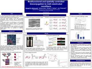
- 1. Extracellular matrix (ECM) inspired biomimetic materials The extra cellular matrix is a complex environment that provides chemical and physical support cells. A key component of biologically functional scaffolds is the need to introduce chemical or biochemical cues that allow for cellular adhesion proliferation, and differentiation, In biological systems, multiple cues are needed to direct cell biology. Continuous Processing of PCLNanofibers through Extrusion Multifunctional and spatially controlled bioconjugation to melt coextruded nanofibers Abigail A. Advincula1, Si-Eun Kim1, Emily C. Harker1, Jon Pokorski1* 1Department of Macromolecular Science & Engineering, Case Western Reserve University 2100 Adelbert Road, Cleveland, Ohio,d 44106 jon.pokorski@case.edu Our goal for this project is to introduce multiple orthogonal chemical modifications onto nanofibers in order to produce a surface capable of attaching various bioactive cues. We have recently reported a new class of fibrous biomaterials using coextrusion and a photochemical modification procedure to introduce functional groups onto the fibers. This project extends our methodology to control surface modification density, describe methods to synthesize multifunctional fibers, and provide methods to spatially control functional group modification. We investigate the effect of fiber modification with multiple cell-responsive peptides, the RGD sequence and osteogenic growth peptide (OGP), on substrates for osteoblast differentiation. Goal Results Introduction Acknowledgements The bioavailability of multiple peptides by our substrate was sufficient to induce both bone differentiation and cellular adhesion. Utilizing multiple ‘click’ types of chemistry allows for the covalent attachment of several different types of molecules. This is a useful strategy if the CuAAC reaction is not appropriate or if orthogonal chemistry may be needed. We report a simple method for surface modification that enables the manipulation of surface concentration based on UV fluence. We have demonstrated control over modulus, fiber alignment, and the deposition of multiple chemical functionalities in a spatially controlled manner. Results Experimental Conclusions Synthetic scheme for multiple functional fiber variants Oxime coupling of doxorubicin Coextruded SEM images of coextruded PCL fibers • Produce 1,024 (256×4) layer system with PEO surface layer No organic solvent Aligned fibers Scalable High Surface Area (A) Digital images of fibers functionalized with AF488 under varying UV irradiation times. (B) UV-Vis spectra of AF488 functionalized fibers following dissolution. Dual gradient modification results Fiber modification with AF488 (A) Bifunctional gradient modification scheme. (B) Fluorescent images of PCL bundles. Top: red channel to visualize TAMARA, Middle: green channel to visualize AF488, Bottom: overlaid green and red channels. (A) Schematic for reversible oxime coupling of DOX. (B) ATR FT-IR spectra of PCL (black) and PCL-Alkoxyamine (red). (C) Doxorubicin kinetic release profile, measured using UV-Vis absorbance at 490 nm in pH 4.5 MES buffer in increments of 10 minutes. Relative ALP activity for peptide modified substrates XPS spectra of peptide modified fiber scaffolds Dual indicates modification with both OGP and RGD. I would like to thank Professor Jonathan Pokorski for his guidance on this project. I would also like to thank Si-Eun Kim and Emily Harker for their help with sample preparation and characterization. Scanning electron micrograph of aligned fibers following PEO dissolution. Scale bar – 10 μm.