
Recommended
More Related Content
Similar to Republic of Gamers
Similar to Republic of Gamers (20)
Recently uploaded
Recently uploaded (20)
Republic of Gamers
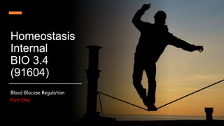
- 1. Homeostasis Internal BIO 3.4 (91604) Blood Glucose Regulation Part One
- 2. Strategies for Success MAKE SURE YOU READ THE PRESENTER NOTES BELOW EACH SLIDE. USE THE FEEDBACK LOOP MODEL. USE THE ESSAY STRUCTURE TEMPLATE TO HELP ORGANIZE YOUR NOTES AND REPORT. SEND ME QUESTIONS VIA CHAT AND SUBMIT YOUR DRAFT WORK VIA ASSIGNMENT. COLLABORATE AND ASK FOR HELP. DON’T PLAGIARISE! = details for Achieve Must Read Should Do = details for Merit and Excellence
- 3. Know the components of the feedback loop and how they work together as a system. This is where the scenario comes in Must Read
- 4. Image from Wikicommons The most crucial energy source between meals is glucose. Many critical organs such as the brain and the nervous system are totally reliant on having a certain level of glucose to function properly so of all the nutrients available to the body the levels of glucose are the most tightly regulated by homeostatic mechanisms and glucose is the only carbohydrate we store in any significant amount. Oxygen Food provides the energy we need to live All of our ATP comes from the food we eat and the vast majority of this will be in the form of carbohydrates, fats or proteins. Most carbohydrate in our diet is either starch (which is long chains of glucose), lactose (disaccharide in milk composed of glucose linked to galactose) and sucrose (dimer of glucose and fructose) or just plain fructose or glucose. Must Read
- 5. Metabolism of Glucose is Via Glycolysis and the Krebs Cycle KREBS CYCLE (Lots of ATP via respiration) ATP ATP ATP ATP ATP ATP ATP ATP ATP ATP ATP ATP ATP ATP ATP ATP ATP ATP ATP ATP ATP ATP ATP ATP ATP ATP ATP ATP ATP ATP ATP ATP ATP ATP Basic glycolysis scheme above from Sciencemusicvideos Must Read
- 6. The Scenario and Report Template (1) Select two parts of Tania’s day to link the scenario to two different manageable disruptions. The scenario helps provide a context to how the homeostatic system works in response to changes in the external and internal environments. (2) Using a hypothetical situation involving Tania is perfectly acceptable, especially when you are relating to a breakdown of the homeostatic system. (3) Use the homeostasis essay structure template (in TEAMS) to help you research and plan your essay. Make sure your report is concise and easy-to-follow. (4) Read the Thermoregulation exemplar (low excellence) and teacher comments to get a feel for the level of detail required at Level 3. Must Read
- 7. Basic Report Structure – FOLLOW THE GLUCOSE MOLECULES!!! Must Read Introduction Level What are the normal levels of blood glucose throughout the day. Achievement Reasons for homeostasis and blood glucose regulation. Achievement Manageable Disruption 1 - Increase in blood glucose negative feedback system Scenario 1 linked to feedback loop. Achievement Overview of what each component is in this system. Achievement Details of what happens at each component (what triggers what) and significance. Merit Managable Disruption 2 - Decrease in blood glucose negative feedback system Scenario 2 linked to feedback loop. Achievement Overview of what each component is in this system overview of system Achievement Details of what happens at each component (what triggers what) ) and significance. Merit Breakdown – Diabetes Scenario 3 linked to breakdown of feedback loop. Merit Overview of what causes diabetes and symptoms. Details of what happens during breakdown and significance. Excellence
- 8. Key biological ideas to discuss at Merit/Excellence : Facilitated diffusion and GLUT transporters Concentration gradient Depolarisation in Beta cells Exocytosis Glycogenolysis, Glycogenesis, Gluconeogenesis Hormones and Signal transduction pathway Phosphorylation Hyperglycemia and Hypoglycemia Ketoacidosis Should Do
- 9. Key Terms You need to Know (and define) Homeostasis Alpha cells Glycolysis Set point Negative feedback loop Beta cells Glycogen Hypoglycemic Receptor Insulin Glucagon Hyperglycemic Controller Hormone Glycogenolysis Type 1 & Type 2 Diabetes Effector GLUT Transporter Glycogenesis Ketoacidosis Variable Facilitated diffusion Gluconeogenesis Manageable disruption Exocytosis Phosphorylation Breakdown Ion channels Must Read
- 10. Normal range of glucose concentration Value range Unit 0.07 -0.10 percent (%) 70 – 100 milligrams per hundred cubic centimetres (mg 100 cm-3) = milligrams per decilitre (mg dL-1) 3.9 – 5.4 millimoles per litre (mmol L-1 or mM) Must Read
- 11. Regulating blood glucose levels Why it’s important not to let blood glucose levels get too high or low (adaptive advantage)
- 12. Glucose Homeostasis Why is it important to not let it drop too low? Some tissues can use a range of energy sources such as fats and even amino acids but several important tissues in the body can only really use glucose These tissues include: red blood cells and immune cells Brain and the nervous system also Therefore, maintaining a certain level of glucose is a matter of life and death. Must Read
- 13. PET (Positron Emission Tomography) imaging is a technique using radioactively labeled glucose to identify tissues in the body that use lots of glucose A Graphic Demonstration of How Much Glucose the Brain Actually Uses Brain Tumors Interesting Extra Information Liver
- 14. Glucose Homeostasis Why is it important to not let it get too high? Do not want to waste glucose obtained by letting it leave the body through urine So we need glucose all the time but we can’t eat all the time so we need a system for storing glucose. The main place we store glucose is in the liver and we can store enough there to keep us going for 2-3 days. Smaller amounts are taken into muscle and fat cells and either used immediately or stored. This is all stimulated by insulin and is the way blood glucose levels are kept low . © Peter Shepherd 2012 – Permission granted for use for teaching purposes Liver Must Read
- 15. But high blood glucose levels are dangerous as they seriously damages some of our major organs over time Kidneys Eye Blood vessels Glucose can react with and damage proteins in blood stream and vessels so tissues don’t get the nutrients and oxygen needed. Immune systems weakens. Must Read
- 16. Why it’s important not to let blood glucose levels to get too high or low (adaptive advantage) If glucose levels get too low: Cells and organs, such as the brain, nerves, red blood cells, immune cells which rely on glucose will stop functioning = go into shock and eventually death If glucose levels get too high: Don’t want to lose glucose through urine as cells are reliant to have some glucose all the time If glucose levels stay too high for a long time, it can cause damage to key organs - won’t cause death straight away but can cause damage that can lead to lower quality of life and then premature death. Must Read
- 17. Glucose is absorbed into cells though facilitated diffusion • Recall what is facilitated diffusion? Add an annotated diagram here. Why can’t glucose simply diffuse through the cell membrane? Should Do
- 18. Glucose How Glucose Uptake Works in some tissues Including Brain and Liver Outside Cell Inside Cell © Peter Shepherd 2012 – Permission granted for use for teaching purposes Glucose transporters are always present in the plasma membrane in most cells Glucose cannot cross the plasma membrane unless the membrane has specific proteins in the membrane called glucose transporters (GLUT proteins) These have pores allowing glucose to cross membranes by facilitated diffusion and flow from areas of high glucose to areas of low glucose concentration until an equilibrium is reached. If glucose is continually being used as in the brain and nerve cells then there will always be low glucose levels in the cell so it will keep flowing into the cell. THIS IS FOR CELLS USING GLUCOSE NOT STORING IT Should Do
- 19. Glucose Transporters (GLUTs in the Body) Please note: There are many more kinds of glucose transporters but GLUT4 is the most important for BG regulation Glucose is a hydrophilic molecule so cannot pass easily through the cell membrane without the help of these protein channels (via facilitated diffusion). Should Do
- 20. The storing and releasing of glucose Involves a balance of two hormones: Insulin and Glucagon • cells involved in insulin pathway • cells involved in glucagon pathway Must Read
- 21. What are hormones? Insulin Should Do Do you recall protein synthesis and exocytosis?
- 22. How do hormones work? Should Do
- 24. When blood glucose levels are high (after a meal) Feedback loop - increase in variable (blood glucose)
- 25. The liver is where our body sorts all the food and also where it stores glucose Must Read
- 26. Stomach All our food goes firstly into stomach…. Must Read
- 27. Small intestines …..then the small intestine…… Polymers (e.g. starch) must be broken down by enzymes before they can diffuse through intestinal cells into the blood stream. Must Read
- 28. LIVER ……then absorbed into special blood vessels that take it directly to the liver before it gets anywhere else Must Read
- 29. Insulin is made in only one place in the body Pancreas 5% of the cells in pancreas are called beta-cells and the main job of these is to make insulin Less that 1 gram of beta cells in your whole body. Stomach Liver Must Read
- 30. LIVER …. Which stimulates the release of insulin along the way Must Read
- 31. As n Cys Ty r As n Glu Leu Gln Ty r Leu Se r Cys Ile Se r Th r Cys Cys Gln Glu Val Ile Gly Th r Lys Pro Th r Ty r Ph e Ph e Gly Ar g Glu Gly Cys Val Leu Ty r Leu Ala Glu Val Leu His Se r Cys Gly Leu His Gln As n Val Ph e S S S S S S High Glucose Stores glucose and so lowers blood glucose levels Insulin is protein released by β-cells when blood glucose levels rise and it lowers blood glucose levels by stimulating uptake into 3 tissues Must Read
- 32. By being able to take up and store glucose when insulin is high and later release it when insulin is low the liver is the major organ responsible for regulating blood glucose levels 32 Must Read
- 33. What happens to glucose when it gets to the liver if there is no insulin present ? Glucose from the gut Glucose Without insulin all the glucose goes through the liver to the rest of the body and is not stored Must Read
- 34. Glucose Most important thing insulin does is to stimulate storage of glucose in the liver after a meal. This lowers blood glucose and provides a store of glucose for period between meals Some of the glucose stored as glycogen Insulin Glucose Some of the glucose goes through into main bloodstream Must Read
- 35. Glucose Glucose is released into the blood stream so organs like the brain can keep functioning Glucose stored as glycogen The glucose stored in the liver is released between meals to keep blood glucose levels stable Must Read
- 36. Two other important targets of insulin are muscle and fat tissues Must Read
- 37. Without insulin not much glucose gets into muscle and fat tissues Glucose Glucose Must Read
- 38. When insulin is present lots of glucose gets into muscle and fat tissues and this helps lower blood glucose after a meal Insulin Glucose Insulin Glucose Must Read
- 39. What Happens After a Meal Glucose levels in the portal vein (i.e the vein that comes directly from gut to the liver) rise rapidly so glucose levels in the pancreas rise and insulin is released from β-cells. Insulin binds to receptors that are found on cells in the liver, in muscle and in fat cells This stimulates the uptake of glucose into these tissues so blood glucose levels go down Glucose taken up by liver is mostly stored as glycogen Some of the glucose going into fat cells is turned into glycerol and so contribute to the accumulation of fat in these cells. Some of the glucose going into muscle is stored as glycogen but tends to be used very rapidly afterwards Must Read
- 40. Blood Glucose Feedback VARIABLE – Blood Sugar Levels Increase RECEPTOR – Receptors (Beta Cells in the Islets of Langerhans) in Pancreas identify increase of blood sugar CONTROLLER – Beta Cells in the Pancreas secrete the hormone Insulin EFFECTOR – Insulin receptors in the liver, muscle or fat cells uptakes and converts Glucose and stores it RESPONSE – Blood sugar levels decrease FEEDBACK – Return to homeostasis when blood sugar levels fall returns to steady state Must Read
- 41. Blood Glucose and Insulin The next set of slides are graphs that shows changes in blood glucose and insulin levels after a meal. Keep track of the time things take to change. A good way to link in the scenario. Must Read
- 42. 1 5 3 4 2 6 7 8 9 30 60 90 120 150 180 Time after a meal (in minutes) After a meal blood glucose levels rise as glucose is taken up by the gut but rapidly falls back to a set point (i.e a homeostatic mechanism is in action). How is this achieved and why is it important ? Blood Glucose Concentration (mM) © Peter Shepherd 2012 – Permission granted for use for teaching purposes Must Read
- 43. 1 5 3 4 2 6 7 8 9 30 60 90 120 150 180 Time after a meal (in minutes) Blood Glucose Concentration (mM) To be able to compare blood glucose levels we usually assess this after an overnight fast as after this amount of time every ones glucose has come to their homeostatic set point which we call their “Fasting blood glucose levels”. This is about 5 mM in humans Blood Glucose Changes After a Meal Must Read
- 44. 1 5 3 4 2 6 7 8 9 30 60 90 120 150 180 Time after a meal (in minutes) Blood Glucose Concentration (mM) Insulin secretion starts Blood glucose levels start to rise after a meal Must Read
- 45. 1 5 3 4 2 6 7 8 9 30 60 90 120 150 180 Time after a meal (in minutes) Blood Glucose Concentration (mM) Insulin secretion starts Insulin starts to work Must Read
- 46. 1 5 3 4 2 6 7 8 9 30 60 90 120 150 180 Time after a meal (in minutes) Feedback mechanisms act to lower blood glucose Blood Glucose Concentration (mM) Insulin secretion starts Insulin starts to work Peak blood glucose level after a meal Must Read
- 47. 1 5 3 4 2 6 7 8 9 30 60 90 120 150 180 Time after a meal (in minutes) Fasting blood glucose level Blood Glucose Concentration (mM) Insulin secretion starts Insulin starts to work Insulin feedback mechanisms act to lower blood glucose Peak blood glucose level after a meal Must Read
- 48. 1 5 3 4 2 6 7 8 9 30 60 90 120 150 180 Time after a meal (in minutes) Peak blood glucose level after a meal Feedback mechanisms act to lower blood glucose Fasting blood glucose level is reached again Blood Glucose Concentration (mM) Insulin secretion starts Insulin starts to work Must Read
- 49. 1 5 3 4 2 6 7 8 9 30 60 90 120 150 180 Time after a meal (in minutes) Peak blood glucose level after a meal Feedback mechanisms act to lower blood glucose Fasting blood glucose level Blood Glucose Concentration (mM) Insulin secretion starts Insulin starts to work Must Read
- 50. 1 5 3 4 2 6 7 8 9 30 60 90 120 150 180 Time after a meal (in minutes) With all this insulin coming into the system how come the insulin doesn’t just make the blood glucose levels fall below 5mM Blood Glucose Concentration (mM) ? Must Read
- 51. 1 5 3 4 2 6 7 8 9 30 60 90 120 150 180 Time after a meal (in minutes) 1. As blood glucose levels fall less insulin is secreted so insulin receptors on cells shut off quickly. This is an example of feedback regulation Blood Glucose Concentration (mM) 2. If blood glucose levels do fall below 5 mM then a second hormone called glucagon is secreted which causes glucose to be released from stores held in the liver and so this raises blood glucose levels again Must Read
- 52. 1 5 3 4 2 6 7 8 9 30 60 90 120 150 180 Time after a meal (in minutes) 1. As blood glucose levels fall less insulin is secreted. This is an example of feedback regulation Blood Glucose Concentration (mM) 2. If blood glucose levels do fall below 5 mM then a second hormone called glucagon is secreted which causes glucose to be released from stores held in the liver and so this raises blood glucose levels again Why Do Glucose Levels Stabilise Must Read
- 53. Watch these videos to help understand the basics of blood glucose regulation: • https://www.youtube.com/watch?v=X78C5ajmKJs&ab_chann el=TamerShabaan
- 54. Zooming in… … on insulin pathway
- 55. Zooming in to role of Pancreas Watch this video to find out the role the pancreas plays in our bodies: What does the pancreas do?
- 56. The Islets of Langerhans are the Central Regulator of Insulin and Glucagon Levels Islet cell structure Insulin and glucagon are both hormones made only in a special group of cells in the pancreas called the Islets of Langerhans (or islets for short). • The islets contain several cell types but the most important are the α-cells - the only cells in the body to make glucagon. These cells sense when blood glucose falls below 5 mM and release glucagon. • β-cells are the only cells in the body that make insulin. They sense when blood glucose rises above 5 mM and release insulin. The pancreas is adjacent to the gut and on the portal vein which drains nutrients from gut to liver. This means islets are very well placed to sense an influx of nutrients and to respond by releasing hormones. Must Read
- 57. Glucose We need to release insulin very quickly (within a few minutes) but it takes 30-60 minutes for the insulin gene to be switched on and for new insulin protein to be made. This is too slow so the -cells make insulin in advance and package it in vesicles just waiting for the increase in glucose levels to occur. β-cells Store Insulin in Prepackaged Granules Ready For Instant Release When Blood Glucose levels Rise Outside Cell Inside Cell © Peter Shepherd 2012 – Permission granted for use for teaching purposes Should Do
- 58. Glucose The extra glucose coming into the -cell triggers the insulin containing vesicle to move the the plasma membrane and to fuse with the plasma membrane , so releasing the contents (i.e the insulin) to the bloodstream How is Insulin Released from β-cells Outside Cell Inside Cell © Peter Shepherd 2012 – Permission granted for use for teaching purposes Should Do
- 59. Outside Cell Inside Cell © Peter Shepherd 2012 – Permission granted for use for teaching purposes How is Insulin Released from β-cells The extra glucose coming into the -cell triggers the insulin containing vesicle to move the the plasma membrane and to fuse with the plasma membrane , so releasing the contents (i.e the insulin) to the bloodstream Should Do
- 60. How do Beta cells detect glucose levels? Should Do
- 61. FYI: this is how vesicles translocate (move) to the cell membrane. Aren’t they just the coolest little robots you have ever seen. YouTube ‘motor proteins’ and watch an animation of them in action. Obviously not necessary for this internal. For interest sake only!
- 62. Signal Transduction - Insulin binding with the receptor Insulin moves through the blood stream until it finds its specific receptor on the surface of the liver cells, muscle cells and fat cells. The binding of insulin causes change in the shape of the intracellular portion of the receptor which activates an enzymatic activity. The receptor is now said to be activated and this brings about changes inside the cell. Should Do
- 63. How insulin works on target cells (effector) Should Do
- 64. Insulin Receptor Insulin How Does insulin Stimulate Glycogen Synthesis in Liver O O H CH2OH O H O H O H O O H CH2OH O H O H O H O O H CH2OH O H O O H O O H CH2OH O H O H O H Glycogen Synthase Glucose Outside Cell Inside Cell O O H CH2OH O H O H O H O O H CH2OH O H O H O H Should Do
- 65. Insulin Receptor How Does insulin Stimulate Glycogen Synthesis in Liver Glycogen Synthase Outside Cell Inside Cell O O H CH2OH O H O H O H O O H CH2OH O H O H O O H CH2OH O H O O H CH2OH O H O H O O O Glycogen O O H CH2OH O H O H O H O O H CH2OH O H O H O H Should Do
- 66. Insulin Receptor How Does insulin Stimulate Glycogen Synthesis in Liver Glycogen Synthase Outside Cell Inside Cell O O H CH2OH O H O H O H O O H CH2OH O H O H O O H CH2OH O H O O H CH2OH O H O H O O O Glycogen O O H CH2OH O H O H O H O O H CH2OH O H O H O H Should Do
- 67. Insulin Receptor How Does insulin Stimulate Glycogen Synthesis in Liver Glycogen Synthase Outside Cell Inside Cell O O H CH2OH O H O H O H O O H CH2OH O H O H O O H CH2OH O H O O H CH2OH O H O H O O O Glycogen O O H CH2OH O H O H O H O O H CH2OH O H O H O H Should Do
- 68. Insulin Receptor Glucose Vesicles containing GLUT4 glucose transporter Insulin How Does insulin Stimulate Glucose Uptake Into Muscle And Fat Cells Outside Cell Inside Cell Should Do
- 69. Glucose How Does insulin Stimulate Glucose Uptake Into Muscle And Fat Cells Outside Cell Inside Cell Should Do
- 70. Glucose How Does insulin Stimulate Glucose Uptake Into Muscle And Fat Cells Outside Cell Inside Cell Should Do
- 71. Glucose How Does insulin Stimulate Glucose Uptake Into Muscle And Fat Cells Outside Cell Inside Cell Should Do
- 72. Insulin Receptor Insulin How Does Insulin Work and How is the Signal Shut Off Outside Cell Inside Cell Receptor’s Enzyme Activated Should Do
- 73. Outside Cell Inside Cell How Does Insulin Work and How is the Signal Shut Off Insulin receptor starts to internalise into a vesicle pinched off from the membrane Should Do
- 74. Outside Cell Inside Cell H+ H+ H+ H+ H+ H+ H+ How Does Insulin Work and How is the Signal Shut Off • The vesicle containing the insulin receptor acidifies • This changes the protein structure (see year 12 Cells chapter) • Insulin can no longer bind to its receptor • Insulin signal shut off Should Do
- 75. When blood glucose levels are low (between meals)
- 76. Antagonistic Hormones The effects of one hormone are often counteracted by an opposing hormone, this is known as antagonistic hormones. Feedback mechanisms adjust the balance of the two hormones to maintain physiological function. Example: insulin decreases blood glucose and glucagon raises it. Insulin secretion Glucagon secretion Raises blood glucose level Lowers blood glucose level Decrease in blood glucose stimulates glucagon release Increase in blood glucose stimulates insulin release Must Read
- 77. Antagonistic Hormones Insulin Glucagon Produced by β-cells of the Pancreas Produced by α-cells of the Pancreas Released into circulatory system when blood glucose is high Released into the circulatory system when blood glucose is low Facilitates the transport of glucose into target cells Signals the liver to break down glycogen into simple glucose Must Read
- 78. Blood Glucose Feedback INPUT – Blood Sugar Levels Decrease RECEPTOR – Receptors (Alpha Cells in the Islets of Langerhans) in Pancreas identify decrease of blood sugar CONTROLLER – Alpha Cells in the Pancreas secrete the hormone Glucagon EFFECTOR – The Liver breaks down Glycogen converts the Glycogen into Glucose and releases it back into the bloodstream RESPONSE – Blood sugar levels increase FEEDBACK – Return to homeostasis when blood sugar levels rise returns to steady state Must Read
- 79. How does glucagon stimulate glycogen breakdown and release of glucose from the liver Glucagon binds to its receptor on the surface of liver cells and activates signaling pathways that result in the activation of an enzyme called glycogen phosphorylase which catalyses the breakdown of glycogen polymers back into glucose. Glucose levels build up in the liver cells and when they exceed the concentration outside the cells then the glucose moves out into the bloodstream by facilitated diffusion through the glucose transporters. Since glucagon is stimulating the breakdown of lots of glycogen this means there is a constant flow of glucose out of the liver as long as glucagon is around. Should Do
- 80. Glucagon Receptor Glucagon How Does Glucagon Stimulate Glycogen Breakdown in Liver and Release Glucose Into the Blood O O H CH2OH O H O H O H O O H CH2OH O H O H O O H CH2OH O H O O H CH2OH O H O H O O O Glycogen Glycogen Phosphorylase Outside Cell Inside Cell Should Do
- 81. Glucagon Receptor Glucagon How Does Glucagon Stimulate Glycogen Breakdown in Liver and Release Glucose Into the Blood O O H CH2OH O H O H O H O O H CH2OH O H O H O O H CH2OH O H O O H CH2OH O H O H O O O Glycogen Glycogen Phosphorylase O O H CH2OH O H O H O H O O H CH2OH O H O H O H O O H CH2OH O H O O H O O H CH2OH O H O H O H Glucose Outside Cell Inside Cell O O H CH2OH O H O H O H Should Do
- 82. 100grams of glucose Muscle 25g Brain 17g Kidney 9g Used immediately Muscle 26g Adipocytes 2g Liver 21g Stored for later Where does the glucose go ? (exact figures depend on a person muscle and fat mass but a typical example is shown below) Must Read