
Recommended
More Related Content
Similar to Microcytic Anemia Causes and Thalassemia Traits
Similar to Microcytic Anemia Causes and Thalassemia Traits (20)
More from AbdulKaderSouid
More from AbdulKaderSouid (15)
Recently uploaded
Recently uploaded (20)
Microcytic Anemia Causes and Thalassemia Traits
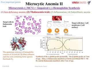
- 1. Microcytic Anemia II Microcytosis (↓MCV) = Impaired (↓) Hemoglobin Synthesis Abdul-Kader Souid9/26/2018 (1) Iron deficiency anemia; (2) Thalassemia trait; (3) Inflammation; (4) Sideroblastic anemia Target cells in thalassemia trait The quaternary structure of hemoglobin consists of 2α (red) & 2β (blue) subunits, each has iron-containing heme (green) that binds O2. Five important points Synthesis of the a globin peaks prenatally. The b globin peaks at 3 mo of age. Thus, a-thalassemia manifests at birth (cord blood MCV <94 fL) and b-thalassemia manifests after 3 mo of age. Target cells have ↑cell membrane (↑cell surface)
- 2. 2 b 3’5’ b 3’5’ Chromosomes 11 (two alleles) a2 a1 3’5’ Chromosomes 16 (two alleles) a2 a1 3’5’ Normal Hemoglobin Variants The four globins (a, b, , ) form three normal hemoglobin variants: A (>93%) = 2a + 2b A2 (<3.5%) = 2a + 2 (↑ in b-thalassemia trait) F (<3.5%) = 2a + 2 (↑ in b-thalassemia trait) Must know (5 points) Tetramers of “” or “b“ are abnormal variants and indicate a-thalassemia. Hemoglobin Barts = 4 (newborn) Hemoglobin H = 4b (≥1 y of age)
- 3. a-Thalassemia Trait a2 a1 3’5’ Chromosomes 16 (two alleles) a2 a1 3’5’ When there is production of the α gene on a chromosome 16 (only one of the two α genes on a chromosome is deleted; trans deletion; (-,a/-,a), the designation for that chromosome is α+ thalassemia (α+). When both α genes on a single chromosome 16 are deleted (cis deletion; (-,-/a,a) ), the designation α° (null allele) is used for that chromosome. Must know (2 points) α° thalassemia (α°) is prevalent in China, Thailand, Malaysia, and Philippines (Asian & Mediterranean). α+ thalassemia (α+) is benign and common in the UAE, occurring in >20% of the population
- 4. b-Thalassemia Trait b 3’5’ b 3’5’ Chromosomes 11 (two alleles) One b-globin gene mutation (heterozygous bo or b+; only one abnormal allele) → b-thalassemia trait → mild anemia, ↓MCV, ↑RBC count + normal RDW + ↑A2 (2a, 2) >3.5%. Two b-globin gene mutations (homozygous bo or b+; two abnormal alleles) → b-thalassemia major → severe anemia (transfusion dependent after 3 mo of age). Must know (2 point)
- 5. Thalassemia Trait: Summary • The term “thalassemia” refers to the inherited disorders characterized by decreased (“trait”) or absent (“major”) a or b globin. • a-thalassemia trait (a deletion allele) refers to ↓ a-globin (usually gene deletion). – Biomarkers for a-thalassemia trait include: (1) Persistent microcytosis (MCV at birth <94 fL and MCV at adulthood <80 fL), (2) Hemoglobin Bart (4) at birth (2.5% to 5%) and H (4b) at adulthood, (3) Normal A2 (2a2), (4) Two a gene deletions. • b-thalassemia trait (bo or b+ allele) refers to ↓ b-globin (usually gene mutation). – OMIM#613985 – Biomarkers for b-thalassemia trait include: (1) Microcytosis after 3 mo of age, (2) ↑ hemoglobin A2 (2a2), (3) HBB (hemoglobin beta) gene mutation. 9/26/2018 5
- 6. This Emirati family has a-thalassemia trait • Father: Hemoglobin 127 g/L, MCV 68 fL, A = 95%, F = 2.0%, A2 = 3.0% (a-thalassemia trait). • Mother: Hemoglobin 115 g/L, MCV 72 fL, A = 95%, F = 3.0%, A2 = 2.0% (a-thalassemia trait). • Daughter (22 mo): Hemoglobin 115 g/L, MCV 61 fL, A = 96%, F = 2.0%, A2 = 2.0% (a-thalassemia trait). 9/26/2018 6 trans deletion (-,a/-,a) α+ thalassemia (α+) Frequency (f) of the silent allele (-,a) in UAE is about 50% (= 0.5). Frequency (f2) of a-thalassemia (-,a/-,a) in UAE is 25% (0.5 x 0.5 = 0.25). Must know
- 7. A 27-year-old woman presents with mild microcytosis, ↑A2, and normal serum ferritin Alpha-Thalassemia PCR Beta-Thalassemia PCR https://www.omim.org/ IVS1-5 (G→C) mutation is the most common β-thalassemia mutation. https://www.ncbi.nlm.nih.gov/clinvar/variation/15447/ NMD, nonsense-mediated mRNA decay; HBB gene (OMIM#141900), hemoglobin beta locus; IVS-1 5, InterVening Sequence (intron).
- 8. Required Reading • Harteveld CL, Higgs DR. Alpha-thalassaemia. Orphanet J Rare Dis. 2010;5:13. doi:10.1186/1750-1172-5-13. • Danckwardt S, Neu-Yilik G, Thermann R, Frede U, Hentze MW, Kulozik AE. Abnormally spliced beta-globin mRNAs: a single point mutation generates transcripts sensitive and insensitive to nonsense-mediated mRNA decay. Blood. 2002;99:1811-6. PubMed PMID: 11861299. • Kukreti R, Dash D, E VK, Chakravarty S, Das SK, De M, Talukder G. Spectrum of beta-thalassemia mutations and their association with allelic sequence polymorphisms at the beta-globin gene cluster in an Eastern Indian population. Am J Hematol. 2002;70:269-77. PubMed PMID: 12210807. 9/26/2018 8