Knee Diagnostic MSK Ultrasound.pptx
•Download as PPTX, PDF•
0 likes•2 views
Basics for Advance Practitioners, orthopaedic, registrar who wanted to learn the diagnostic ultrasound, implement and improve musculoskeletal practice as POCUS (point of care ultrasound). Other healthcare professionals can also benefit such as sonographers, osteopath, chiropractors.
Report
Share
Report
Share

Recommended
Role of Ultrasound in shoulder pathologies.
In this presentation we will discuss the rotator cuff pathologies.Role of ultrasound in clinical evaluation of shoulder Dr. Muhammad Bin Zulfiqar
Role of ultrasound in clinical evaluation of shoulder Dr. Muhammad Bin ZulfiqarDr. Muhammad Bin Zulfiqar
WOUND CARE WORKSHOP 2018 AUGUST
Venous Anatomy
INvestigations NON INVASIVE AND INVASIVE
Duplex Scan
Investigations for varicose veins Dr Joel Arudchelvam MBBS (COL), MD (SUR),...
Investigations for varicose veins Dr Joel Arudchelvam MBBS (COL), MD (SUR),...Joel Arudchelvam MBBS, MD, MRCS, FCSSL
Mtp kit in kuwait௹+918133066128....) @abortion pills for sale in Kuwait City ✒Abortion CLINIC In Kuwait ?Kuwait pills +918133066128௵) safe Abortion Pills for sale in Salmiya, Kuwait city,Farwaniya-cytotec pills for sale in Kuwait city. Kuwait pills +918133066128WHERE I CAN BUY ABORTION PILLS IN KUWAIT, CYTOTEC 200MG PILLS AVAILABLE IN KUWAIT, MIFEPRISTONE & MISOPROSTOL MTP KIT FOR SALE IN KUWAIT. Whatsapp:+Abortion Pills For Sale In Mahboula-abortion pills in Mahboula-abortion pills in Kuwait City- .Kuwait pills +918133066128)))abortion pills for sale in Mahboula …Mtp Kit On Sale Kuwait pills +918133066128mifepristone Tablets available in Kuwait?Zahra Kuwait pills +918133066128Buy Abortion Pills Cytotec Misoprostol 200mcg Pills Brances and now offering services in Sharjah, Abu Dhabi, Dubai, **))))Abortion Pills For Sale In Ras Al-Khaimah(((online Cytotec Available In Al Madam))) Cytotec Available In muscat, Cytotec 200 Mcg In Zayed City, hatta,Cytotec Pills௵+ __}Kuwait pills +918133066128}— ABORTION IN UAE (DUBAI, SHARJAH, AJMAN, UMM AL QUWAIN, ...UAE-ABORTION PILLS AVAILABLE IN DUBAI/ABUDHABI-where can i buy abortion pillsCytotec Pills௵+ __}Kuwait pills +918133066128}}}/Where can I buy abortion pills in KUWAIT , KUWAIT CITY, HAWALLY, KUWAIT, AL JAHRA, MANGAF , AHMADI, FAHAHEEL, In KUWAIT ... pills for sale in dubai mall and where anyone can buy abortion pills in Abu Dhabi, Dubai, Sharjah, Ajman, Umm Al Quwain, Ras Al Khaimah ... Abortion pills in Dubai, Abu Dhabi, Sharjah, Ajman, Fujairah, Ras Al Khaimah, Umm Al Quwain…Buy Mifepristone and Misoprostol Cytotec , Mtp KitABORTION PILLS _ABORTION PILLS FOR SALE IN ABU DHABI, DUBAI, AJMAN, FUJUIRAH, RAS AL KHAIMAH, SHARJAH & UMM AL QUWAIN, UAE ❤ Medical Abortion pills in ... ABU DHABI, ABORTION PILLS FOR SALE ----- Dubai, Sharjah, Abu dhabi, Ajman, Alain, Fujairah, Ras Al Khaimah FUJAIRAH, AL AIN, RAS AL KHAIMAMedical Abortion pills in Dubai, Abu Dhabi, Sharjah, Al Ain, Ajman, RAK City, Ras Al Khaimah, Fujairah, Dubai, Qatar, Bahrain, Saudi Arabia, Oman, ...Where I Can Buy Abortion Pills In Al ain where can i buy abortion pills in #Dubai, Exclusive Abortion pills for sale in Dubai ... Abortion Pills For Sale In Rak City, in Doha, Kuwait.௵ Kuwait pills +918133066128₩ Abortion Pills For Sale In Doha, Kuwait,CYTOTEC PILLS AVAILABLE Abortion in Doha, ꧁ @ ꧂ ☆ Abortion Pills For Sale In Ivory park,Rabie Ridge,Phomolong. ] Abortion Pills For Sale In Ivory Park, Abortion Pills+918133066128In Ivory Park, Abortion Clinic In Ivory Park,Termination Pills In Ivory Park,. *)][(Abortion Pills For Sale In Tembisa Winnie Mandela Ivory Park Ebony Park Esangweni Oakmoor Swazi Inn Whats'app...In Ra al Khaimah,safe termination pills for sale in Ras Al Khaimah. | Dubai.. @Kuwait pills +918133066128Abortion Pills For Sale In Kuwait, Buy Cytotec Pills In Kuwait.Cytotec Pills௵ __}Kuwait pills +918133066128}}}/Where can I buy abortion pills in KUWAIT ,@Safe Abortion pills IN Jeddah(+918133066128) Un_wanted kit Buy Jeddah
@Safe Abortion pills IN Jeddah(+918133066128) Un_wanted kit Buy JeddahAbortion pills in Kuwait Cytotec pills in Kuwait
More Related Content
Similar to Knee Diagnostic MSK Ultrasound.pptx
WOUND CARE WORKSHOP 2018 AUGUST
Venous Anatomy
INvestigations NON INVASIVE AND INVASIVE
Duplex Scan
Investigations for varicose veins Dr Joel Arudchelvam MBBS (COL), MD (SUR),...
Investigations for varicose veins Dr Joel Arudchelvam MBBS (COL), MD (SUR),...Joel Arudchelvam MBBS, MD, MRCS, FCSSL
Similar to Knee Diagnostic MSK Ultrasound.pptx (20)
Knee clinical examination for orthopaedic residents.dr mohamed ashraf TD medi...
Knee clinical examination for orthopaedic residents.dr mohamed ashraf TD medi...
Hip Joint anatomy, surgical approches & AVN review
Hip Joint anatomy, surgical approches & AVN review
Investigations for varicose veins Dr Joel Arudchelvam MBBS (COL), MD (SUR),...
Investigations for varicose veins Dr Joel Arudchelvam MBBS (COL), MD (SUR),...
Presentation1.pptx. ultrasound examination of the ankle joint.
Presentation1.pptx. ultrasound examination of the ankle joint.
Hip Arthroscopy in 2013: Inova Annual Sports Medicine Program
Hip Arthroscopy in 2013: Inova Annual Sports Medicine Program
Varicose veins:A never ending problem if mistreated!
Varicose veins:A never ending problem if mistreated!
Abdominal Trauma and FAST scan (Dr. Derhim Afaqeeh , Yemen )
Abdominal Trauma and FAST scan (Dr. Derhim Afaqeeh , Yemen )
Recently uploaded
Mtp kit in kuwait௹+918133066128....) @abortion pills for sale in Kuwait City ✒Abortion CLINIC In Kuwait ?Kuwait pills +918133066128௵) safe Abortion Pills for sale in Salmiya, Kuwait city,Farwaniya-cytotec pills for sale in Kuwait city. Kuwait pills +918133066128WHERE I CAN BUY ABORTION PILLS IN KUWAIT, CYTOTEC 200MG PILLS AVAILABLE IN KUWAIT, MIFEPRISTONE & MISOPROSTOL MTP KIT FOR SALE IN KUWAIT. Whatsapp:+Abortion Pills For Sale In Mahboula-abortion pills in Mahboula-abortion pills in Kuwait City- .Kuwait pills +918133066128)))abortion pills for sale in Mahboula …Mtp Kit On Sale Kuwait pills +918133066128mifepristone Tablets available in Kuwait?Zahra Kuwait pills +918133066128Buy Abortion Pills Cytotec Misoprostol 200mcg Pills Brances and now offering services in Sharjah, Abu Dhabi, Dubai, **))))Abortion Pills For Sale In Ras Al-Khaimah(((online Cytotec Available In Al Madam))) Cytotec Available In muscat, Cytotec 200 Mcg In Zayed City, hatta,Cytotec Pills௵+ __}Kuwait pills +918133066128}— ABORTION IN UAE (DUBAI, SHARJAH, AJMAN, UMM AL QUWAIN, ...UAE-ABORTION PILLS AVAILABLE IN DUBAI/ABUDHABI-where can i buy abortion pillsCytotec Pills௵+ __}Kuwait pills +918133066128}}}/Where can I buy abortion pills in KUWAIT , KUWAIT CITY, HAWALLY, KUWAIT, AL JAHRA, MANGAF , AHMADI, FAHAHEEL, In KUWAIT ... pills for sale in dubai mall and where anyone can buy abortion pills in Abu Dhabi, Dubai, Sharjah, Ajman, Umm Al Quwain, Ras Al Khaimah ... Abortion pills in Dubai, Abu Dhabi, Sharjah, Ajman, Fujairah, Ras Al Khaimah, Umm Al Quwain…Buy Mifepristone and Misoprostol Cytotec , Mtp KitABORTION PILLS _ABORTION PILLS FOR SALE IN ABU DHABI, DUBAI, AJMAN, FUJUIRAH, RAS AL KHAIMAH, SHARJAH & UMM AL QUWAIN, UAE ❤ Medical Abortion pills in ... ABU DHABI, ABORTION PILLS FOR SALE ----- Dubai, Sharjah, Abu dhabi, Ajman, Alain, Fujairah, Ras Al Khaimah FUJAIRAH, AL AIN, RAS AL KHAIMAMedical Abortion pills in Dubai, Abu Dhabi, Sharjah, Al Ain, Ajman, RAK City, Ras Al Khaimah, Fujairah, Dubai, Qatar, Bahrain, Saudi Arabia, Oman, ...Where I Can Buy Abortion Pills In Al ain where can i buy abortion pills in #Dubai, Exclusive Abortion pills for sale in Dubai ... Abortion Pills For Sale In Rak City, in Doha, Kuwait.௵ Kuwait pills +918133066128₩ Abortion Pills For Sale In Doha, Kuwait,CYTOTEC PILLS AVAILABLE Abortion in Doha, ꧁ @ ꧂ ☆ Abortion Pills For Sale In Ivory park,Rabie Ridge,Phomolong. ] Abortion Pills For Sale In Ivory Park, Abortion Pills+918133066128In Ivory Park, Abortion Clinic In Ivory Park,Termination Pills In Ivory Park,. *)][(Abortion Pills For Sale In Tembisa Winnie Mandela Ivory Park Ebony Park Esangweni Oakmoor Swazi Inn Whats'app...In Ra al Khaimah,safe termination pills for sale in Ras Al Khaimah. | Dubai.. @Kuwait pills +918133066128Abortion Pills For Sale In Kuwait, Buy Cytotec Pills In Kuwait.Cytotec Pills௵ __}Kuwait pills +918133066128}}}/Where can I buy abortion pills in KUWAIT ,@Safe Abortion pills IN Jeddah(+918133066128) Un_wanted kit Buy Jeddah
@Safe Abortion pills IN Jeddah(+918133066128) Un_wanted kit Buy JeddahAbortion pills in Kuwait Cytotec pills in Kuwait
TEST BANK For Little and Falace's Dental Management of the Medically Compromised Patient, 10th Edition by Craig Miller, Verified Chapters 1 - 30, Complete Newest VersionTEST BANK For Little and Falace's Dental Management of the Medically Compromi...
TEST BANK For Little and Falace's Dental Management of the Medically Compromi...rightmanforbloodline
💚Mature Women / Aunty Call Girl Chandigarh Escorts Service 💯Call Us 🔝 9352988975 🔝💃Top Class Call Girl Service Available
In BEST CALL GIRL ESCORTS SERVICE IN CALL WATTSAPP 7014168258 THE MOST BEAUTIFUL INDEPENDENT ESCORT CALL GIRL SERVICE In WE ARE PROVIDING GENUINE CALL GIRL SERVICE
I AM A a NATURAL BRUNETTES, SLIM BODY, NATURAL LONG HAIR AND ALL TYPE OF HAIR IS A NATURAL BRUNETTE IN THE MOST BEAUTIFUL MODELS INDEPENDENT ESCORT GIRL I AM A NATURAL BRUNETTE WITH ROOM AND HOTEL AND A NATURAL BRUNETTE WITH A BODY MADE FOR SIN AND ALL TYPE OF ME ALL THE TIME
I SEND YOU A HAIR, VERY SOCIABLE AND FUNNY, READY TO ENTERTAIN TO ENTERTAIN U AND MAKE FORGET ABOUT TO AGET ENTERTAINMENT YOU AND MAKE FORGET ABOUT ALL THE PROBLEMS. LET'S HAVE A WONDERFUL TIME TOGETHER AND FORGET ABOUT EVERYTHING ALL TYPE SERVICE ENJOYMENT SAFE AND SECURE IN CALL OUT CALL HOME AND HOTEL ANYTIME AVAILABLE
AND ALL TYPE SERVICE ENJOYMENT INDEPENDENT BEST CALL GIRL ESCORTS SERVICE IN INDEPENDENT CALL GIRLS Call Girls In BEST Call Girls in Escort Service provide Cute Nice sweet and Sexy Models in beautiful city cash in hand to hand call girl in and escorts. HOT & SEXY MODELS // COLLEGE GIRLS IN AVAILABLE FOR COMPLETE ENJOYMENT WITH HIGH PROFILE INDIAN MODEL AVAILABLE HOTEL & HOME ★ SAFE AND SECURE HIGH CLASS SERVICE AFFORDABLE RATE ★ 100% SATISFACTION,UNLIMITED ENJOYMENT. ★ All Meetings are confidential and no information is provided to any one at any cost.
★ EXCLUSIVE Profiles Are Safe and Consensual with Most Limits Respected
★ Service Available In: - HOME & 24x7 :: 3 * 5 *7 *Star Hotel Service .In Call & Out call
Services : #G05
★ A-Level (5 star escort)
★ Strip-tease
★ BBBJ (Bareback Blowjob)Receive advanced sexual techniques in different mode make their life more pleasurable.
★ Spending time in hotel rooms
★ BJ (Blowjob Without a Condom)
★ Completion (Oral to completion)
★ Covered (Covered blowjob Without a Condom) Call Girls💚Mature Women / Aunty Call Girl Chandigarh Escorts Service 💯Call Us 🔝 9352988...
💚Mature Women / Aunty Call Girl Chandigarh Escorts Service 💯Call Us 🔝 9352988...Chandigarh Call Girls
Spesialis Penggugur Kandungan 081466799220 Jual Obat Aborsi Cytotec Gastrul Peluntur Janin 1 2 3 4 5 6 7 Bulan, Metode Cara Aborsi Menggugurkan Kandungan Aman Cepat & Tuntas 100% Berhasil, Obat Aborsi, Obat Penggugur Kandungan, Jual Obat Misotrol, Cytotec, dan Cyprostol, Cyrux Pendorong Janin, Pil Aborsi, Pil Cytotec Aborsi, Jual Obat Penggugur Kandungan, Klinik Konsultasi, Obat Telat Bulan, Obat Pelancar Haid, Jual Cytotec Asli, Cara Menggugurkan Kandungan, Penjual Obat Aborsi Terpercaya, Obat Aborsi Ampuh, Obat Aborsi Manjur, Obat Aborsi Tuntas, Obat Aborsi Aman, Obat Aborsi Asli, Obat Aborsi Bergaransi, Obat Aborsi 6 Bulan, Obat Aborsi 5 Bulan, Obat Aborsi 4 Bulan, Obat Aborsi 3 Bulan, Obat Aborsi 2 Bulan, Obat Aborsi 1 Bulan, Apotik Jual Obat Aborsi, Apotik Jual Cytotec, Cara Penggunaan Obat Aborsi Cytotec, Klinik Aborsi, Pil Penggugur Kandungan, Cara Pakai Cytotec, Cara Pemakian Cyrux Pendorong Janin
KAMI SIAP BANTU ANDA YANG INGIN MENGGUGURKAN KANDUNG
1.TERJAMIN BERHAS
2.TERJAMIN TUNTA
3.TERJAMIN AMA
4.TERJAMIN TANPA EFEK SAMPIN
5.TERJAMIN 100% TERBUKT
6.TERJAMIN REAL & TERPERCAY
NB: HIMBAUHAN BAGI KONSUMEN JANGAN TERPENGARUH DENGAN PENAWARAN HARGA MURAH. KARENA OBAT ASLI TIDAK DI JUAL DENGAN HARGA MURAH. PASTIKAN ANDA MEMBELI DI TEMPAT KAMI DI JAMIN ASLI & BERGARANSI CUKUP 1X PEMAKIAN JANIN LANGSUNG GUGUR 100% AMAN TANPA EFEK SAMPI
Cytotec ® adalah nama merek dagang Misoprostol yang di produksi oleh PFIZER (searle) yang di setujui US FDA. Obat cytotec botol digunakan untuk mengobati masalah yang berkaitan dengan NSAID (Non Steroidal Anti Inflammatory Drugs) atau obat anti inflamasi non steroid (AINS). Selain itu, proses cytotec menyebabkan kontraksi otot rahim yang mengarah pada penggunaannya sebagai obat penggugur kandungan untuk menginduksi rahim gugur secara bertahap dan bersih hingga TUNTA
Penjual Obat Aborsi Penggugur Kandungan Cytotec 400 mg dan Cyrux Pendorong Janin, Kami akan membantu serta memberikan panduan cara pakai / menggunakan obat cytotec mcg sampai gugur dan ama
Obat Aborsi Penggugur Kandungan Cytotec 400 mg dan Cyru
Beli Obat Cytotec Tanpa Resep Dokter hanya Pil Aborsi Asli untuk kehamilan 1 Bulan, 2 Bulan, 3 Bulan, 4 Bulan, 5 Bulan, 6 Bulan, 7 Bulan Jika Kehamilan Anda Kuat atau Bandel susah digugurkan Perlu menggunakan dosis tinggi khusus janin kuat bisa menggunakan 20 Butir Cytotec / Paket Tuntas 8 Bulan
Cytotec 400 mcg: Fungsi, Dosis Tinggi, Aturan Pakai, dan 100% Berhas
Jual Obat Aborsi Cytotec 400 Mcg Asli. Sedia Obat Penggugur Kandungan Gastrul dan Cytotec 400 mcg Asli Di Apotek
Obat Cytotec Pfizer 400 mcg adalah obat aborsi yang mengandung zat aktif misoprostol. Kandungan cytotec asli ini dapat menurunkan kadar asam lambung yang berlebihan dan mencegah kerusakan lambung yang disebabkan obat OAINS (obat antiinflamasi nonsteroid)
Fungsi Cytotec 400 m
Obat Cytotec Asli bekerja untuk haid yang tidak teratur atau untuk mengatur yang tidak diinginkanOBAT PENGGUGUR KANDUNGAN 081466799220 PIL ABORSI CYTOTEC PELUNTUR JANIN
OBAT PENGGUGUR KANDUNGAN 081466799220 PIL ABORSI CYTOTEC PELUNTUR JANINJUAL OBAT GASTRUL MISOPROSTOL 081466799220 PIL ABORSI CYTOTEC 1 2 3 4 5 6 7 BULAN TERPERCAYA
CALL GIRLS IN GOA & ESCORTS SERVICE 9316020077 Door Step Delivery We Offering You 100% Genuine Completed Body And Mind Relaxation With Happy Ending ServiCe Done By Most Attractive Charming Soft Spoken Bold Beautiful Full Cooperative Independent Escort Girls ServiCe In All Star Hotel And Home ServiCe In All Over North Goa-Baga , Calangute , Anjuna , Candolim , Arpora , Vagator , Morjim , Arambol , Mandrem , Mapusa , Siolim , Porvorim , Panaji , Miramar , Dona Paula ,Etc. Goa Also …,
I Have Extremely Beautiful Broad Minded Cute Sexy & Hot Call Girls and Escorts, We Are Located in 3* 4* 5* Hotels in GOA. Safe & Secure High Class Services Affordable Rate 100% Satisfaction, Unlimited Enjoyment. Any Time for Model/Teens Escort in GOA High Class luxury and Premium Escorts ServiCe.
★ CALL US High Class Luxury and Premium Escorts ServiCe We Provide Well Educated, Royal Class Female, High-Class Escorts Offering a Top High Class Escorts Service In the & Several Nearby All Places Of .
★ Get The High Profile, Bollywood Queens , Well Educated , Good Looking , Full Cooperative Model Services. You Can See Me at My Comfortable Hotels or I Can Visit You In hotel Our Service Available IN All SERVICE, 3/4/5 STAR HOTEL , In Call /Out Call Services.24 hrs ,
★ To Enjoy With Hot and Sexy Girls .
★ We Are Providing :-
• Models
• Vip Models
• Russian Models
• Foreigner Models
• TV Actress and Celebrities
• Receptionist
• Air Hostess
• Call Center Working Girls/Women
• Hi-Tech Co. Girls/Women
• Housewife
• Collage Going Girls.
• Travelling Escorts.
• Ramp-Models
• Foreigner And Many More.. Incall & Outcall Available…
• INDEPENDENT GIRLS / HOUSE WIFES
No Advance 931~602~0077 Goa ✂️ Call Girl , Indian Call Girl Goa For Full nig...
No Advance 931~602~0077 Goa ✂️ Call Girl , Indian Call Girl Goa For Full nig...Real Sex Provide In Goa
Recently uploaded (20)
@Safe Abortion pills IN Jeddah(+918133066128) Un_wanted kit Buy Jeddah
@Safe Abortion pills IN Jeddah(+918133066128) Un_wanted kit Buy Jeddah
RIMJHIM $ best call girls in Agra Call Girls Service 👉📞 7014168258 👉📞 Just📲 C...
RIMJHIM $ best call girls in Agra Call Girls Service 👉📞 7014168258 👉📞 Just📲 C...
Call Girls In Kharar 💯Call Us 🔝 9915851334🔝 💃 Top Class ☎️ Call Girl Service ...
Call Girls In Kharar 💯Call Us 🔝 9915851334🔝 💃 Top Class ☎️ Call Girl Service ...
VIP Just Call 9548273370 Lucknow Top Class Call Girls Number | 8630512678 Esc...
VIP Just Call 9548273370 Lucknow Top Class Call Girls Number | 8630512678 Esc...
Independent Call Girl in 😋 Goa +9316020077 Goa Call Girl
Independent Call Girl in 😋 Goa +9316020077 Goa Call Girl
TEST BANK For Little and Falace's Dental Management of the Medically Compromi...
TEST BANK For Little and Falace's Dental Management of the Medically Compromi...
💚Mature Women / Aunty Call Girl Chandigarh Escorts Service 💯Call Us 🔝 9352988...
💚Mature Women / Aunty Call Girl Chandigarh Escorts Service 💯Call Us 🔝 9352988...
Leading large scale change: a life at the interface between theory and practice
Leading large scale change: a life at the interface between theory and practice
❤️ Chandigarh Call Girls ☎️99158-51334☎️ Escort service in Chandigarh ☎️ Chan...
❤️ Chandigarh Call Girls ☎️99158-51334☎️ Escort service in Chandigarh ☎️ Chan...
👉 Solapur Call Girls Service 👉📞 7014168258 👉📞 Just📲 Call Ruhi Call Girl Near ...
👉 Solapur Call Girls Service 👉📞 7014168258 👉📞 Just📲 Call Ruhi Call Girl Near ...
Cara Menggugurkan Kandungan Secara Alami 3 Jam Tuntas
Cara Menggugurkan Kandungan Secara Alami 3 Jam Tuntas
OBAT PENGGUGUR KANDUNGAN 081466799220 PIL ABORSI CYTOTEC PELUNTUR JANIN
OBAT PENGGUGUR KANDUNGAN 081466799220 PIL ABORSI CYTOTEC PELUNTUR JANIN
Pathways to Equality: The Role of Men and Women in Gender Equity
Pathways to Equality: The Role of Men and Women in Gender Equity
Call Girls Service In Jalandhar💯Call Us 🔝 8146719683🔝 💃 Top Class ☎️ Call Gir...
Call Girls Service In Jalandhar💯Call Us 🔝 8146719683🔝 💃 Top Class ☎️ Call Gir...
Obat aborsi Jakarta Timur Wa 081225888346 Jual Obat aborsi Cytotec asli Di Ja...
Obat aborsi Jakarta Timur Wa 081225888346 Jual Obat aborsi Cytotec asli Di Ja...
No Advance 931~602~0077 Goa ✂️ Call Girl , Indian Call Girl Goa For Full nig...
No Advance 931~602~0077 Goa ✂️ Call Girl , Indian Call Girl Goa For Full nig...
Making change happen: learning from "positive deviancts"
Making change happen: learning from "positive deviancts"
Knee Diagnostic MSK Ultrasound.pptx
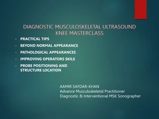
- 1. • PRACTICAL TIPS • BEYOND NORMAL APPEARANCE • PATHOLOGICAL APPEARANCES • IMPROVING OPERATORS SKILS • PROBE POSITIONING AND STRUCTURE LOCATION AAMIR SAFDAR-KHAN Advance Musculoskeletal Practitioner Diagnostic & Interventional MSK Sonographer
- 2. QUADRICEPS TENDON And SYNOVIAL RECESS Evaluation in LS (longitudinal) Importance of Starting Position Three layers Attachment Enthestitis Dynamic Test Central Tendon
- 3. Evaluating Medial and Lateral in TS (transverse) Fluid collection in dependent recesses Doppler (power) Pressure during Doppler Can track tendon as far as rectus femoris Window for injection - I prefer medial
- 4. PATELLAR TENDON INSERTIONS (patellar and tibial tuberosity) Tibial Insertion: Enthesitis? Patellar Insertion: Enthesitis? Bursitis: One small fluid? Hoffa Fat Pad: Impingement – what are the signs (Doppler?)
- 5. IT Band Gerdy;s tubercle Probe positioning otherwise can get confused with patellar tendon Tracking to femoral condyle for bursitis
- 6. PES ANSERINUS TENDON AND BURSA Groove with neurovasuclar bundle underneath Doppler may be helpful in identifying Tracking MCL is technique to identify it
- 7. CHECK PES ANSERINUS BURSITIS
- 8. MCL AND LCL BICEPS FEMORIS Angulation and cross position For LCL head of fibula Biceps femoris head of fibula Fibular pattern of all of them Relevance patellar groove and tendon to LCL Common injury of MCL (proximal attachment) Common injury of LCL (distal attachment) Tracking biceps femoris to conjoint tendon (posterior hip if having a time)
- 9. MCL relationship LCL relationship Biceps femoris
- 10. Baker’s cyst Gastrocnemius and Semi-Membranosus Lets open the link and learn more about https://www.linkedin.com/posts/aamir-safdar-khan- 35238816_importance-of-ultrasound-calf-tear-is-a-activity- 6773968991934410752-JXcE Thank you……………. AAMIR SAFDAR KHAN Advance Practice Physiotherapist Diagnostic & Interventional MSK Sonographer