More Related Content
Similar to Ultrasound-Guided Lower Extremity Nerve Block Techniques
Similar to Ultrasound-Guided Lower Extremity Nerve Block Techniques (20)
Ultrasound-Guided Lower Extremity Nerve Block Techniques
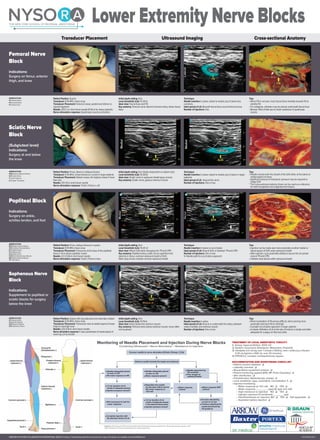
- 1. Transducer Placement Ultrasound Imaging Cross-sectional Anatomy
Femoral Nerve
Block
Indications:
Surgery on femur, anterior
thigh, and knee
Sciatic Nerve
Block
(Subgluteal level)
Indications:
Surgery at and below
the knee
Popliteal Block
Indications:
Surgery on ankle,
achilles tendon, and foot
Saphenous Nerve
Block
Indications:
Supplement to popliteal or
sciatic blocks for surgery
below the knee
Patient Position: Supine
Transducer: 8-16 MHz, linear array
Transducer Placement: Femoral crease, parallel and inferior to
inguinal ligament
Needle: 22G 5 cm short bevel needle (8-10cm for obese patients)
Nerve stimulation response: Quadriceps muscle contraction
Patient Position: Prone, lateral or oblique (shown)
Transducer: 6-16 MHz, Linear (shown) or curved in larger patients
Transducer Placement: Gluteal crease, the highest crease if more
than one
Needle: 21G 10cm short bevel needle
Nerve stimulation response: Twitch of foot or calf
Patient Position: Prone, oblique (shown) or supine.
Transducer: 8-16 MHz, linear array
Transducer Placement: Transverse at the base of the popliteal
fossa 4 -5cm above popliteal crease
Needle: 22 G 5-8cm short bevel needle
Nerve stimulation response: Twitch of foot or toes
Patient Position: Supine with leg abducted and externally rotated
Transducer: 8-16 MHz, linear array
Transducer Placement: Transverse view at medial aspect of lower
thigh to mid-thigh level
Needle: 22G 5-8cm short bevel needle
Nerve stimulation response: If used, paresthesia of medial aspect of
lower leg can be elicited
Initial depth setting: 4cm
Local Anesthetic (LA): 15-20mL
Ideal view: Fascia iliaca and FN
Key anatomy: Femoral nerve lateral to femoral artery, below fascia
iliaca
Initial depth setting: 5cm (highly dependent on patient size)
Local Anesthetic (LA): 15-20mL
Ideal view: Sciatic nerve in epineural sheath (grey arrows)
Key anatomy: Sciatic nerve, gluteus maximus muscle
Initial depth setting: 4cm
Local Anesthetic (LA): 15-25 ml
Ideal view: Where ScN starts diverging into TN and CPN
Key anatomy: Popliteal artery, sciatic nerve superficial and
lateral to it, femur, common epineural sheath of ScN
Note: Gray arrows indicate common epineural sheath
Initial depth setting: 3cm
Local Anesthetic (LA): 10-15mL
Ideal view: Artery below the sartorius muscle
Key anatomy: Femoral artery below sartorius muscle, nerve often
not visualized
Technique:
Needle insertion: In plane, lateral to medial, (out of plane less
common),
Ideal spread of LA: Beneath fascia iliaca around femoral nerve
Number of injections: One
Technique:
Needle insertion: In plane, lateral to medial, (out of plane in larger
patients)
Ideal spread of LA : Around the nerve
Number of injections: One or two
Technique:
Needle insertion: In plane or out of plane
Ideal spread of LA: Around ScN, or between TN and CPN
Number of injections: One or two
X- Needle path for out of plane approach
Technique:
Needle insertion: In plane
Ideal spread of LA: Around or underneath the artery, between
vastus medialis and sartorius muscle
Number of injections: One or two
Tips:
• When FN is not seen, track fascia illiaca medially towards FA to
identify FN
• For analgesia, catheters may be placed underneath fascia iliaca
• Beware: Risk of falls due to motor weakness of quadriceps
muscle
Tips:
• Needle should enter the sheath of the ScN either at the lateral or
medial aspect of nerve.
• Significant amount of transducer pressure may be required to
image ScN
* The cross-sectional anatomy shown can be used as a reference
for both transgluteal and subgluteal techniques.
Tips:
• Injection can be made also more proximally at either medial or
lateral aspect of ScN under epineural sheath
• After injection, scan proximally-distally to assure the LA spread
around TN and CPN
• Catheter best placed within epineural sheath
Tips:
• When localization of FA proves difficult, start scanning more
proximally and trace FA to mid-thigh
• Consider out of plane approach in larger patients
• A simple infiltration of LA at the site of incision is simple and often
adequate for surgery on foot and ankle
TREATMENT OF LOCAL ANESTHETIC TOXICITY
1) Airway, hyperventilation, 100% O2
2) Abolish convulsions (Diazepam, Midazolam, Propofol)
3) Intralipids (1.5 mL/kg over 1 minute (~100mL), then continuous infusion
0.25 mL/kg/min (~500 mL over 30 minutes)
4) CPR/ACLS, consider cardiopulmonary bypass
CREATED BY NYSORA COLLABORATIVE INTERNATIONAL GROUP. A listing of contributing institutions and electronic copy of the poster are available at www.NYSORA.com
NYSO
THE NEW YORK SCHOOL OF REGIONAL ANESTHESIA
R A
ABBREVIATIONS
FA Femoral Artery
FN Femoral Nerve
FV Femoral Vein
ABBREVIATIONS
GMM Gluteus Maximus Muscle
ScN Sciatic Nerve
IT Ischial Tubercle
GT Greater Trochanter
ABBREVIATIONS
FA Femoral Artery
Medialis M. (Vastus)
SaM Sartorius Muscle
SaN Saphenous Nerve
ABBREVIATIONS
BFM Biceps Femoris Muscle
CPN Common Peroneal Nerve
PA Popliteal Artery
PV Popliteal Vein
ScN Sciatic Nerve
SmM Semimembranosus Muscle
StM Semitendinosus Muscle
TN Tibial Nerve
DOCUMENTATION AND MONITORING CHECK-LIST
• Patient consent obtained q
• Laterality checked q
• Resuscitative equipment present q
• Patient monitoring applied (EKG, BP, Pulse Oxymetry) q
• Skin disinfection q
• Premedication: Medication(s), dose(s) q
• Local anesthetic: type, volume(ml), concentration % q
• Injection monitoring:
– Motor response at <0.5 mA: NO q YES q
– Motor response _________(specify type and mA)
– High resistance to injection: NO q YES q
– Injection pressure (if monitored): _______ (psi)
– Pain/Paresthesia on injection: NO q YES q Not applicable q
– Aspiration before injection q
©NYSORA 2012
Lower Extremity Nerve Blocks
Monitoring of Needle Placement and Injection During Nerve Blocks
Combining Ultrasound + Nerve Stimulation + Resistance to Injection
● Needle adequately placed
as seen on US
● No motor response to NS
● Needle adequately placed
as seen on US
● Motor response present
● Needle placement by
US uncertain
● Poor images of
anatomy/needle
● Motor response
present
● Motor response NOT
present
● Increase stimulating
current to 1.5 mA
● Continue adjusting the
needle placement by
US guidance
● Reposition the needle
(or decrease mA) to assure
NO motor response at
<0.5 mA†
● 1-2 mL injection of LA
results in adequate spread
in the desired tissue plane
● Injection pressure normal‡
● 1-2 mL injection of LA
results in adequate spread
● Injection pressure normal?‡
● Not necessary to look for
motor response
● Complete injection with
the planned volume of LA
Advance needle towards the target nerve (plexus)
Legend: US-ultrasound, NS-nerve stimulator, Normal injection pressure defined as <15 psi (pounds per square inch)‡.
†May indicate an intraneural/intrafascicular needle placement
Connect needle to nerve stimulator (0.5mA, 0.1msec, 2 Hz)