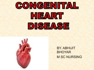
Congenital heart diseases
•Download as PPTX, PDF•
7 likes•3,407 views

The document provides information on congenital heart defects, including their causes, types, signs and symptoms, diagnosis, and treatment. It discusses several specific defects in detail, including aortic stenosis, coarctation of the aorta, pulmonary stenosis, and patent ductus arteriosus. The key points are: 1. Congenital heart defects can involve the heart's chambers, valves, or vessels and have various causes including genetic syndromes. 2. Specific defects like aortic stenosis and pulmonary stenosis can cause obstruction to blood flow while others like patent ductus arteriosus allow extra blood flow to the lungs. 3. Symptoms depend on the severity of the defect but may include heart failure,

Recommended
More Related Content
What's hot
What's hot (20)
Similar to Congenital heart diseases
Similar to Congenital heart diseases (20)
More from ABHIJIT BHOYAR
More from ABHIJIT BHOYAR (20)
Recently uploaded
Recently uploaded (20)
Congenital heart diseases
- 1. BY. ABHIJIT BHOYAR M SC NURSING
- 2. Anatomy & physiology Of heart
- 3. INTRODUCTION Congenital heart disease (or defects) (CHD) is one of the most common forms of congenital anomalies. It involves Chambers, Valves, Great vessels arising from the heart The heart is a muscular organ about the size of a fist, located just behind and slightly left of the breastbone. The heart pumps blood through the network of arteries and veins called the cardiovascular system.
- 4. In most cases, the cause of CHD is not known. Some infants and children with CHD may appear perfectly healthy, whereas others may be critically ill. Most infants and children with CHD can be successfully managed with medications and surgeries.
- 5. ETIOLOGY AND INCIDENCE CHD affects 8 to 12 of every 1,000 neonates. Exact cause of CHD is unknown in 90% of cases. The heart begins as a single cell and develops into a four-chambered pumping system during the third to eighth weeks of gestation
- 6. Conti… Associated factors for CHD include: ◦ Fetal or maternal infection during the first trimester (rubella). ◦ Chromosomal abnormalities (trisomy 21, 18, 13). ◦ Maternal insulin-dependent diabetes. ◦ Teratogenic effects of drugs and alcohol Teratogens are drugs, chemicals, or even infections that can cause abnormal fetal
- 7. Conti… Syndromes that include CHD: ◦ Marfan's syndrome: mitral valve prolapse (MVP), dilated aortic root. ◦ Turner's syndrome: aortic valve stenosis (AVS), coarctation of the aorta (CoA). ◦ Noonan's syndrome: dysplastic pulmonary valve. ◦ William's syndrome: supravalvular pulmonary stenosis (PS).
- 8. Conti.. ◦ DiGeorge syndrome: interrupted aortic arch (IAA), truncus arteriosus, transposition of great arteries (TGA), tetralogy of Fallot (TOF). ◦ Down syndrome (trisomy 21): atrioventricular (AV) canal defect, ventricular septal defect (VSD). About 50% of children with Down syndrome have a CHD.
- 9. CLASSIFICATION Congenital heart defects can be classified into three categories: Obstruction to blood flow Increased pulmonary blood flow (acyanotic lesions) Decreased pulmonary blood flow (cyanotic lesions). OBSTRUCTIVE ACYNOTIC CYNOTIC
- 10. CONTI.. Increased pulmonary blood flow (acyanotic): • PDA. • ASD. • VSD. • AV canal. • PAPVR Decreased pulmonary blood flow (cyanotic): • TOF. • TA. • TAPVR. • TGA. • Truncus arteriosus. • HLHS. • DORV. Obstructive lesions • AS, • CoA. • PS
- 11. AORTIC STENOSIS Congenital AS may be caused by a bicuspid aortic valve with fused commissures that does not open completely. The result is turbulent blood flow across the aortic valve and into the ascending aorta. Aortic stenosis is a narrowing of the aortic valve opening
- 12. Conti.. AS is the most common form of left ventricular outflow tract obstruction. It accounts for 3% to 6% of congenital heart defects. AS may occur at any age, and it occurs more commonly in boys than in girls. In most children, it is a progressive lesion that creates LVOTO. Left ventricular outflow tract obstruction (LVOTO)
- 13. Pathophysiology Blood flows at an increased velocity across the obstructive valve or stenotic area and into the aorta. During systole, left ventricular pressure rises dramatically to overcome the increased resistance at the aortic valve
- 14. Conti.. Myocardial ischemia may occur because of an imbalance between the increased oxygen requirements related to the hypertrophied left ventricle (LV) and the amount of oxygen that can be supplied. Left-sided heart failure results in an increased LV end diastolic pressure that is reflected back to the left atrium and pulmonary veins
- 15. Clinical Manifestations IN NEONATE Severe congestive heart failure (CHF). Metabolic acidosis. Tachypnea. Faint peripheral pulses, poor perfusion, poor capillary refill, cool skin. Poor feeding and feeding intolerance
- 16. Conti.. CHILD AND ADOLESCENT Chest pain on exertion, Decreased exercise tolerance. Dyspnea, Fatigue, Shortness of breath. Syncope, Light-headedness. Palpitations. Sudden death Syncope (pronounced “sin ko pea”) is the medical term for fainting or passing out.
- 17. Diagnostic Evaluation Auscultation. ◦ Systolic ejection murmur heard best at right upper sternal border, radiates to neck. ◦ Ejection click. ◦ S2 splits normally or narrowly. Electrocardiogram (ECG): left ventricular hypertrophy (LVH) with a strain pattern may be seen in severe cases. Ejection clicks are high- pitched sounds that occur at the moment of maximal opening of the aortic or pulmonary valves.
- 18. Conti.. Chest X-ray: increased cardiac silhouette, increased pulmonary vascular markings. A prominent aortic knob may be seen occasionally from poststenotic dilatation with valvular AS. Echocardiogram: two-dimensional echocardiogram with Doppler study and color flow mapping to visualize the anatomy and to estimate the gradient across the valve and through the aorta.
- 19. Management NEONATE Stabilize with prostaglandin E1 (PGE1) infusion to maintain cardiac output through the PDA. Inotropic support as needed. Intubation and ventilation as needed. Infective endocarditis prophylaxis (lifelong). Cardiac catheterization: aortic balloon valvuloplasty or aortic balloon angioplasty. Surgical valvotomy, commissurotomy, or myectomy/myotomy.
- 20. Child and Adolescent Medical management with close follow-up to monitor increasing gradient across the aortic valve or through the aorta. Restrict strenuous exercise and anaerobic exercise (eg, weight lifting). Restrict participation in competitive sports
- 21. Aortic balloon valvuloplasty or aortic balloon angioplasty. Infective endocarditis prophylaxis (lifelong). Surgical intervention. ◦ Surgical valvotomy, commissurotomy, or myectomy/ myotomy. ◦ Aortic valve replacement. Mechanical prosthesis (St. Jude valve). Ross procedure (pulmonary autograft).
- 22. COMPLICATIONS - CHF - Pulmonary edema - Dizziness - Light- headedness - Syncope -Palpitations -Arrhythmias - Infective endocarditis. - Sudden death.
- 23. COARCTATION OF THE AORTA CoA is a discrete narrowing or a long segment hypoplasia of the aortic arch, usually in the juxtaductal position. It accounts for 8% to 10% of congenital heart defects
- 24. Pathophysiology The discrete narrowing or hypoplastic segment of the aorta increases the workload of the left ventricle (increased LV systolic pressure). In a neonate with critical CoA, lower body blood flow occurs through the PDA (right-to-left shunting). In the older child, collateral vessels grow and bypass the coarctation to perfuse the lower body.
- 25. Clinical Manifestations The neonate with critical CoA (ductal dependent lesion): ◦ Asymptomatic until the PDA begins to close. ◦ After PDA closure: severe CHF, poor lower body perfusion, tachypnea, acidosis, progressive circulatory shock, absent femoral and pedal pulses.
- 26. The child or adolescent with CoA: ◦ Usually asymptomatic normal growth and development. ◦ Hypertension in the upper extremities, with absent or weak femoral pulses. ◦ Nosebleeds, headaches, leg cramps.
- 27. Diagnostic Evaluation Auscultation - varies; nonspecific systolic ejection murmur. Chest X-ray- cardiomegaly and pulmonary edema or pulmonary venous congestion. ECG varies; normal or right ventricular hypertrophy (RVH) in infants and LVH in older children.
- 28. Two-dimensional echocardiogram with Doppler study and color flow mapping identifies area of aortic arch narrowing and associated lesions (bicuspid aortic valve, VSD, PDA). Invasive studies (cardiac catheterization) usually not needed to make the initial diagnosis; may need aortic angiography to identify collateral vessels before surgery. Cardiac magnetic resonance imaging may be done to noninvasively assess the location and degree of narrowing and identify collateral vessels.
- 29. Management Critical Coarctation in the Neonate Medical management: ◦ Resuscitation and stabilization with PGE1 infusion: monitor for complications related to PGE1 therapy (fever, apnea). ◦ Intubation and ventilation as needed. ◦ Infective endocarditis prophylaxis (lifelong). ◦ Anticongestive therapy (digoxin and Lasix) and inotropic support as needed. ◦ Assess renal, hepatic, and neurologic function.
- 30. Balloon angioplasty may be indicated for infants who are a high surgical risk. Surgical intervention: usually performed as soon as the diagnosis is made. ◦ Subclavian flap repair (Waldhausen procedure). ◦ End-to-end anastomosis. ◦ Dacron patch repair
- 31. Coarctation in the Child or Adolescent Surgical intervention. ◦ End-to-end anastomosis. ◦ Dacron patch. Medical management for hypertension (beta- adrenergic blockers). Infective endocarditis prophylaxis (lifelong).
- 32. Recurrent Coarctation in the Neonate or Child Balloon angioplasty in the cardiac catheterization laboratory. Redo surgical intervention
- 33. Complications Systemic hypertension. CHF. Cerebral hemorrhage. Infective endocarditis. Left ventricular failure. Aortic aneurysm
- 34. PULMONARY STENOSIS The pulmonary valve opens during systole to let blood flow from the right ventricle into the main PA. Obstruction to flow can occur at three levels: subvalvular, valvular, or supravalvular level. The most common cause of RV outflow tract obstruction is pulmonary valve stenosis. PS accounts for 8% to 12% of CHDs.
- 35. Pathophysiology • Critical PS in the neonate: blood flows into the right atrium, across a patent foramen ovale (PFO) into the left heart; • pulmonary blood flow comes from a left-to-right shunt through a PDA. • Right ventricular pressure increases to pump blood across the obstructive pulmonary valve. • RVH develops in response to the increased pressure gradient across the pulmonary valve. Signs of right-sided heart failure include hepatic congestion, neck vein distention, elevated central venous pressure (CVP).
- 36. C L I N I C A L M A N I F E S T A T I O N S • Hypoxia. • Tachypnea. • RV failure. Critical PS in the Neonate • Asymptomatic. • Decreased exercise tolerance, fatigue, dyspnea. • Chest pain. Mild to Moderate PS in the Child and Adolescent
- 37. Auscultation: systolic ejection murmur heard best at the left upper sternal border; ejection click. ECG: varies; normal in mild cases and RVH in moderate to severe cases. Chest X-ray: varies; may show right ventricular enlargement; poststenotic dilatation of PA. Two-dimensional echocardiography with Doppler study and color flow mapping to visualize the sites of obstruction, observe the degree of RVH, and estimate the pressure gradient across the valve. Cardiac catheterization is usually not needed for the initial diagnosis. D I A G N O S T I C E V A L U A T I O N
- 38. NEONATE WITH CRITICAL PS Medical management: ◦ Stabilize and improve oxygen saturations with PGE1 infusion. ◦ Intubation and ventilation as needed. ◦ Inotropic support as needed. ◦ Infective endocarditis prophylaxis (lifelong). Balloon pulmonary valvuloplasty. Blalock-Taussig shunt (Gore-Tex graft between the subclavian artery and the PA to supply pulmonary blood flow). M A N A G E M E N T
- 39. Child and Adolescent with PS Medical management: ◦ Close follow-up to monitor and record RV to PA gradient and assess RV function. ◦ Restrict strenuous exercise and participation in competitive sports. ◦ Infective endocarditis prophylaxis (lifelong). ◦ Refer for intervention when RV pressure is greater than two-thirds of the systemic pressures. M A N A G E M E N T
- 40. Balloon pulmonary valvuloplasty in the cardiac catheterization laboratory. Surgical intervention: ◦ Valvotomy or valvectomy for dysplastic pulmonary valve. ◦ Patch repair of the right ventricular outflow tract. ◦ Placement of an RV-to-PA conduit. M A N A G E M E N T
- 42. PATENT DUCTUS ARTERIOSUS The ductus arteriosus is a normal fetal connection between the left PA and the descending aorta. During fetal life, blood flow is shunted away from the lungs through the ductus arteriosus and directly into the systemic circulation. PDAs are common in premature neonates who weigh less than 1,500 g. They account for 5% to 10% of CHDs, excluding premature neonates.
- 43. P A T H O P H Y S I O L O G Y During fetal life, the ductus arteriosus allows blood to bypass the pulmonary circulation (fetus receives oxygen from the placenta) and flow directly into the systemic circulation After birth, the ductus arteriosus is no longer needed. Functional closure usually occurs within 48 hours after birth. Anatomic closure is completed by age 2 to 3 weeks. When the ductus arteriosus fails to close, blood from the aorta (high pressure) flows into the low-pressure PA, resulting in pulmonary overcirculation. Increased pulmonary blood flow leads to a volume- loaded LV.
- 44. Small to Moderate-Sized PDA Usually asymptomatic. Large PDA CHF, Tachypnea, Frequent respiratory tract infections. Poor weight gain, failure to thrive. Feeding difficulties. Decreased exercise tolerance. C L I N I C A L M A N I F E S T A T I O N
- 45. Auscultation: continuous murmur heard best at left upper sternal border. Hyperactive precordium with large PDAs. Wide pulse pressure; bounding pulses. Chest X-ray: varies; normal or cardiomegaly with increased pulmonary vascular markings. ECG: varies; normal or LVH. Two-dimensional echocardiogram with Doppler study and color flow mapping to visualize the PDA with left-to-right blood flow. Cardiac catheterization is not needed for the initial diagnosis. D I A G N O S T I C E V A L U A T I O N
- 46. In the symptomatic premature neonate: indomethacin given I.V. Medical management: ◦ Monitor growth and development. ◦ Reassess for spontaneous PDA closure. ◦ Increase caloric intake as needed for normal weight gain. ◦ Diuretics: furosemide (Lasix), spironolactone (Aldactone). ◦ Infective endocarditis prophylaxis for 6 months after surgery or coil occlusion M A N A G E M E N T
- 47. Cardiac catheterization: ◦ For small PDAs coil occlusion. ◦ For larger PDAs a closure device may be used. Surgical management through PDA ligation M A N A G E M E N T
- 48. C O M P L I C A T I O N CHF, pulmonary edema. Infective endocarditis. Pulmonary hypertension/pulmonary vascular occlusive disease. Recurrent pneumonia.
- 49. ATRIAL SEPTAL DEFECT ASD is an abnormal communication between the left and right atrias. ASDs account for 9% of CHDs.
- 50. T Y P E S O F A S D Ostium secundum • The most common type of ASD; abnormal opening in the middle of the atrial septum Ostium primum • Abnormal opening at the bottom of the atrial septum; increased association with cleft mitral valve and atrioventricular defects. Sinus venosus • Abnormal opening at the top of the atrial septum; increased association with partial anomalous pulmonary venous return.
- 51. Blood flows from the higher-pressure left atrium across the ASD into the lower-pressure right atrium (left-to-right shunt). Increased blood return to the right heart leads to right ventricular volume overload and right ventricular dilation. Increased pulmonary blood flow leads to elevated pulmonary artery pressures. P A T H O P H Y S I O L O G Y
- 52. Clinical Manifestations Usually asymptomatic. Clinical symptoms vary depending on type of associated defects: ◦ CHF ◦ Frequent upper respiratory infections (URIs). ◦ Poor weight gain. ◦ Decreased exercise tolerance
- 53. Diagnostic Evaluation Auscultation: soft systolic ejection murmur heard best at the left upper sternal border; widely split, fixed second heart sound. Chest X-ray: varies; normal to right atrial and ventricular dilation, increased pulmonary markings. ECG: varies; right axis deviation and mild RVH or right bundle-branch block.
- 54. Two-dimensional echocardiogram with Doppler study and color flow mapping to identify the site of the ASD and associated lesions and document left-to- right flow across the atrial septum. Cardiac catheterization usually not needed for initial diagnosis; performed if defect can be closed using an atrial occlusion device (device can be used only in ostium secundum defects).
- 55. Management Medical management: ◦ Monitor and reassess (spontaneous closure rate is small but may occur up to age 2). ◦ Treatment with anticongestive therapy (digoxin and Lasix) may be necessary if signs of CHF are present. ◦ Infective endocarditis prophylaxis for 6 months after surgery or atrial occlusion devise is used.
- 56. Cardiac catheterization for placement of an atrial occlusion device for ostium secundam defects. Surgical intervention: ◦ Primary repair: suture closure of the ASD. ◦ Patch repair of the ASD.
- 57. Complications CHF (rare). Infective endocarditis. Embolic stroke. Pulmonary hypertension. Atrial arrhythmias.
- 58. VENTRICULAR SEPTAL DEFECT A VSD is an abnormal communication between the right and left ventricles. It is the most common type of congenital heart defect, accounting for approximately 25% of all CHDs. VSDs vary in the size (small and restrictive to large and nonrestrictive defect), number (single versus multiple), and type (perimembranous or muscular).
- 59. Pathophysiology Blood flows from the high-pressure left ventricle across the VSD into the low-pressure right ventricle and into the PA, resulting in pulmonary overcirculation. A left-to-right shunt because of a VSD results in increased right ventricular pressure and increased PA pressure. The increased pulmonary venous return to the left side of the heart results in left atrial dilation.
- 60. Long-standing pulmonary overcirculation causes a change in the pulmonary arterial bed, leading to increased pulmonary vascular resistance. High pulmonary vascular resistance (PVR) can reverse the blood flow pattern that leads to a right-to- left shunt across the VSD (Eisenmenger's syndrome), resulting in cyanosis. Once this develops, the child is no longer a candidate for surgical repair.
- 61. Clinical Manifestations Small VSD”usually asymptomatic; high spontaneous closure rate during the first year of life. Large VSDs. ◦ CHF: tachypnea, tachycardia, excessive sweating associated with feeding, hepatomegaly. ◦ Frequent URIs. ◦ Poor weight gain, failure to thrive. ◦ Feeding difficulties. ◦ Decreased exercise tolerance
- 62. Diagnostic Evaluation Auscultation: harsh systolic regurgitant murmur heard best at the lower left sternal border (LLSB); systolic thrill felt at LLSB, narrowly split S2. Chest X-ray: varies; normal or cardiomegaly and increased pulmonary vascular markings. Pulmonary vascular markings are directly proportionate to the amount of left-to-right shunting. ECG: varies; normal to biventricular hypertrophy.
- 63. Two-dimensional echocardiogram with Doppler study and color flow mapping to identify the size, number, and sites of the defects, estimate pulmonary artery pressure, and identify associated lesions. Cardiac catheterization usually not needed for initial diagnosis; may be needed to calculate the size of the shunt or to assess PVR. May be performed if defect can be closed using a ventricular occlusion device (device can be used only in muscular
- 64. Management Small VSD Medical management: ◦ Usually no anticongestive therapy is needed. ◦ Infective endocarditis prophylaxis for 6 months after surgical implantation of a ventricular occlusion device. Cardiac catheterization for placement of a ventricular occlusion device for muscular defects. Surgical intervention is usually not necessary.
- 65. Moderate to Large VSD Medical Management: ◦ CHF management: digoxin and diuretics (furosemide, spironolactone) and afterload reduction. ◦ Avoid oxygen; oxygen is a potent pulmonary vasodilator and will increase blood flow into the PA. ◦ Increase caloric intake: fortify formula or breast milk to make 24 to 30 cal/oz formula; supplemental nasogastric feeds as needed. ◦ Infective endocarditis prophylaxis for 6 months after surgery/ventricular device occluder.
- 66. Cardiac catheterization for placement of a ventricular occlusion device for muscular defects.). Refer for surgical intervention. ◦ Usually repaired before age 1. ◦ One-stage approach: preferred surgical plan; patch closure of VSD. ◦ Two-stage approach: first surgery is to band the PA to restrict pulmonary blood flow; second surgery is to patch close the VSD and remove the PA band.
- 68. TETRALOGY OF FALLOT TOF is the most common complex congenital heart defect; it accounts for 6% to 10% of all CHDs. The four abnormalities of TOF include the following: A large, nonrestrictive VSD. Aortic override. Pulmonary stenosis (right ventricular outflow tract obstruction). Right ventricular hypertrophy.
- 69. Pathophysiology • Degree of cyanosis depends on the size of the VSD and the degree of right ventricular outflow tract obstruction (RVOTO). • Obstruction of blood flow from the right ventricle to the PA results in deoxygenated blood being shunted across the VSD and into the aorta (right-to-left shunt causes cyanosis). • RVOTO can occur at any or all of the following three levels: pulmonary valve stenosis, infundibular stenosis, or supravalvular stenosis.
- 70. • The right ventricle becomes hypertrophied as a result of the increased gradient across the RVOT. • Minimal RVOTO results in a pink TOF variant, with the physiology behaving more like a large, nonrestrictive VSD.
- 71. Clinical Manifestations Cyanosis. ◦ Neonate may have normal oxygen saturations; as the infant grows, the RVOTO increases and the oxygen saturation falls. ◦ Neonate with unacceptably low oxygen saturation needs PGE1 infusion to maintain ductal patency and adequate oxygen saturation. ◦ Cyanosis may initially be observed only with crying and with exertion.
- 72. Polycythemia. Decreased exercise tolerance. A common clinical manifestation years ago was squatting, a posture characteristically assumed by older children to increase systemic vascular resistance and to encourage increased pulmonary blood flow. Squatting is rarely seen currently because TOF is now surgically repaired during the first year of life.
- 73. Hypercyanotic spells (formerly known as Tet spells): a life-threatening hypoxic event with a dramatic decrease in oxygen saturations. ◦ Typical hypoxic spells occur in the morning soon after awakening; during or after a crying episode; during or after a feeding; during painful procedures such as blood draws. ◦ Typical scenario includes tachypnea, irritability, and increasing cyanosis, followed by flaccidity and loss of consciousness.
- 74. ◦ Home treatment for the caregiver: soothe the infant and place him or her in a knee-chest position; notify the health care provider immediately. ◦ Hospital treatment includes knee-chest position, sedation (morphine), oxygen, beta-adrenergic blockers (propranolol, esmolol) to relax the infundibulum, and administration of medications to increase systemic vascular resistance (phenylephrine). ◦ Hypercyanotic spells usually prompt the cardiologist to refer for surgical intervention.
- 75. Diagnostic Evaluation Auscultation: harsh systolic ejection murmur heard best at the upper left sternal border (RVOT murmur); single second heart sound; during a hypercyanotic spell the murmur disappears. Chest X-ray: varies; normal or decreased pulmonary vascular markings. The heart may appear “boot shaped”• because of a concave main PA with an upturned apex resulting from RVH. ECG: varies; normal or RVH.
- 76. Two-dimensional echocardiogram with Doppler study and color flow mapping to identify the structural abnormalities, estimate the degree of RVOTO and assess the coronary artery pattern. Cardiac catheterization is usually not needed for the initial diagnosis. May be performed before surgical intervention to identify the location and number of VSDs, the PVR, the degree of RVOTO, and the presence of any coronary abnormalities.
- 77. Management Medical Management: ◦ Monitor oxygen saturation level. ◦ Monitor growth and development. ◦ Monitor for hypercyanotic spells (many spells go unnoticed by parents). ◦ Infective endocarditis prophylaxis (lifelong). ◦ Restrict strenuous activity and participation in competitive sports. Balloon pulmonary angioplasty (rarely).
- 78. Surgical intervention: Palliative versus definitive repair. ◦ Potential obstacles for one-stage repair: abnormal coronary artery distribution (left anterior descending arises from right coronary artery and crosses RVOT); multiple VSDs; hypoplastic branch pulmonary arteries; small infant weighing less than 5.5 lb (2.5 kg).
- 79. ◦ Palliative surgery: modified Blalock-Taussig shunt (BT shunt); tube Gore-Tex graft between the left subclavian artery and the PA: increased pulmonary blood flow results in higher oxygen saturations. ◦ Definitive surgery: patch closure of VSD, relief of right ventricular outflow tract obstruction; with or without transannular patch across the pulmonary valve.
- 80. Long-Term Follow-Up Assess RV outflow tract, monitor degree of pulmonary insufficiency. Monitor RV function and exercise tolerance. Monitor for arrhythmias.
- 81. Complications Hypoxia. Hypercyanotic spells. Polycythemia. CHF: rare; associated with pink TOF. Right ventricular dysfunction. Ventricular arrhythmias. Infective endocarditis
- 82. TRANSPOSITION OF THE GREAT ARTERIES Transposition of the great arteries (TGA) occurs when the PA arises off the left ventricle and the aorta arises off the right ventricle. It accounts for 5% to 10% of CHDs. Associated lesions include VSD, ASD, PDA, PS, and CoA
- 83. Pathophysiology This defect results in two parallel circulations: ◦ The right atrium receives deoxygenated blood from the inferior vena cava (IVC) and superior vena cava (SVC); blood flow continues through the tricuspid valve into the right ventricle and is pumped back to the aorta. ◦ The left atrium receives richly oxygenated blood from the pulmonary veins; blood flow continues through the mitral valve into the left ventricle and is pumped
- 84. To sustain life, there must be an accompanying defect that allows mixing of deoxygenated blood and oxygenated blood between the two circuits, such as PDA, ASD, PFO, or VSD. Neonates born with tga with an intact ventricular system are usually more cyanotic and sicker than neonates born with TGA with a VSD.
- 85. Clinical Manifestations Symptoms evident soon after birth; clinical scenario is influenced by the extent of intercirculatory mixing. Cyanosis. Tachypnea. Metabolic acidosis. CHF. Feeding difficulties.
- 86. Diagnostic Evaluation Auscultation: varies; no murmur, or a murmur related to an associated defect. Chest X-ray: varies; neonate chest X-ray usually normal; cardiomegaly with a narrow mediastinum (egg-shaped cardiac silhouette) and increased pulmonary markings; or decreased pulmonary markings with pulmonary stenosis. ECG: RVH or biventricular hypertrophy. Two-dimensional echocardiogram.
- 87. Management Medical Management Stabilize with PGE1 infusion. Treat pulmonary overcirculation with digoxin and diuretics as needed. Intubate and ventilate as needed. Inotropic support as needed. Infective endocarditis prophylaxis (lifelong).
- 88. Cardiac catheterization Balloon atrial septostomy (Rashkind) is indicated for severe hypoxia to create or improve atrial level mixing. Surgical Management Arterial switch operation procedure of choice: ◦ Ideally performed during the first week of life. ◦ The aorta and PA are switched back to their anatomically correct ventricle above the level of the valve.
- 89. ◦ Coronary arteries are transferred to the new aorta. ◦ Associated lesions are also repaired at this time. Rastelli operation”performed for TGA, VSD, and PS. ◦ Repaired during the first year of life. ◦ VSD patch repaired to include LV to aortic outflow continuity with pulmonary blood flow provided via an RV to PA homograft.
- 90. Atrial switch operation: Mustard or Senning procedure: ◦ Rerouting of atrial blood flow: RA, mitral valve, LV, PA and LA tricuspid valve,’ RV, Ao ◦ Restores oxygenated blood into the systemic system and deoxygenated blood guided to the pulmonary system. ◦ Disadvantages: RV is left as the systemic ventricle”will develop RV dysfunction. Increased incidence of atrial dysrhythmias and baffle obstruction
- 91. Complications Severe hypoxia. Multiorgan ischemia. Arrhythmias. RV dysfunction. Coronary artery obstruction leading to myocardial ischemia or death
- 92. TRICUSPID ATRESIA Tricuspid atresia involves the absence of the tricuspid valve and hypoplasia of the right ventricle. Associated defects such as an ASD, VSD, or PDA are necessary for survival. Tricuspid atresia accounts for 1% to 3% of CHDs.
- 93. Pathophysiology With TA, systemic venous return enters the right atrium and cannot continue into the RV; blood flows across an atrial septal opening into the left atrium. Pulmonary blood flow occurs through a PDA or VSD.
- 94. Clinical Manifestations Cyanosis. Tachypnea. Feeding difficulties.
- 95. Diagnostic Evaluation Auscultation: murmurs vary depending on the associated lesions; single second heart sound. Chest X-ray: pulmonary vascular markings related to the amount of pulmonary blood flow (usually decreased); normal to slightly increased cardiac silhouette. ECG: superior axis; right and left atrial hypertrophy; LVH. echocardiogram Cardiac catheterization may be necessary to delineate anatomy.
- 96. Management Medical Management Stabilize with PGE1 infusion. Intubate and ventilate as needed. Inotropic support as needed. Infective endocarditis prophylaxis (lifelong).
- 97. Surgical Management First surgery neonate: ◦ BT shunt indicated if pulmonary blood flow is insufficient. ◦ Pulmonary artery band indicated if pulmonary blood flow is excessive. ◦ No treatment is required if pulmonary blood flow is balanced. Second surgery: ages 6 to 9 months: ◦ Bidirectional Glenn shunt: end-to-side anastomosis of the SVC to the right PA. Third surgery: ages 18 months to 3 years: ◦ Fontan completion: IVC to PA connection (extracardiac conduit or intracardiac baffle). A Blalock-Taussig (BT) shunt is a small tube, only a few millimeters wide, that is used to create a pathway for blood to go from the arterial circulation to the lungs. It is used to treat congenital heart defects that affect blood flow to the lungs.
- 98. Complications CHF. Persistent pleural effusion (especially after Stage II and Stage III repairs). Thrombus formation in the systemic venous system. Infective endocarditis. Rarely, heart block.
- 99. NURSING CARE OF THE CHILD WITH CONGENITAL HEART DISEASE Nursing Assessment Obtain a thorough nursing history. Discuss the care plan with the health care team (cardiologist, cardiac surgeon, nursing case manager, social worker, nutritionist). Discuss the care plan with the patient, parents, and other caregivers. Measure and record height and weight. Plot on a growth chart. Record vital signs and oxygen saturations. ◦ Measure vital signs at a time when the infant/child is quiet. ◦ Choose appropriate-size blood pressure (BP) cuff.
- 100. Assess and record: ◦ Skin color: pink, cyanotic, mottled. ◦ Mucous membranes: moist, dry, cyanotic. ◦ Extremities: check peripheral pulses for quality and symmetry; dependent edema; capillary refill; color and temperature. Assess for clubbing (cyanotic heart disease). Assess chest wall for deformities; prominent precordial activity.
- 101. Assess respiratory pattern. ◦ Before disturbing the child, stand back and count the respiratory rate. ◦ Loosen or remove clothing to directly observe chest movement. ◦ Assess for signs of respiratory distress: increased respiratory rate, grunting, retractions, nasal flaring. ◦ Auscultate for crackles, wheezing, congestion, stridor. Assess heart sounds. ◦ Determine rate (bradycardia, tachycardia, or normal for age) and rhythm (regular or irregular).
- 102. Assess fluid status. ◦ Daily weights. ◦ Strict intake and output (number of wet diapers; urine output). Assess and record the child's level of activity. ◦ Observe the infant while feeding. Does the infant need frequent breaks or does he or she fall asleep during feeding? Assess for sweating, color change, or respiratory distress while feeding. ◦ Observe the child at play. Is play interrupted to rest? Ask the parent if the child keeps up with peers while at play. ◦ Assess and record findings relevant to the child's developmental level: age-appropriate behavior, cognitive
- 103. Nursing Diagnoses Impaired Gas Exchange related to altered pulmonary blood flow or pulmonary congestion Decreased Cardiac Output related to decreased myocardial function Activity Intolerance related to hypoxia or decreased myocardial function Imbalanced Nutrition: Less Than Body Requirements related to excessive energy demands required by increased cardiac workload Risk for Infection related to chronic illness
- 104. Nursing Interventions Goal- Relieving Respiratory Distress Position the child in a reclining, semi-upright position. Suction oral and nasal secretions as needed. Identify target oxygen saturations and administer oxygen as prescribed. Administer prescribed medications and document response to medications (improved, no change, or worsening respiratory status). ◦ Diuretics. ◦ Bronchodilators. May need to change oral feedings to nasogastric feedings because
- 105. Improving Cardiac Output Organize nursing care and medication schedule to provide periods of uninterrupted rest. Provide play or educational activities that can be done in bed with minimal exertion. Maintain normothermia. Administer medications as prescribed. ◦ Diuretics (furosemide, spironolactone): Give the medication at the same time each day. For older children, do not give a dose right before bedtime.
- 106. Monitor the effectiveness of the dose: measure and record urine output. ◦ Digoxin: Check heart rate for 1 minute. Withhold the dose and notify the physician for bradycardia (heart rate less than 90 beats/minute [bpm]). Lead II rhythm strip may be ordered for PR interval monitoring. Prolonged PR interval indicates first-degree heart block (dose of digoxin may be withheld). Give medication at the same time each day. For infants and children, digoxin is usually divided and given twice per day.
- 107. Monitor serum electrolytes. Increased incidence of digoxin toxicity associated with hypokalemia. ◦ Afterload-reducing medications (captopril, enalapril): When initiating medication for the first time: check BP immediately before and 1 hour after dose. Monitor for signs of hypotension: syncope, light- headedness, faint pulses. Withhold medication and notify the physician according to ordered parameters.
- 108. Improving Oxygenation and Activity Tolerance Place pulse oximeter probe (continuous monitoring or measure with vital signs) on finger, earlobe, or toe. Administer oxygen as needed. Titrate amount of oxygen to reach target oxygen saturations. Assess response to oxygen therapy: increase in baseline oxygen saturations, improved work of breathing, and change in patient comfort. Explain to the child how oxygen will help. If possible, give the child the choice for face mask oxygen or nasal cannula oxygen.
- 109. Providing Adequate Nutrition For the infant: ◦ Small, frequent feedings. ◦ Fortified formula or breast milk (up to 30 cal/oz). ◦ Limit oral feeding time to 15 to 20 minutes. ◦ Supplement oral feeds with nasogastric feedings as needed to provide weight gain (ie, continuous nasogastric feedings at night with ad-lib by- mouth feeds during the day). For the child: ◦ Small, frequent meals. ◦ High-calorie, nutritional supplements. ◦ Determine child's likes and dislikes and plan meals accordingly.
- 110. ◦ Allow the parents to bring the child's favorite foods to the hospital. Report feeding intolerance: nausea, vomiting, diarrhea. Document daily weight (same time of day, same scale, same clothing). Record accurate inputs and outputs; assess for fluid retention. Fluid restriction not usually needed for children; manage excess fluid with diuretics.
- 111. Preventing Infection Maintain routine childhood immunization schedule. Administer yearly influenza vaccine. Administer RSV immunization for children younger than age 2 with complex CHD and those at risk for CHF or pulmonary hypertension. Prevent exposure to communicable diseases. Good hand washing. Report fevers. Report signs of URI: runny nose, cough, increase in nasal
- 112. THANK YOU TO TAKE MY CARE