SlideShare a Scribd company logo
1 of 12
Download to read offline
Iron Metabolism - Distribution
Anemias of Disordered Iron
Metabolism and Heme Synthesis

Iron-containing compounds
Functional compounds – metabolic
(hemoglobin) or enzymatic functions
Transport (transferrin) or storage (ferritin)

Total iron concentration = 40-50 mg/kg
body weight (60-75% in hemoglobin)
Hemoglobin from degrading RBC is
degraded in liver, spleen – iron is released.
85% of this iron is recycled – delivered to
bone marrow bound to transferrin

Iron Metabolism – Absorption
Primarily in mucosa of proximal small intestine –
Amount absorbed depends on
Amount of iron ingested
Form of ingested iron - ferric iron not easily absorbed
Tissue iron stores – inversely related to amount
absorbed
Condition of mucosal cells in GI tract
Hematopoietic activity of bone marrow
Intralumunial factors (parasites, toxins, etc)

Iron Metabolism - Transport
Major iron transport protein is transferrin.
Each gram of transferrin will bind 1.4 mg
of iron.
Total transferrin present in plasma to bind
253-435 µg of iron/dL of plasma – Total
iron-binding capacity (TIBC)
Serum iron concentration is 70 – 201
µg/dL – 95% is complexed with transferrin

1
Iron metabolism - Storage
Transferrin is about 1/3 saturated with
iron (% saturation = serum iron/TIBC X 100%)
The reserve iron-binding capacity of
transferrin is called the unsaturated
iron-binding capacity or UIBC (UIBC =
TIBC – serum iron)
Most iron bound to transferrin comes
from the breakdown of hemoglobin
Excess iron deposited in tissues

Major storage depot is liver
Stored as ferritin and hemosiderin
Ferritin
primary storage compound for body’s need
Readily released for heme synthesis
Small amounts found in blood – parallels
the storage iron in the body

Iron Metabolism - Requirements
Hemosiderin
Major long term storage form of iron
Slow release
Estimation of hemosiderin made on bone
marrow tissue sections

Body iron conserved through reutilization – only
about 1 mg lost per day
Iron deficiency occurs when negative iron balance
Increased requirements
Inadequate diet
Malabsorption

Iron overload when
Increased absorption
Multiple transfusions
Iron injections

2
Increased Iron Requirements
Menstruation – menstruating females
have twice the daily requirements as
males (2 mg/day)
Pregnancy – 3.4 mg/day
Infancy/childhood – rapid growth
From:
http://www.cdc.gov/hemochromatosis/training/
pathophysiology/iron_cycle_popup.htm

At birth, enough iron stores for 4-5 months
Milk is poor source of iron
Iron supplementation recommended

Laboratory Assessment of Iron
Serum iron levels
Total iron binding capacity
Calculation of % saturation of transferrin
Serum ferritin

Ferrokinetic studies (not done routinely)monitor movement of 59Fe from plasma to
bone marrow and into circulating RBCs.
Rate at which iron leaves plasma is PIT (plasma
iron turnover) – good indication of total
erythropoiesis
Are at which iron incorporated into circulating
erythrocytes is EIT (erythrocyte iron turnover) –
measure of effective erythropoiesis- correlates with
RPI

3
Iron deficiency Anemia (IDA)
Normal Values
iron: 60-170 µg/dl
TIBC: 240-450 µg/dl
transferrin saturation: 20-50%

Most common nutritional deficiency in the
world
Symptoms
General anemia symptoms –
Shortness of breath on exertion
Lethargy
Heart palpitations

Mucosal atrophy -glossitis, cheilitis, dysphagia
“Spoon nails” - koilonychia
Pica

Etiology vs. Pathophysiology
Etiology is the cause or origin of a
disorder.
Pathophysiology is the functional
alteration in a disease or a defect.

Etiology of IDA
Dietary Deficiency – rarely the cause in
developed countries (except in infancy,
pregnancy, adolescence)
Blood Loss –
Adult males have about 8 years of storage
iron. Deficiency if chronic blood loss – e.g.
gastrointestinal or genitourinary bleeding
Intravascular hemolysis

Malabsorption

4
Pathophysiology of IDA
Diminished total body iron content,
developing in stages over a period of
negative iron balance
Iron depletion – Stage One
Iron deficient erthyropoiesis – Stage Two
Iron deficiency anemia – Stage Three

Stage Two
Insufficient iron to insert into protoporphyrin
ring to form heme –
Protoporphyrin accumulates in cell and
complexes with zinc to form ZPP
No anemia, no hypochromia, but slight
microcytosis
RPI may be decreased

Stage One
Iron storage is exhausted - indicated by
decrease in serum ferritin levels
No anemia – RBC morphology is normal
May have elevated RDW

Stage Three
All laboratory tests for iron status
become abnormal
Most significant finding is microcytic,
hypochromic anemia

5
Laboratory findings – Fe deficiency
Microcytic/hypochromic anemia –
microcytosis first
MCV = 55-74 fl
MCHC = 22-31 %
MCH = 14-26 pg

Hgb < 10 g/dL
Increased RDW

RPI < 2.0 (ineffective erythropoiesis)
Poikilocytosis (target cells, elliptocytes,
dacryocytes)
May be thrombocytosis
serum ferritin
Serum iron (<30 µg/dL); TIBC; %
saturation (<16 %)

Therapy
Treat underlying disorder
Administer iron and observe response
Oral administration of ferrous sulfate
Increase of 1 gm/hemoglobin per month
Reticulocyte count peaks at about 8 to 10
days (4-10%)

Anemias Caused by
Abnormal Iron Metabolism
Sideroblastic anemias
Anemia of chronic disease

6
Sideroblastic anemia
Characterized by
Increase in total body iron
Presence of ringed sideroblasts in bone
marrow
Hypochromic anemia

Classification
Hereditary form
Acquired form (more common)
Idiopathic – Refractory anemia with ringed
sideroblasts (RARS)
secondary

Hereditary Sideroblastic Anemia
Pathophysiology
Disturbances of enzymes regulating heme
synthesis
Ringed sideroblasts form when nonferritin
iron accumulated in the mitochondria that
circle the normoblast nucleus

Most common form is sex-linked and due
to an abnormal δ-aminolevulinate
synthase enzyme (ALAS)
Decreased heme synthesis due to block
in iron utilization –
perceived by body as increased need
for iron
Increased iron absorption results in iron
overload

7
Acquired Sideroblastic Anemia
RARS (Refractory anemia with ringed
sideroblasts) – acquired stem cell disorder
Secondary
Lead poisoning (plumbism)
Associated with hyperactivity, low IQ, concentration
disorders in children
Inhibits cellular enzymes involved in heme
synthesis

Alcoholism – alcoholics frequently have
anemias due to one or more causes
Malignancy

Bone marrow
Erythroid hyperplasia - megaloblastosis
Ringed sideroblasts in 50% of
normoblasts

Laboratory findings in SA –
Peripheral Blood
Moderate to severe anemia – dimorphic
(mild macrocytosis prevalent in RARS and
alcoholism)
Target cells
Pappenheimer bodies
Basophilic stippling – coarse basophilic
stippling characteristic in lead poisoning
↑ Fe, N to ↓ TIBC, ↑ % saturation, ↑ ferritin

Therapy
Pyroxidine – hereditary form (50%
response)
Folic acid if megaloblastoid features
Phlebotomy/chelation therapy to remove
excess iron

8
Anemia of Chronic Disease
Anemia that occurs in patients with
chronic infections, chronic inflammatory
disorders, or neoplastic disorders not
due to bleeding, hemolysis, or marrow
involvement (renal, endocrine, or hepatic
insufficiencies are excluded)
Low serum iron, normal iron stores

Laboratory findings
Mild anemia (not less than 9 g/dL)
Normocytic, normochromic (some are
normocytic, hypochromic)
RPI<2
↑ Fe (10-70 µg/dL), N to ↓ TIBC (100300 µg/dL), N to ↓ % saturation (1025%), N or ↑ ferritin

Pathophysiology
Impaired marrow response to anemia –
cytokines produced as result of immune
response inhibit EPO production
Block in iron release from macrophage
Shortened erythrocyte survival

Hemochromatosis
Not an anemia – but will be discussed
because of abnormal iron studies
Parenchymal tissue damage from iron
overload – excess iron stored in
macrophages and hepatocytes, cardiac
cells, endocrine cells

9
Recessive genetic disorder
Prevalence of 1 in 200-250 persons
1:10 Caucausians in US are carriers
Abnormal protein produced that binds to
transferrin receptor on cells –
Clinical findings
Chronic fatigue
Diabetes
Cirrhosis
Hyperpigmentation of skin

Laboratory findings
Criteria for diagnosis: >50% transferrin
saturation in females; >60% in males

Treatment
Phlebotomy –each unit of blood removes
about 250 mg of iron

Porphyrias
Group of inherited disorders characterized
by block in porphyrin synthesis
Lack of enzyme in pathway of heme synthesis
Heme precursors accumulate in tissues –
excreted in urine and/or feces

Symptoms

2 forms depending on primary site of
defective porphyrin metabolism
Heptatic
Erythropoietic – (we will discuss these
because they affect the erythrocytes)

Photosensitivity
Abdominal pain
neuropathy

10
Pathophysiology of EP
Abnormality of heme biosynthetic
pathway within the normoblasts
2 types
Congenital erythropoietic porphyria (CEP)
Erythropoietic protoporphyria (EEP)

Porphyrinogens are the precursors or
porphyrins
Series III – precursors of heme
Series I - functionless

Erythropoeitic Protoporphyria
Overproduction of protoporphyrin
No anemia present – excess
protoporphyrin IX builds up – leaks into skin
dermal capillaries

Congenital Erythropoietic Porphyria
Excessive amounts of series I porpyrins
? Enzyme defect in uroporpyringoen III
cosynthase so porphyrinogens are
channeled to series I isomer
Excess porpyrins deposited in body
tissues –
Hemolytic anemia - ? Result of excess
porphyrins in RBC

Clinical Features
CEP – rare autosomal recessive disorder (only
~100 cases reported)
Pink to reddish brown urine
Excess porphyrins- extreme photosensitivity
Vesicular eruptions on skin exposed to sunlight
Hypertrichosis

EEP – autosomal dominant (some autosomal
recessive inheritance) – ~300 cases reported

11
Laboratory Features
CEP
Mild to severe N/N anemia with anisocytosis
and poikilocytosis
Polychromasia with ↑ reticulocytes – RBCs
fluoresce with UV light

EEP
No abnormalities on routine testing

12

More Related Content

What's hot

Iron metabolism in neonates and role of hepcidin
Iron metabolism in neonates and role of hepcidinIron metabolism in neonates and role of hepcidin
Iron metabolism in neonates and role of hepcidinkamalarora21
 
Iron metabolism final
Iron metabolism finalIron metabolism final
Iron metabolism finalsarojben
 
Role of protein in iron metabolism
Role of protein in iron metabolismRole of protein in iron metabolism
Role of protein in iron metabolismMuni Venkatesh
 
Iron Homeostasis
Iron  HomeostasisIron  Homeostasis
Iron Homeostasisbuddhi271
 
Iron metabolism by Dr Anurag Yadav
Iron metabolism by Dr Anurag YadavIron metabolism by Dr Anurag Yadav
Iron metabolism by Dr Anurag YadavDr Anurag Yadav
 
Iron metabolism, iron deficiency
Iron metabolism, iron deficiencyIron metabolism, iron deficiency
Iron metabolism, iron deficiencyGuvera Vasireddy
 
Hematinic I ARF: Iron
Hematinic I ARF: IronHematinic I ARF: Iron
Hematinic I ARF: IronAditiaFitri
 
Pharmacology anemia and its treatment
Pharmacology   anemia and its treatmentPharmacology   anemia and its treatment
Pharmacology anemia and its treatmentMBBS IMS MSU
 

What's hot (20)

Hepcidin
HepcidinHepcidin
Hepcidin
 
Iron metabolism in neonates and role of hepcidin
Iron metabolism in neonates and role of hepcidinIron metabolism in neonates and role of hepcidin
Iron metabolism in neonates and role of hepcidin
 
Iron deficiency anemia
Iron deficiency anemiaIron deficiency anemia
Iron deficiency anemia
 
Iron metab PART 2
Iron metab PART 2Iron metab PART 2
Iron metab PART 2
 
Iron Metabolism
Iron MetabolismIron Metabolism
Iron Metabolism
 
Iron metabolism final
Iron metabolism finalIron metabolism final
Iron metabolism final
 
Iron metabolism
Iron metabolismIron metabolism
Iron metabolism
 
Role of protein in iron metabolism
Role of protein in iron metabolismRole of protein in iron metabolism
Role of protein in iron metabolism
 
Starry Starry Night - Vincent
Starry Starry Night - VincentStarry Starry Night - Vincent
Starry Starry Night - Vincent
 
Iron kinetics part 1
Iron kinetics part 1Iron kinetics part 1
Iron kinetics part 1
 
Iron Homeostasis
Iron  HomeostasisIron  Homeostasis
Iron Homeostasis
 
Fe metabolism
Fe metabolismFe metabolism
Fe metabolism
 
Iron metabolism by Dr Anurag Yadav
Iron metabolism by Dr Anurag YadavIron metabolism by Dr Anurag Yadav
Iron metabolism by Dr Anurag Yadav
 
Iron metabolism, iron deficiency
Iron metabolism, iron deficiencyIron metabolism, iron deficiency
Iron metabolism, iron deficiency
 
Haematinics
HaematinicsHaematinics
Haematinics
 
Iron metabolism
Iron metabolism Iron metabolism
Iron metabolism
 
Hematinic I ARF: Iron
Hematinic I ARF: IronHematinic I ARF: Iron
Hematinic I ARF: Iron
 
Iron Metabolism
Iron MetabolismIron Metabolism
Iron Metabolism
 
Pharmacology anemia and its treatment
Pharmacology   anemia and its treatmentPharmacology   anemia and its treatment
Pharmacology anemia and its treatment
 
11hematinics
11hematinics11hematinics
11hematinics
 

Viewers also liked

International Finance Corporation (IFC in India)
International Finance Corporation (IFC in India)International Finance Corporation (IFC in India)
International Finance Corporation (IFC in India)Melvin Mathew
 
indian dental association and dental council of india Submitted by – Dr.Risha...
indian dental association and dental council of india Submitted by – Dr.Risha...indian dental association and dental council of india Submitted by – Dr.Risha...
indian dental association and dental council of india Submitted by – Dr.Risha...rishisarkar
 
49317076 ppt-on-international-financial-institutions
49317076 ppt-on-international-financial-institutions49317076 ppt-on-international-financial-institutions
49317076 ppt-on-international-financial-institutionsKIIT University
 
International Monetary Fund
International Monetary FundInternational Monetary Fund
International Monetary FundCitibank N.A.
 
Ppt on role of international financial institutions
Ppt on role of international financial institutionsPpt on role of international financial institutions
Ppt on role of international financial institutionsnainagupta
 
02.09.12: A GI Smorgasbord - Common GI Problems part II
02.09.12: A GI Smorgasbord - Common GI Problems part II02.09.12: A GI Smorgasbord - Common GI Problems part II
02.09.12: A GI Smorgasbord - Common GI Problems part IIOpen.Michigan
 
International Finance Corporation (IFC) - investing in the mining sector in e...
International Finance Corporation (IFC) - investing in the mining sector in e...International Finance Corporation (IFC) - investing in the mining sector in e...
International Finance Corporation (IFC) - investing in the mining sector in e...Karsten Fuelster
 
Dentist Act of India, DCI & IDA
Dentist Act of India, DCI & IDADentist Act of India, DCI & IDA
Dentist Act of India, DCI & IDAJippy Jack
 
IDA Presentation
IDA PresentationIDA Presentation
IDA Presentationimran_mattu
 
Who oral-health-surveys-basic-methods-5th-edition-2013
Who oral-health-surveys-basic-methods-5th-edition-2013Who oral-health-surveys-basic-methods-5th-edition-2013
Who oral-health-surveys-basic-methods-5th-edition-2013LE HAI TRIEU
 
Finance In Dental Care
Finance In Dental CareFinance In Dental Care
Finance In Dental CareNazakat Ali
 

Viewers also liked (20)

Ida, dci
Ida, dciIda, dci
Ida, dci
 
International Finance Corporation (IFC in India)
International Finance Corporation (IFC in India)International Finance Corporation (IFC in India)
International Finance Corporation (IFC in India)
 
International Finance Corporation (IFC)
International Finance Corporation (IFC)International Finance Corporation (IFC)
International Finance Corporation (IFC)
 
indian dental association and dental council of india Submitted by – Dr.Risha...
indian dental association and dental council of india Submitted by – Dr.Risha...indian dental association and dental council of india Submitted by – Dr.Risha...
indian dental association and dental council of india Submitted by – Dr.Risha...
 
49317076 ppt-on-international-financial-institutions
49317076 ppt-on-international-financial-institutions49317076 ppt-on-international-financial-institutions
49317076 ppt-on-international-financial-institutions
 
Role and function of imf
Role and function of imfRole and function of imf
Role and function of imf
 
International Monetary Fund
International Monetary FundInternational Monetary Fund
International Monetary Fund
 
Ppt on role of international financial institutions
Ppt on role of international financial institutionsPpt on role of international financial institutions
Ppt on role of international financial institutions
 
Ibrd
IbrdIbrd
Ibrd
 
02.09.12: A GI Smorgasbord - Common GI Problems part II
02.09.12: A GI Smorgasbord - Common GI Problems part II02.09.12: A GI Smorgasbord - Common GI Problems part II
02.09.12: A GI Smorgasbord - Common GI Problems part II
 
International Finance Corporation (IFC) - investing in the mining sector in e...
International Finance Corporation (IFC) - investing in the mining sector in e...International Finance Corporation (IFC) - investing in the mining sector in e...
International Finance Corporation (IFC) - investing in the mining sector in e...
 
Dentist Act of India, DCI & IDA
Dentist Act of India, DCI & IDADentist Act of India, DCI & IDA
Dentist Act of India, DCI & IDA
 
Miga
MigaMiga
Miga
 
IDA Presentation
IDA PresentationIDA Presentation
IDA Presentation
 
Anemias
AnemiasAnemias
Anemias
 
IDA part one & two
IDA part one & twoIDA part one & two
IDA part one & two
 
Who oral-health-surveys-basic-methods-5th-edition-2013
Who oral-health-surveys-basic-methods-5th-edition-2013Who oral-health-surveys-basic-methods-5th-edition-2013
Who oral-health-surveys-basic-methods-5th-edition-2013
 
Finance In Dental Care
Finance In Dental CareFinance In Dental Care
Finance In Dental Care
 
ICSID
ICSIDICSID
ICSID
 
Anemias i
Anemias iAnemias i
Anemias i
 

Similar to Ida

5. Iron deficiency anemia (1).ppt66666666
5. Iron deficiency anemia (1).ppt666666665. Iron deficiency anemia (1).ppt66666666
5. Iron deficiency anemia (1).ppt66666666JamesAmaduKamara
 
Iron deficiency
Iron deficiencyIron deficiency
Iron deficiencyethan
 
HAEMATINICS.pptx
HAEMATINICS.pptxHAEMATINICS.pptx
HAEMATINICS.pptxRupaSingh83
 
haematinics-160713185907.pptx
haematinics-160713185907.pptxhaematinics-160713185907.pptx
haematinics-160713185907.pptxRupaSingh83
 
Iron deficiency anaemia (for v year mbbs)
Iron deficiency anaemia (for v year mbbs)Iron deficiency anaemia (for v year mbbs)
Iron deficiency anaemia (for v year mbbs)mona aziz
 
Iron deficiency anemia
Iron deficiency anemiaIron deficiency anemia
Iron deficiency anemiaSnehil Agrawal
 
Approach to a case of iron defciency anaemia
Approach to a case of iron defciency anaemiaApproach to a case of iron defciency anaemia
Approach to a case of iron defciency anaemiaSachin Adukia
 
Iron deficiency anemia
Iron deficiency anemia  Iron deficiency anemia
Iron deficiency anemia Asif Zeb
 
Fluorosis Anemia Iodine deficincey disorder_relation
Fluorosis Anemia Iodine deficincey disorder_relationFluorosis Anemia Iodine deficincey disorder_relation
Fluorosis Anemia Iodine deficincey disorder_relationdrdduttaM
 
CLASS IRON METAB.ppt
CLASS IRON METAB.pptCLASS IRON METAB.ppt
CLASS IRON METAB.pptSneha Manjul
 
MINERALS-REVISION - 27-05-2017
MINERALS-REVISION - 27-05-2017MINERALS-REVISION - 27-05-2017
MINERALS-REVISION - 27-05-2017YESANNA
 
外文讲义5
外文讲义5外文讲义5
外文讲义5Deep Deep
 

Similar to Ida (20)

5. Iron deficiency anemia (1).ppt66666666
5. Iron deficiency anemia (1).ppt666666665. Iron deficiency anemia (1).ppt66666666
5. Iron deficiency anemia (1).ppt66666666
 
Iron deficiency
Iron deficiencyIron deficiency
Iron deficiency
 
HAEMATINICS.pptx
HAEMATINICS.pptxHAEMATINICS.pptx
HAEMATINICS.pptx
 
haematinics-160713185907.pptx
haematinics-160713185907.pptxhaematinics-160713185907.pptx
haematinics-160713185907.pptx
 
Haematinics
HaematinicsHaematinics
Haematinics
 
Iron deficiency anaemia (for v year mbbs)
Iron deficiency anaemia (for v year mbbs)Iron deficiency anaemia (for v year mbbs)
Iron deficiency anaemia (for v year mbbs)
 
Iron deficiency anemia
Iron deficiency anemiaIron deficiency anemia
Iron deficiency anemia
 
Iron deficiency anemia
Iron deficiency anemiaIron deficiency anemia
Iron deficiency anemia
 
Approach to a case of iron defciency anaemia
Approach to a case of iron defciency anaemiaApproach to a case of iron defciency anaemia
Approach to a case of iron defciency anaemia
 
Iron deficiency anemia
Iron deficiency anemia  Iron deficiency anemia
Iron deficiency anemia
 
Fluorosis Anemia Iodine deficincey disorder_relation
Fluorosis Anemia Iodine deficincey disorder_relationFluorosis Anemia Iodine deficincey disorder_relation
Fluorosis Anemia Iodine deficincey disorder_relation
 
Ida by asif
Ida by asifIda by asif
Ida by asif
 
Rbc disorders 2
Rbc disorders 2Rbc disorders 2
Rbc disorders 2
 
CLASS IRON METAB.ppt
CLASS IRON METAB.pptCLASS IRON METAB.ppt
CLASS IRON METAB.ppt
 
Iron Deficiency Anemia
Iron Deficiency AnemiaIron Deficiency Anemia
Iron Deficiency Anemia
 
Lecture 6 .iron deficiency anemia
Lecture 6 .iron deficiency anemiaLecture 6 .iron deficiency anemia
Lecture 6 .iron deficiency anemia
 
anemia clasification.pdf
anemia clasification.pdfanemia clasification.pdf
anemia clasification.pdf
 
MINERALS-REVISION - 27-05-2017
MINERALS-REVISION - 27-05-2017MINERALS-REVISION - 27-05-2017
MINERALS-REVISION - 27-05-2017
 
外文讲义5
外文讲义5外文讲义5
外文讲义5
 
Heamatinics
HeamatinicsHeamatinics
Heamatinics
 

More from soundar rajan

More from soundar rajan (20)

urinary tract infection;pediatric
urinary tract infection;pediatricurinary tract infection;pediatric
urinary tract infection;pediatric
 
Mcq ped neuro
Mcq ped neuroMcq ped neuro
Mcq ped neuro
 
tuberculosis
tuberculosistuberculosis
tuberculosis
 
Acute cns infection in children
Acute cns infection in childrenAcute cns infection in children
Acute cns infection in children
 
Liver function tests
Liver function testsLiver function tests
Liver function tests
 
meslaes,mumps,rubella
meslaes,mumps,rubellameslaes,mumps,rubella
meslaes,mumps,rubella
 
mumps,measles,rubella
mumps,measles,rubellamumps,measles,rubella
mumps,measles,rubella
 
persisitent diarrhea in children
persisitent diarrhea in childrenpersisitent diarrhea in children
persisitent diarrhea in children
 
Japanese B encephalitis
Japanese B encephalitisJapanese B encephalitis
Japanese B encephalitis
 
Encephalitis
EncephalitisEncephalitis
Encephalitis
 
Pyogenic meningitis in child
Pyogenic meningitis in childPyogenic meningitis in child
Pyogenic meningitis in child
 
neuro cutaneous markers
neuro cutaneous markersneuro cutaneous markers
neuro cutaneous markers
 
Febrile seizures
Febrile seizuresFebrile seizures
Febrile seizures
 
Cnsinfection
CnsinfectionCnsinfection
Cnsinfection
 
Cns infection in chidren
Cns infection in chidrenCns infection in chidren
Cns infection in chidren
 
neural tube defects
neural tube defectsneural tube defects
neural tube defects
 
Iem
IemIem
Iem
 
Ict
IctIct
Ict
 
Hematuria
HematuriaHematuria
Hematuria
 
Growth monitor
Growth monitorGrowth monitor
Growth monitor
 

Ida

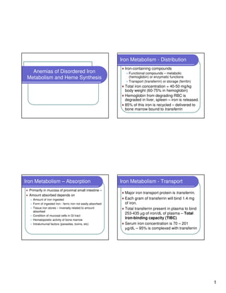
  • 1. Iron Metabolism - Distribution Anemias of Disordered Iron Metabolism and Heme Synthesis Iron-containing compounds Functional compounds – metabolic (hemoglobin) or enzymatic functions Transport (transferrin) or storage (ferritin) Total iron concentration = 40-50 mg/kg body weight (60-75% in hemoglobin) Hemoglobin from degrading RBC is degraded in liver, spleen – iron is released. 85% of this iron is recycled – delivered to bone marrow bound to transferrin Iron Metabolism – Absorption Primarily in mucosa of proximal small intestine – Amount absorbed depends on Amount of iron ingested Form of ingested iron - ferric iron not easily absorbed Tissue iron stores – inversely related to amount absorbed Condition of mucosal cells in GI tract Hematopoietic activity of bone marrow Intralumunial factors (parasites, toxins, etc) Iron Metabolism - Transport Major iron transport protein is transferrin. Each gram of transferrin will bind 1.4 mg of iron. Total transferrin present in plasma to bind 253-435 µg of iron/dL of plasma – Total iron-binding capacity (TIBC) Serum iron concentration is 70 – 201 µg/dL – 95% is complexed with transferrin 1
  • 2. Iron metabolism - Storage Transferrin is about 1/3 saturated with iron (% saturation = serum iron/TIBC X 100%) The reserve iron-binding capacity of transferrin is called the unsaturated iron-binding capacity or UIBC (UIBC = TIBC – serum iron) Most iron bound to transferrin comes from the breakdown of hemoglobin Excess iron deposited in tissues Major storage depot is liver Stored as ferritin and hemosiderin Ferritin primary storage compound for body’s need Readily released for heme synthesis Small amounts found in blood – parallels the storage iron in the body Iron Metabolism - Requirements Hemosiderin Major long term storage form of iron Slow release Estimation of hemosiderin made on bone marrow tissue sections Body iron conserved through reutilization – only about 1 mg lost per day Iron deficiency occurs when negative iron balance Increased requirements Inadequate diet Malabsorption Iron overload when Increased absorption Multiple transfusions Iron injections 2
  • 3. Increased Iron Requirements Menstruation – menstruating females have twice the daily requirements as males (2 mg/day) Pregnancy – 3.4 mg/day Infancy/childhood – rapid growth From: http://www.cdc.gov/hemochromatosis/training/ pathophysiology/iron_cycle_popup.htm At birth, enough iron stores for 4-5 months Milk is poor source of iron Iron supplementation recommended Laboratory Assessment of Iron Serum iron levels Total iron binding capacity Calculation of % saturation of transferrin Serum ferritin Ferrokinetic studies (not done routinely)monitor movement of 59Fe from plasma to bone marrow and into circulating RBCs. Rate at which iron leaves plasma is PIT (plasma iron turnover) – good indication of total erythropoiesis Are at which iron incorporated into circulating erythrocytes is EIT (erythrocyte iron turnover) – measure of effective erythropoiesis- correlates with RPI 3
  • 4. Iron deficiency Anemia (IDA) Normal Values iron: 60-170 µg/dl TIBC: 240-450 µg/dl transferrin saturation: 20-50% Most common nutritional deficiency in the world Symptoms General anemia symptoms – Shortness of breath on exertion Lethargy Heart palpitations Mucosal atrophy -glossitis, cheilitis, dysphagia “Spoon nails” - koilonychia Pica Etiology vs. Pathophysiology Etiology is the cause or origin of a disorder. Pathophysiology is the functional alteration in a disease or a defect. Etiology of IDA Dietary Deficiency – rarely the cause in developed countries (except in infancy, pregnancy, adolescence) Blood Loss – Adult males have about 8 years of storage iron. Deficiency if chronic blood loss – e.g. gastrointestinal or genitourinary bleeding Intravascular hemolysis Malabsorption 4
  • 5. Pathophysiology of IDA Diminished total body iron content, developing in stages over a period of negative iron balance Iron depletion – Stage One Iron deficient erthyropoiesis – Stage Two Iron deficiency anemia – Stage Three Stage Two Insufficient iron to insert into protoporphyrin ring to form heme – Protoporphyrin accumulates in cell and complexes with zinc to form ZPP No anemia, no hypochromia, but slight microcytosis RPI may be decreased Stage One Iron storage is exhausted - indicated by decrease in serum ferritin levels No anemia – RBC morphology is normal May have elevated RDW Stage Three All laboratory tests for iron status become abnormal Most significant finding is microcytic, hypochromic anemia 5
  • 6. Laboratory findings – Fe deficiency Microcytic/hypochromic anemia – microcytosis first MCV = 55-74 fl MCHC = 22-31 % MCH = 14-26 pg Hgb < 10 g/dL Increased RDW RPI < 2.0 (ineffective erythropoiesis) Poikilocytosis (target cells, elliptocytes, dacryocytes) May be thrombocytosis serum ferritin Serum iron (<30 µg/dL); TIBC; % saturation (<16 %) Therapy Treat underlying disorder Administer iron and observe response Oral administration of ferrous sulfate Increase of 1 gm/hemoglobin per month Reticulocyte count peaks at about 8 to 10 days (4-10%) Anemias Caused by Abnormal Iron Metabolism Sideroblastic anemias Anemia of chronic disease 6
  • 7. Sideroblastic anemia Characterized by Increase in total body iron Presence of ringed sideroblasts in bone marrow Hypochromic anemia Classification Hereditary form Acquired form (more common) Idiopathic – Refractory anemia with ringed sideroblasts (RARS) secondary Hereditary Sideroblastic Anemia Pathophysiology Disturbances of enzymes regulating heme synthesis Ringed sideroblasts form when nonferritin iron accumulated in the mitochondria that circle the normoblast nucleus Most common form is sex-linked and due to an abnormal δ-aminolevulinate synthase enzyme (ALAS) Decreased heme synthesis due to block in iron utilization – perceived by body as increased need for iron Increased iron absorption results in iron overload 7
  • 8. Acquired Sideroblastic Anemia RARS (Refractory anemia with ringed sideroblasts) – acquired stem cell disorder Secondary Lead poisoning (plumbism) Associated with hyperactivity, low IQ, concentration disorders in children Inhibits cellular enzymes involved in heme synthesis Alcoholism – alcoholics frequently have anemias due to one or more causes Malignancy Bone marrow Erythroid hyperplasia - megaloblastosis Ringed sideroblasts in 50% of normoblasts Laboratory findings in SA – Peripheral Blood Moderate to severe anemia – dimorphic (mild macrocytosis prevalent in RARS and alcoholism) Target cells Pappenheimer bodies Basophilic stippling – coarse basophilic stippling characteristic in lead poisoning ↑ Fe, N to ↓ TIBC, ↑ % saturation, ↑ ferritin Therapy Pyroxidine – hereditary form (50% response) Folic acid if megaloblastoid features Phlebotomy/chelation therapy to remove excess iron 8
  • 9. Anemia of Chronic Disease Anemia that occurs in patients with chronic infections, chronic inflammatory disorders, or neoplastic disorders not due to bleeding, hemolysis, or marrow involvement (renal, endocrine, or hepatic insufficiencies are excluded) Low serum iron, normal iron stores Laboratory findings Mild anemia (not less than 9 g/dL) Normocytic, normochromic (some are normocytic, hypochromic) RPI<2 ↑ Fe (10-70 µg/dL), N to ↓ TIBC (100300 µg/dL), N to ↓ % saturation (1025%), N or ↑ ferritin Pathophysiology Impaired marrow response to anemia – cytokines produced as result of immune response inhibit EPO production Block in iron release from macrophage Shortened erythrocyte survival Hemochromatosis Not an anemia – but will be discussed because of abnormal iron studies Parenchymal tissue damage from iron overload – excess iron stored in macrophages and hepatocytes, cardiac cells, endocrine cells 9
  • 10. Recessive genetic disorder Prevalence of 1 in 200-250 persons 1:10 Caucausians in US are carriers Abnormal protein produced that binds to transferrin receptor on cells – Clinical findings Chronic fatigue Diabetes Cirrhosis Hyperpigmentation of skin Laboratory findings Criteria for diagnosis: >50% transferrin saturation in females; >60% in males Treatment Phlebotomy –each unit of blood removes about 250 mg of iron Porphyrias Group of inherited disorders characterized by block in porphyrin synthesis Lack of enzyme in pathway of heme synthesis Heme precursors accumulate in tissues – excreted in urine and/or feces Symptoms 2 forms depending on primary site of defective porphyrin metabolism Heptatic Erythropoietic – (we will discuss these because they affect the erythrocytes) Photosensitivity Abdominal pain neuropathy 10
  • 11. Pathophysiology of EP Abnormality of heme biosynthetic pathway within the normoblasts 2 types Congenital erythropoietic porphyria (CEP) Erythropoietic protoporphyria (EEP) Porphyrinogens are the precursors or porphyrins Series III – precursors of heme Series I - functionless Erythropoeitic Protoporphyria Overproduction of protoporphyrin No anemia present – excess protoporphyrin IX builds up – leaks into skin dermal capillaries Congenital Erythropoietic Porphyria Excessive amounts of series I porpyrins ? Enzyme defect in uroporpyringoen III cosynthase so porphyrinogens are channeled to series I isomer Excess porpyrins deposited in body tissues – Hemolytic anemia - ? Result of excess porphyrins in RBC Clinical Features CEP – rare autosomal recessive disorder (only ~100 cases reported) Pink to reddish brown urine Excess porphyrins- extreme photosensitivity Vesicular eruptions on skin exposed to sunlight Hypertrichosis EEP – autosomal dominant (some autosomal recessive inheritance) – ~300 cases reported 11
  • 12. Laboratory Features CEP Mild to severe N/N anemia with anisocytosis and poikilocytosis Polychromasia with ↑ reticulocytes – RBCs fluoresce with UV light EEP No abnormalities on routine testing 12