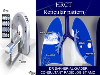
HRCT Reticular pattern
•Download as PPT, PDF•
39 likes•8,541 views

Describes the basic radiology of diffuse interstitial disease ,with differential diagnosis of reticular interstitial pattern and how to approach HRCT findings .

Recommended
More Related Content
What's hot
What's hot (20)
Similar to HRCT Reticular pattern
Similar to HRCT Reticular pattern (20)
More from Sakher Alkhaderi
More from Sakher Alkhaderi (18)
Recently uploaded
Recently uploaded (20)
HRCT Reticular pattern
- 1. HRCT Reticular pattern DR SAKHER-ALKHADERI CONSULTANT RADIOLOGIST AMC
- 2. INTRODUCTIONINTRODUCTION • HRCT -- Use of thin section CT images (0.625 to 2 mm slice thickness) often with a high-spatial- frequency reconstruction algorithm to detect and characterize disease affecting the pulmonary parenchyma and airways. • Superior to chest radiography for detection of lung disease, points a specific diagnosis and helps in identification of reversible disease. 2
- 3. 3
- 4. Thin section produces better contrast between lung parenchyma and bronchus and pulmonary vessel. A scan obtained with increased slice thickness, produces volume averaging with blurring of pathological details.
- 5. The division of trachea gives rise to the left and right mainstream bronchi, which further divides into lobar and segmental bronchi. Segmental bronchi divides after 6 to 20 division they no longer contain cartilage in their walls and are referred to as bronchioles.
- 6. There are approximately 23 generation of dichotomous branching From trachea to the alveolar sac HRCT can identify upto 8th order central bronchioles 6
- 8. SUBJECTS Anatomy of the secondary lobule Basic HRCT patterns Distribution of abnormalities Differential diagnosis of interstitial lung diseases
- 9. Secondary lobule • The secondary lobule is the basic anatomic unit of pulmonary structure and function. Interpretation of interstitial lung diseases is based on the type of involvement of the secondary lobule. It is the smallest lung unit that is surrounded by connective tissue septa. It measures about 1-2 cm and is made up of 5-15 pulmonary acini, that contain the alveoli for gas exchange.
- 10. Secondary lobule Basic anatomic unit of pulmonary structure and function. 1-2 cm and is made up of 5-15 pulmonary acini Supplied by a small bronchiole (terminal bronchiole) in the center, that is parallelled by the centrilobular artery. Pulmonary veins and lymphatics run in the periphery Two lymphatic systems: central network peripheral network
- 11. Secondary lobule
- 12. • The secondary lobule is supplied by a small bronchiole (terminal bronchiole) in the center, that is parallelled by the centrilobular artery. Pulmonary veins and lymphatics run in the periphery of the lobule within the interlobular septa. Under normal conditions only a few of these very thin septa will be seen.
- 13. There are two lymphatic systems: a central network, that runs along the bronchovascular bundle towards the centre of the lobule and a peripheral network, that is located within the interlobular septa and along the pleural linings.
- 14. The terminal bronchiole in the center divides into respiratory bronchioli with acini that contain alveoli. Lymphatics and veins run within the interlobular septa
- 15. Centrilobular area It is the central part of the secondary lobule. It is usually the site of diseases, that enter the lung through the airways ( i.e. hypersensitivity pneumonitis, respiratory bronchiolitis, centrilobular emphysema ).
- 16. Centrilobular area in blue perilymphatic area in yellow
- 17. Perilymphatic area Perilymphatic areais the peripheral part of the secundary lobule. It is usually the site of diseases, that are located in the lymphatics of in the interlobular septa ( i.e. sarcoid, lymphangitic carcinomatosis, pulmonary edema). These diseases are usually also located in the central network of lymphatics that surround the bronchovascular bundle.
- 18. Raoof, S. , CHEST 2006; 129:805
- 20. 20
- 21. A group of terminal bronchioles 21
- 23. Surrounded by lymph vessels 23
- 26. 26 Connective Tissue StromaConnective Tissue Stroma
- 28. 28
- 30. In chest radiology, reticular and linear opacification refers to a broad sub-group ofpulmonary opacification caused by a decrease in the gas to soft tissue ratio caused by a pathological process centred in and around the pulmonary interstitium. This includes thickening of any of the interstitial compartments by blood, water, tumour, cells, fibrous disease or any combination fine "ground-glass" (1-2 mm): seen in processes that thicken the pulmonary interstitium to produce a fine network of lines, e.g. interstitial pulmonary oedema medium "honeycombing" (3-10 mm): commonly seen in pulmonary fibrosis with involvement of the parenchymal and peripheal interstitium coarse (> 10 mm): cystic spaces caused by parenchymal descruction, e.g. usual interstitial pneumonia, pulmonary sarcoidosis, pulmonary Langerhans cell histiocytosis
- 32. Focal irregular septal thickening in lymphangitic carcinomatosis Lymphangitic Carcinomatosis results from hematogenous spread to the lung, with subsequent invasion of interstitium and lymphatics. The presenting symptoms are dyspnea and cough and can predate the radiographic abnormalities. In many cases however the patients are asymptomatic. Lymphangitic Carcinomatosis is seen in carcinoma of the lung, breast, stomach, pancreas, prostate, cervix, thyroid and metastatic adenocarcinoma from an unknown primary.
- 37. usual interstitial pneumonia / idiopathic pulmonary fibrosis (UIP/IPF) non-specific interstitial pneumonia (NSIP) cryptogenic organizing pneumonia (COP): formerly bronchiolitis obliterans organizing pneumonia (BOOP) respiratory bronchiolitis–associated interstitial lung disease (RB-ILD) desquamative interstitial pneumonia (DIP) lymphoid interstitial pneumonia (LIP) acute interstitial pneumonia (AIP): the only acute process in the list
- 38. Typical UIP
- 39. Usual interstitial pneumonia Usual interstitial pneumonia (UIP) is a form of lung disease characterized by progressive scarring of both lungs.[1] The scarring (fibrosis) involves the supporting framework (interstitium) of the lung. UIP is thus classified as a form of interstitial lung disease. The term "usual" refers to the fact that UIP is the most common form of interstitial fibrosis. "Pneumonia" indicates "lung abnormality", which includes fibrosis and inflammation. A term previously used for UIP in the British literature is cryptogenic fibrosing alveolitis, a term that has fallen out of favor since the basic underlying pathology is now thought to be fibrosis, not inflammation. Location: distribution The distribution of UIP on CT images is typically characteristically with an apico-basal gradient with basal and peripheral predominance, although it is often patchy. Typical features include 1,5 : the presence of reticular opacities in the immediate subpleural lung, often associated with honeycombing and/ or traction bronchiectasis,
- 42. Traction bronchiectasis Bronchial dilatation occurring as a consequence of interstitial fibrosis is referred to as traction bronchiectasis (Figure 5). The bronchi often appear irregular (corkscrewed) and are not associated with radiologic evidence of bronchial inflammation (gross bronchial wall thickening or mucous impaction). Traction bronchiectasis is often accompanied by other signs of lung fibrosis (honeycombing or irregular reticulation). While traction bronchiectasis is quite specific for fibrosis, the differential diagnosis is broader than that of honeycombing. Idiopathic pulmonary fibrosis (IPF) is commonly associated with traction bronchiectasis. However, in the absence of honeycombing, other diseases are more likely (Chart 3). In patients with known collagen vascular disease, bibasilar, peripheral, traction bronchiectasis accompanied by ground-glass attenuation can be considered diagnostic of NSIP. When the circumstances are less diagnostic, a surgical biopsy might be required.
- 44. Honeycombing Honeycomb lung remodeling (honeycombing) reflects the end stage of a number of diseases that cause parenchymal destruction. It presents a characteristic HRCT pattern, with subpleural, thick- walled cysts that share walls and, when advanced, are often stacked in multiple layers (Figure 6). It is typically accompanied by other signs of fibrosis (traction bronchiectasis and reticulation). Honeycombing is highly suggestive of a pathologic diagnosis of usual interstitial pneumonia (UIP), although it can be attributable to other diseases (Chart 3). Honeycombing seen on HRCT scans is often considered diagnostic of UIP in patients presenting the appropriate clinical profile, and the majority of such patients will not be subjected to surgical lung biopsy. Because bilateral honeycombing on HRCT scans is considered diagnostic under these conditions, it is vitally important for the radiologist to be confident that honeycombing is truly present before describing it.
- 45. NSIP
- 46. Non-specific interstitial pneumonia -fibrotic non specific interstitial pneumonia: more common -cellular non specific interstitial pneumonia: less common Prognosis is much better when compared with UIP with 90% 5 years survival rate for cellular and 45-90 % 5 years survival in fibrotic subtype. Common manifestations include: ground-glass opacities combined with irregular linear or reticular opacities tends to be a dominant feature: can be symmetrically or diffusely distributed in all zones or display a basal predominance there can be relative subpleural sparing 11 - relatively specific sign reticular opacities (sometimes - minor subpleural reticulation) irregular linear opacities: with NSIP with fibrosis 6-7 thickening of bronchovascular bundles: with NSIP with fibrosis 6 scattered micronodules in advanced disease traction bronchiectasis consolidation microcystic honeycombing
- 48. Cardiogenic pulmonary edema (CPE) is defined as pulmonary edema due to increased capillary hydrostatic pressure secondary to elevated pulmonary venous pressure. CPE reflects the accumulation of fluid with a low-protein content in the lung interstitium and alveoli as a result of cardiac dysfunction .
- 54. Drug toxicity disease can result in DILD, with histopathologic reactions ranging from acute injury to UIP-like fibrotic patterns.(36) The mechanisms of drug-induced lung injury vary from cytotoxicity to hypersensitivity A wide variety of therapy-related reactions have been described as a consequence of chemotherapeutic agents (bleomycin, busulfan, chlorambucil, cyclophosphamide, 1,3- bis(2-chloroethyl)-1-nitrosourea, and 1-(2-chloroethyl)-3- cyclohexyl-1-nitrosourea), statins, amiodarone, nitrofurantoin, methotrexate
- 55. All of the named rheumatic diseases can produce lung fibrosis. Rheumatoid arthritis and scleroderma are predominately implicated in cases where a UIP HRCT pattern is seen, and with similar functional abnormalities.
- 56. Radiation-induced lung disease (RILD) is a frequent complication of radiotherapy to the chest for chest wall or intrathoracic malignancies and can have a variety of appearances, especially depending on when the patient is imaged. Acute and late phases are described, corresponding to radiation pneumonitis and radiation fibrosisrespectively. These occur at different times after completion of radiotherapy and have different imaging features and differential diagnoses.
- 57. -Hypersensitivity pneumonitis -Lymphoid pulmonary lesions -Fibrosing sarcoidosis -Asbestosis DDX
- 58. THE END
Editor's Notes
- Thin section produces better contrast between lung parenchyma and bronchus and pulmonary vessel. A scan obtained with increased slice thickness, produces volume averaging with blurring of pathological details.
- The division of trachea gives rise to the left and right mainstream bronchi, which further divides into lobar and segmental bronchi. Segmental bronchi divides after 6 to 20 division they no longer contain cartilage in their walls and are referred to as bronchioles.
- Unit of lung (0.5-3 cm) Irregularly polyhedral متعدد السطوح Supplied by a group of terminal bronchioles and accompanying pulmonary arterioles surrounded by lymph vessels Demarcated by “interlobular septa” pulmonary veins pulmonary lymphatics connective tissue stroma
- Unit of lung (0.5-3 cm) Irregularly polyhedral متعدد السطوح Supplied by a group of terminal bronchioles and accompanying pulmonary arterioles surrounded by lymph vessels Demarcated by “interlobular septa” pulmonary veins pulmonary lymphatics connective tissue stroma
- Unit of lung (0.5-3 cm) Irregularly polyhedral متعدد السطوح Supplied by a group of terminal bronchioles and accompanying pulmonary arterioles surrounded by lymph vessels Demarcated by “interlobular septa” pulmonary veins pulmonary lymphatics connective tissue stroma
- Unit of lung (0.5-3 cm) Irregularly polyhedral متعدد السطوح Supplied by a group of terminal bronchioles and accompanying pulmonary arterioles surrounded by lymph vessels Demarcated by “interlobular septa” pulmonary veins pulmonary lymphatics connective tissue stroma
- Unit of lung (0.5-3 cm) Irregularly polyhedral متعدد السطوح Supplied by a group of terminal bronchioles and accompanying pulmonary arterioles surrounded by lymph vessels Demarcated by “interlobular septa” pulmonary veins pulmonary lymphatics connective tissue stroma
- Unit of lung (0.5-3 cm) Irregularly polyhedral متعدد السطوح Supplied by a group of terminal bronchioles and accompanying pulmonary arterioles surrounded by lymph vessels Demarcated by “interlobular septa” pulmonary veins pulmonary lymphatics connective tissue stroma
- Unit of lung (1 cm to 1 inch) Irregularly polyhedral Supplied by a group of terminal bronchioles and accompanying pulmonary arterioles surrounded by lymph vessels Demarcated by “interlobular septa” pulmonary veins pulmonary lymphatics connective tissue stroma