Radiotherapy in the Treatment of Sarcomas in Adolescents and Young Adults
•
9 likes•1,671 views

Daniel Indelicato, MD, University of Florida, Jacksonville, FL Presented at the 2010 Texas Adolescent and Young Adult Oncology Conference, Methodist Healthcare-San Antonio

Recommended
More Related Content
What's hot
What's hot (20)
Viewers also liked
Viewers also liked (13)
Similar to Radiotherapy in the Treatment of Sarcomas in Adolescents and Young Adults
Similar to Radiotherapy in the Treatment of Sarcomas in Adolescents and Young Adults (20)
More from Methodist HealthcareSA
More from Methodist HealthcareSA (20)
Recently uploaded
Recently uploaded (20)
Radiotherapy in the Treatment of Sarcomas in Adolescents and Young Adults
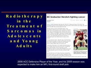
- 1. Radiotherapy in the Treatment of Sarcomas in Adolescents and Young Adults 2008 ACC Defensive Player of the Year, and his 2009 season was expected to make him an NFL first-round draft pick
- 2. No conflicts of interest to disclose
- 3. Scope of Problem in AYAs Sarcomas 15% Leukemia 14% Germ Cell Tumors 13% Brain Tumors 10% Thyroid Carcinoma 8% Melanoma 8% Other 9% Lymphoma 23% Bone sarcoma 8% Soft tissue sarcoma 7%
- 4. How Radiation is Perceived Seminars in Oncology, 2009 Number of times “radiation” is mentioned relative to side effects: 11 Number of times “radiation” is mentioned as a treatment modality: 7
- 8. Value of RT for Adolescent and Young Adult Bone Sarcomas Remember: Sarcoma type determines radiosensitivity, disease control outcomes
- 13. Value of RT for Adolescent and Young Adult Soft Tissue Sarcomas
- 14. “ The management of this condition should be radical surgery and that no help should be expected from radiotherapy either as a pre- or postoperative procedure” - Dr. Ralston Paterson (1963) Holt Radium Institute at the Christie Cancer Hospital, founded by Ralston Patterson Soft Tissue Sarcoma: Then
- 15. Annals of Surgery, 1982 27 pts WLE+RT 16 pts Amputation 43 pts High grade STS Extremity
- 16. Level I Data: General Population
- 22. Bone Tumors Soft Tissue Sarcoma
- 23. Challenges of Using Radiation for AYA Sarcoma Remember: Sarcoma location dictates treatment side effects and complications
- 26. Local Management of Lower Extremity Ewing Tumors at the University of Florida: 1970-2006 (Indelicato 2007)
- 27. 1987 1969 65 Gy 60 Gy + 20 years
- 31. Musculoskeletal Complications of Radiation Treatment
- 32. Bone Complications of Radiation Treatment Traditional risk factors for fracture: ● radiation dose (mean dose >37 Gy) ● periosteal stripping ● female gender Pathogenesis: microvascular supply; osteoclast alteration? Rate of fracture in patients with Ewing sarcoma of weight bearing bone: 30% Slipped femoral capital epiphysis radiation injury to the proximal femoral growth apparatus + weight-bearing stress After only 12 Gy
- 42. Guidelines: Minimizing Toxicity in AYA Sarcoma Male with synovial sarcoma of the proximal medial thigh Protons Photons
- 44. "He hasn't lost anything“ - ESPN commentator Curt Warner 9/6/2010
Editor's Notes
- In 1970’s, 30-40% extremity sarcoma’s were treated with amputation Now, its <15%
- AP view of a knee in a teenager treated with 54.8 Gy for a synovial sarcoma Transverse pathologic fracture of the paroximal tibial metaphysis 4 years later, healed fracture but extensive radiation osteitis of the distal femur and proximal tibia 9 years later, osteosarcoma developed in the proximal tibia
- AP view of the knee of a Ewing sarcoma patient shows growth impairment with metasphseal widening and sclerosis 14 months laters, the distal femoral metaphysis is markedly sclerotic and irregegular. The epiphyseal cartilage plate appears hypertrophied.
- Teenager treated for Ewing sarcoma of the right humerus, photo 11 years post RT. Note difference in the muscule development when he started lifting weights.
- Maynard