Chandigarh Call Girls Service ❤️🍑 9809698092 👄🫦Independent Escort Service Cha...
Penile doppler a review
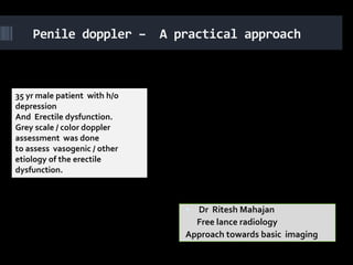
1. Penile doppler – A practical approach
35 yr male patient with h/o
depression
And Erectile dysfunction.
Grey scale / color doppler
assessment was done
to assess vasogenic / other
etiology of the erectile
dysfunction.
Dr Ritesh Mahajan
Free lance radiology
Approach towards basic imaging
3. Penile vascular anatomy……….
Internal pedundle artery
through bulbar artery supplies Venous drainage is through
base of the penis . Penile efferent venules – emisssary
artery divides into two veins - dorsal veins . Base of
cavernosal arteries and the penis through crural
continues as dorsal artery . veins drains into the
There are helicine arteries that periprostatic venous plexus
run through the substance of in to the internal iliac veins .
the corpora .
The glans region has it’s
Cavernosal arteries are drainage into the external
paramedian in location. iliac venous system.
Cavernosal and dorsal
Penile venous system is
arteries show more
variability than venous more constant than the
drainage of the penis . arterial anatomy.
4. Basis of normal erection……….
Flaccid state : Intracavernosal arterial
After neural impulse resistance is high . Cavernosal arterial flow
has low systolic, dampened diastolic flow .
Vasodilatation After giving vasoactive agent : Increased
Increased blood supply dilatation of the cavernosal arterial tree is
there with increased systolic and diastolic
Increased intracavernosal component of the flow and velocities.
pressure There is sinusoidal expansion of the arterial
Efferent venous channel are flow with obstructed venous egress
obstructed by taut tunica Further rise in cavernosal pressure leads to
systolic dampening and loss of diastolic
albuginea. component .
On doppler study With rigid erection – there is near total loss
predictable spectral of diastolic flow and at times reversal .
waveform corrborates with As far as venous flow is concerned : flaccid
changes in the intra state has sluggish flow. With vasoactive
cavernosal pressure . agent there is increase in the dorsal venous
flow and with rigid erection the venous
flow can stop . Retrograde venous flow is
also appreciated in normal individuals.
5. Basis of normal erection……
Phases of erection …………. After neural impulse there is
rise in the intracavernosal
presssure –There is cavernosal
Flaccid arterial dilatation and rise in
the systolic and diastolic flow
Latent . The dorsal venous flow also
rises initially . With rise in the
Tumescent cavernosal pressure –
distended sinusoids abut the
Rigid tunica albuginea and this
Detumescence leads to cessation of the
venous egress and leads to
rigid erection. With rigid
erection ,this diastolic
component of the cavernosal
arterial flow is lost and at
times reverses also .
6. Penile imaging ………………………..
ERECTILE DYSFUNCTION
ETIOLOGY
Psychogenic
Endocrine
Pharmacological
Neurological
Vascular
Organic etiology – Vasogenic
etiology is important and
penile Doppler assessment
can be of use to ascertain
the same .
7. Penile imaging …………………………….
Diagnostic work up for erectile
dysfunction Penile anatomy
Medical / drug history . Three distensible corpora
chambers -
Routine / endocrine blood 1. Corpora spongiosum enveloping
analysis. the urethera. This does not play
significant role in erection.
Non invasive testing 2. Corpora cavernosa – dorsal in
position –paired .
Brachial – penile indices Mid line septum separates the
Nocturnal penile two corpora cavernosa . Thick
fascia (tunica albugenia) encircles
tumescence. the corpora cavernosa and bucks
fascia covers corpora cavernosa
and spongiosa .
8. Basic methodology of penile doppler
Linear transducer parallel Grey scale assessment
to skin surface is used . involves assessment of
Both ventral and dorsal echogenic tunica
transducer position albuginea. Midlevel echoes
approaches can be used. of the corpora cavernosa .
Slow flow detection Assess mid line septum .
settings are to be used. Cavernosal arteries are
Longitudinal and assessed by echogenic
parasagitttal image walls and with paramedian
acquisition is to be done . location.
9. Brief about doppler examination…….
Complete discussion of Velocity measurements are
the examination with the done along the base of the penis
. Angle of assessment <60
patient is to be done . degree.
Assessment of the privacy PSV, EDV, RI , PI is done for
cavernosal arteries on either side
is to be done . .
Quiet examination setting Look for cavernosal artery
stenosis , occlusion, retrograde
is necessary . arterial flow , dampened spectral
flow.
Pharmacological agents :
Cavernosal artery dilatation
papaverine, phentolamine, <75% of the base arterial
prostaglandin E diametre is indirect e/o
vasogenic etiology of erectile
Eye technique : visual dysfunction.
inspection is important .
10. Grey scale sonography…..
Grey scale sonography
Good for assessment of
Peyronie’s disease.
Penile trauma
Penile neoplasm
11. venous insufficiency………
Variations ………………….. Venous insufficiency
Most common form of
Absence of the penile impotence
artery : +_ cause of the EDV > 5cm/sec suggests venous
impotence . incompetence .
PSV > 30 cm/sec helps to rule
Corpora cavernosa - out arterial etiology and search
corpora spongiosum for venous etiology has to be
sorted out .
collaterals , dorsal venous EDV > 2 to 6cm/sec supports
and corpora collaterals venous insufficiency .
should also be assessed. Instead of measuring EDV : RI (
<.8) , PI (<4) also support
venous insufficiency as etiology
of erectile dysfunction.
12. Grey scale and basic
doppler assessment
BASIC COLOR DOPPLER ASSESMENT –
GREY SCALE DONE AT BASE OF THE PENIS
PLAQUE / CALCIFICATION. Imaging especially for
doppler is done along the
MID LEVEL ECHOES OF
base of the penis .
CORPORA CAVERNOSA
The sequence of the
TUNICA ALBUGENIA imaging is as following :
/BUCKS FASCIA 1. Flaccid state
2. Papaverine injection
3. Post injection imaging is
done at 5 , 10,15,20,25
minutes .
13. PARAMETRES TO BE ASSESED IN
THE FLACCID STATE
Dorsal vein diameter
Cavernosal artery ( both left
and left artery )
1. Diametre
2. PSV
3. EDV
4. PI
5. Dorsal cavernosal collaterals
6. Cavernosal spongiosal collaterals
14. PARAMETRES TO BE ASSESED POST
PAPAVERINE INJECTION
Post papaverine
Dorsal vein diameter
injection
Cavernosal artery ( both left
5minutes
10minutes and left artery )
15 minutes 1. Diametre
20 minutes 2. PSV
25 minutes 3. EDV
4. PI
15. INTERPRETATION
PSV Rt cavernosal artery
PSV left cavernosal artery
Difference between the PSV on either side
( should not be more than 10cm/sec).
Diastolic flow loss
DIASTOLIC REVERSAL
Persistence of the dorsal venous flow
16. NORMAL VALUES
Corpora cavernosal artery PSV values :
1. PSV : 35 cm/sec : Normal
2. PSV : 25-35 cm/sec : indeterminate
3. PSV : <25 cm/sec : Abnormal
Venoocclusive incompetence
1. No diastolic flow loss
2. No diastolic flow reversal
3. EDV ( Cavernosal artery): 2 to 6 cm/sec
4. RI ( Cavernosal artery) < .8
5. PI (cavernosal artery) <4
17. PRECAUTIONS
Inject papaverine only once
Keep region of injection pressed
Use insulin syringe
Alcohol swab to clean
Keep watch for priapism ( urologist
/anesthetist support ) .
18. Flaccid state
Flaccid state assessment of the
Dorsal vein diametre dorsal vein
19. Flaccid state
Flacid state assesment of the corpora / Cavernosal artery on either
bucks fascia / intercavernosal connection side diametre assesment
Sagittal / axial images
20. Flaccid state – cavernosal
artery Left cavernosal artery flaccid state –
appreciate relatively high resistance
Rt cavernosal artery flaccid state – appreciate
relatively high resistance flow no diastolic flow no diastolic component
component
21. Ancilliary findings
No e/o dorsal cavernosal collaterals . No
e/o cavernosal spongiosal collaterals
22. Injection of the papaverine injection in the
left corpora cavernosa
INSULIN SYRINGE
USED
INJECTION DONE IN
LEFT CORPORA
CAVERNOSA
GUIDED INJECTION
DONE AVOIDING THE
LEFT SIDE
CAVERNOSAL ARTERY
ANESTHETIST WAS
INVOLVED IN THE
INTERVENTION .
ALCOHOL SWAB WAS
USED .
PRECAUTIONS WERE
TAKEN TO AVOID SPILL.
23. 5 MINUTES AFTER INJECTION
CAVERNOSAL ARTERIES ON EITHER APPRECIATED THE SURGE IN SYSTOLIC
SIDE AFTER PAPAVERINE INJECTION FLOW AND DIASTLOLIC FLOW
24. 5 MINUTES AFTER INJECTION
DORSAL VEIN FLOW AFTER DORSAL VEIN DIAMETRE
FIVE MINUTES AFTER 5 MINUTES
25. 10 MINUTES AFTER INJECTION
CAVERNOSAL ARTERIES ON EITHER SIDE 10 APPRECIATED THE SURGE IN SYSTOLIC
minutes AFTER PAPAVERINE INJECTION FLOW AND DIASTLOLIC FLOW
26. 10 MINUTES AFTER INJECTION
DORSAL VEIN FLOW AFTER DORSAL VEIN DIAMETRE
TEN MINUTES AFTER 10 MINUTES
28. 15 MINUTES AFTER INJECTION
DORSAL VEIN FLOW AFTER DORSAL VEIN DIAMETRE
fifteen MINUTES AFTER 15 MINUTES
29. 15 MINUTES AFTER INJECTION
CAVERNOSAL ARTERIES ON EITHER SIDE AFTER 15
APPRECIATE THE SURGE IN SYSTOLIC
minutes of PAPAVERINE INJECTION FLOW AND DIASTLOLIC FLOW
30. 20 MINUTES AFTER INJECTION
DORSAL VEIN FLOW AFTER DORSAL VEIN DIAMETRE
twenty MINUTES AFTER 20 MINUTES
31. 20 MINUTES AFTER INJECTION
CAVERNOSAL ARTERIES ON EITHER SIDE 20 APPRECIATED THE SURGE IN SYSTOLIC
minutes AFTER PAPAVERINE INJECTION FLOW AND DIASTLOLIC FLOW
32. 25 MINUTES AFTER INJECTION
DORSAL VEIN FLOW AFTER DORSAL VEIN DIAMETRE
twenty five MINUTES AFTER 25 MINUTES
33. 25 MINUTES AFTER INJECTION
CAVERNOSAL ARTERIES ON EITHER SIDE 5
minutes AFTER PAPAVERINE INJECTION Diastolic loss
34. 30 MINUTES AFTER INJECTION
DORSAL VEIN FLOW AFTER DORSAL VEIN DIAMETRE
thirty MINUTES AFTER 30 MINUTES
35. 30 MINUTES AFTER INJECTION
CAVERNOSAL ARTERIES ON EITHER SIDE thirty
minutes AFTER PAPAVERINE INJECTION Diastolic loss