Clinical Cases in Kidney Disease - sample chapter
•
0 likes•825 views

This book explores important clinical problems in general nephrology, dialysis and transplantation, using a case-based approach. Together the 32 chapters form a comprehensive collection of clinical vignettes in renal medicine. Each chapter covers a separate clinical problem, posing multiple-choice questions and providing a detailed explanation for each. Clinical Pearls highlight the key issues. Each graded into three disctinct levels, to provide pertinent information tailored to the reader's knowledge level.

Recommended
More Related Content
What's hot
What's hot (20)
Viewers also liked
Viewers also liked (20)
Similar to Clinical Cases in Kidney Disease - sample chapter
Similar to Clinical Cases in Kidney Disease - sample chapter (20)
Recently uploaded
Recently uploaded (20)
Clinical Cases in Kidney Disease - sample chapter
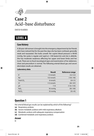
- 1. 25 Case 2 Acid–base disturbance DAVID HARRIS LEVEL A Case history A 28-year-old woman is brought into the emergency department by her friends who have noticed that for the past few days she has been confused, generally weak and nauseated. She looks unwell. Her supine blood pressure is 90/60 mmHg. Her pulse is regular and rapid at >100 beats/min and she is afebrile. She has moderate weakness affecting her upper and lower limbs and her trunk. There are no focal neurological signs and examination of her abdomen, chest and precordium is normal. The following arterial blood gas and serum electrolyte results are obtained. Laboratory data Result Reference range K+ 1.9 mmol/L (3.2–5.5) Na+ 132 mmol/L (136–146) Total CO2 8 mmol/L (24–31) Cl– 113 mmol/L (94–107) pH 7.10 (7.35–7.45) PaO2 95 mmHg (95–100) PaCO2 35 mmHg (35–45) Question 1 Her arterial blood gas results can be explained by which of the following? (a) Respiratory acidosis (b) Severe metabolic acidosis with mild respiratory alkalosis (c) Metabolic acidosis with adequate respiratory compensation (d) Combined metabolic and respiratory acidosis Answer D Harris Ch2.indd 25Harris Ch2.indd 25 13/11/07 11:48:52 AM13/11/07 11:48:52 AM
- 2. PART A FLUIDS AND ELECTROLYTES26A Explanation The patient is acidotic (pH 7.10). Partial pressure of both CO2 (PaCO2) and serum bicarbonate (total CO2) are less than normal, indicating a metabolic acidosis (answer A incorrect). With a pure respiratory acidosis, PaCO2 would be elevated, as would serum bicarbonate in anything but an acute respiratory acidosis (answer A incorrect). The respiratory compensation for a metabolic acidosis is hyperventilation causing reduction in blood PaCO2. PaCO2 should fall by 1 to 1.5 times the drop in serum bicarbonate which, with adequate respiratory compensation in this case, would mean that PaCO2 should fall from the normal value of 40 by 1 to 1.5 times (normal bicarbonate –8), or to between 16 and 24 mmHg. Another rule of thumb is that PaCO2 should fall to 100 times (pH – 7.0) or in this case to 10 mmHg. Thus, her PaCO2 has not fallen sufficiently, and so her respiratory compensation is inadequate (answer C incorrect). If she had combined metabolic acidosis and respiratory alkalosis, her CO2 should have been lower than predicted by the rules of thumb for metabolic acidosis with adequate respiratory compensation (answer B incorrect). The alveolar–arterial (A–a) gradient (FiO2 – PaO2 – PaCO2 × 1.2, which equals 13 in this case) is normal, indicating that her alveolar gas exchange is normal and there is no pulmonary disease. The hypoventilation in this case is probably due to muscle weakness caused by hypokalaemia. Therefore, if this patient did not have any metabolic acidosis, her respiratory acidosis would have been more obvious, and her CO2 would have been higher than normal due to hypoventilation (despite a near normal PaO2, which suggests her alveolar gas exchange is normal) (answer D correct). Clinical pearls • In metabolic acidosis pH, PaCO2 and HCO3 – (total CO2) should all fall. • The normal respiratory compensation for metabolic acidosis is hyperventilation, causing a fall in PaCO2. • PaCO2 should fall by 1 to 1.5 times the fall in serum bicarbonate. • PaCO2 should approximate 100 times (pH – 7.0). • A–a gradient will be normal (less than 15) with hypoventilation and normal lungs. • A–a gradient = FiO2 – PaO2 – PaCO2 ϫ 1.2. Question 2 The plasma anion gap in this case is consistent with: (a) addition of new anions to the body (b) pre-pyloric gastrointestinal fluid loss Harris Ch2.indd 26Harris Ch2.indd 26 13/11/07 11:48:53 AM13/11/07 11:48:53 AM
- 3. CASE 2 ACID–BASE DISTURBANCE 27 A (c) impaired urinary acidification (d) inhibition of carbonic anhydrase. Answer C and D Explanation The plasma anion gap is arbitrarily defined as the difference in plasma concen- tration between the positive ions sodium and potassium and the negative ions chloride and bicarbonate; most of the normal anion gap is comprised of the negative charge on albumin (and so changes in albumin concentration will alter the gap). The normal anion gap is less than 15. The anion gap in this case is 13. (Note that some definitions of anion gap do not include potassium, in which case the normal value would be lower.) The most common causes of normal anion gap metabolic acidosis are loss of bicarbonate from the gut (post-pyloric fluids which are alkaline, not pre-pyloric which are acidic, so answer B is incorrect) and proximal (one example of many is answer D) or distal (answer C correct) renal tubular acidosis. Accumulation of new unmeasured anions in the plasma, giving rise to a raised anion gap (e.g. with renal failure) occurs with addition of new acid from exogenous sources (e.g. toxic alcohols, salicylate) or endogenously (e.g. lactic acid, ketoacid) or failure of renal excretion of the anions of new acids (e.g. with renal failure) (answer A incorrect). This is shown in Figure 2.1. Figure 2.1 The anion gap in patients with normal acid–base balance, and those with metabolic acidosis Na+ ϩ K+ Cl– Na+ ϩ K+ Cl– Na+ ϩ K+ Cl– Na+ ϩ K+ Cl– UC Normal High AG metabolic adidosis Normal AG (hyperchloraemic) UA UC UA UC UA UC UA HCO3 – HCO3 – HCO3 – HCO3 – UC ϭ unmeasured cations UA ϭ unmeasured anions ϭ anion gap (AG) ϭ increased Cl concentration Harris Ch2.indd 27Harris Ch2.indd 27 13/11/07 11:48:53 AM13/11/07 11:48:53 AM
- 4. PART A FLUIDS AND ELECTROLYTES28A Clinical pearls • Plasma anion gap = Na+ + K+ – Cl– – HCO3 – , and is normally <15. • Normal anion gap metabolic acidosis commonly arises due to gastrointestinal bicarbonate loss or renal tubular acidosis. • Hypokalaemia with metabolic alkalosis can occur in many conditions, but when hypokalaemia coexists with metabolic acidosis, the cause is usually either diarrhoea or renal tubular acidosis. • Raised anion gap (>15) metabolic acidosis occurs due to addition of new acid (with a non-chloride anion) or renal failure. Question 3 The body’s defences against this acidosis include: (a) buffering by intracellular proteins (b) respiratory hypoventilation (c) increased renal titratable acid production (d) increased renal ammonium excretion. Answer A and D Explanation Intracellular and extracellular buffering occurs immediately. Extracellular buff- ering is effected predominantly by HCO3 – /CO2. While intracellular buffering likewise involves HCO3 – /CO2, it also involves proteins and phosphates (answer A correct). Asdiscussedpreviously,therespiratorycompensationisthatofhyperventilation, not hypoventilation, to blow off acid in the form of CO2 (answer B incorrect). The kidneys do not produce titratable acids (answer C incorrect); rather, they filter certain ions (mainly phosphates) which titrate H+ in the tubular lumen, to excrete a total of about 30 mmol per day. Normalammoniaproductionandammoniumexcretionbythekidneyisabout 40 mmol per day, and in the case of acidosis this can increase to 200 mmol per day or more (answer D correct). Ammonia is produced from glutamine enzymatically, mainlyintheproximaltubularcell;becauseoftheneedtomakemoreenzyme,there is a delay of several days before maximum ammoniagenesis can occur. Clinical pearl • Body defence against metabolic acidosis includes buffering, hyperventilation and increased renal ammonia production. Harris Ch2.indd 28Harris Ch2.indd 28 13/11/07 11:48:54 AM13/11/07 11:48:54 AM
- 5. CASE 2 ACID–BASE DISTURBANCE 29 A Question 4 At this stage, treatment for this patient should include: (a) artificial ventilation (b) potassium replacement (c) dialysis (d) bicarbonate administration (e) rehydration with normal saline. Answer B and E Explanation The patient’s potassium needs to be corrected to restore muscle power and normal ventilation (answer B correct). The patient is relatively hypotensive; for adequate intracellular buffering, normal tissue perfusion is required to remove CO2 from the periphery to the lungs for exhalation. In the case of poor tissue perfusion, CO2 accumulates within the cells, and so buffering instead involves proteins (‘bad’ buffering) thereby possibly altering their structure and function. Restoration of tissue perfusion with normal saline permits optimal intracellular buffering (answer E correct). Acidosis per se, of this severity, is not life-threatening and so there is no need for immediate correction with bicarbonate; moreover, bicarbonate administration may worsen hypokalaemia by driving potassium into cells (answer D incorrect). Bicarbonate therapy could be used after potassium replacement, especially when acidosis is severe. Artificial ventilation is not required at this stage because the patient is not in frank respiratory failure (answer A incorrect). Dialysis would correct the electrolyte abnormalities quickly, but so too would the simpler measures of potassium replacement (to improve ventilation) and volume resto- ration (to improve tissue buffering by delivering CO2 to the lungs). Moreover, the normal plasma anion gap indicates that there are no new potentially toxic unmeasured anions (of, for example, a toxic alcohol) to be removed by dialysis (answer C incorrect). Clinical pearl • Emergencies in metabolic acidosis relate to associated derangement of potassium, toxic alcohols and severity of acidosis. Harris Ch2.indd 29Harris Ch2.indd 29 13/11/07 11:48:54 AM13/11/07 11:48:54 AM
- 6. PART A FLUIDS AND ELECTROLYTES30B LEVEL B Case history (continued) Additional results are obtained on the patient. These include the following. Plasma creatinine 110 μmol/L (60–125) Plasma urea 15 mmol/L (2.5–6.5) Plasma osmolality 295 mosm/kg (275–295) Plasma glucose 6 mmol/L (3.6–7.7) Urinary pH 6.0 Question 5 Based on these additional results, which of the following is/are likely to explain the pathophysiology of the acidosis? (a) Ingestion of toxic alcohol (b) Diabetic ketoacidosis (c) Rapid metabolism of a toxic alcohol with rapid excretion of its metabolites (d) Rapid metabolism of a toxic alcohol without excretion of its metabolites (e) Treatment with ethanol to inhibit metabolism of toxic alcohol Answer C Explanation Toxic alcohols such as methanol and ethylene glycol are osmotically active and raise the serum osmolar gap. The osmolar gap is the difference between measured and calculated osmolality. The main osmotically active substances in normal serum are electrolytes, urea and glucose. Calculated osmolality is thus the sum of Na+ + K+ multiplied by two (to account for the accompanying negative ions) plus glucose plus urea. Osmolar gap is normally <15 mosm/kg. The osmolar gap in this case is 6 mosm/kg and so there is nothing to suggest additional osmotically active agents (such as toxic alcohols) in the plasma (answer A incorrect). Toxic alcohols are not acidic but are metabolised by alcohol and aldehyde dehydroge- nase to form acidic metabolites that cause the acidosis and can be toxic. If the toxic alcohol were rapidly metabolised, then the osmolar gap might be normal but the anion gap should be raised due to the accumulation of metabolites (new anions); the anion gap is normal in this case (answer D incorrect). However, if the metabolites are rapidly excreted then both the osmolar and the anion gaps could be normal as in this case (answer C correct). Diabetic ketoacidosis is most usually associated with a raised anion gap, though occasionally the plasma anion gap in Harris Ch2.indd 30Harris Ch2.indd 30 13/11/07 11:48:54 AM13/11/07 11:48:54 AM
- 7. CASE 2 ACID–BASE DISTURBANCE 31 B diabetic ketoacidosis can be normal (answer B unlikely). During the recovery phase of diabetic ketoacidosis, patients can develop a normal anion gap metabolic acidosis by an intriguing mechanism. In the early phase of ketoacidosis, some of the hydrogen of ketoacid is buffered by intracellular buffers and some of the ketoanions are lost in urine. Treatment with saline and insulin converts available ketoanions back to bicarbonate. Some of the newly generated bicarbonate is used to replenish the intracellular buffer so that plasma bicarbonate does not return to normal. Since ketoanions have already been lost in urine, a normal anion gap acidosis results. Treatment with ethanol prevents the metabolism of the alcohol to acidic metabolites and so the osmolar gap should be raised but the anion gap should be normal (answer E incorrect). Clinical pearls • Plasma osmolar gap is the difference between measured and calculated osmolality. Calculated osmolality is 2(Na+ + K+ ) + glucose + urea (all in mmol/L). • The normal plasma osmolar gap is <15, as is the normal anion gap and A–a gradient. (The magic gap number is 15!) • Raised osmolar gap suggests toxic alcohols as the cause of metabolic acidosis. Question 6 In this case the urinary pH is 6.0. This can be explained by: (a) bicarbonate wastage (b) impaired secretion of hydrogen ions into the distal tubular lumen (c) impaired sodium reabsorption in the distal nephron (d) impaired potassium secretion in the distal nephron (e) impaired ammoniagenesis (f) superimposed metabolic alkalosis. Answer B and C Explanation The urinary pH is inappropriately high in this case. In proximal renal tubular acidosis due to failure to reabsorb HCO3 – appropriately in the proximal tubule (see Fig. 2.2), urinary pH varies depending on the amount of HCO3 – reaching the distal tubule. If its concentration in plasma and therefore glomerular filtrate is low, then despite the defect sufficient HCO3 – may be reabsorbed in the proximal tubule to preventthelessimportantunimpaireddistaltubularHCO3 – reabsorption(normally Harris Ch2.indd 31Harris Ch2.indd 31 13/11/07 11:48:54 AM13/11/07 11:48:54 AM
- 8. PART A FLUIDS AND ELECTROLYTES32B 15% of total HCO3 – reabsorption) being overwhelmed, thus resulting in no urinary bicarbonate wastage and a low urinary pH. However, if sufficient HCO3 – leaves the proximal tubule (if the filtered HCO3 – is high) then urinary HCO3 – wastage will occur, to buffer H+ secreted into the distal tubule lumen and raise urinary pH. In this case, plasma HCO3 – is low and so there should be no HCO3 – wastage, even in the case of proximal renal tubular acidosis (answer A incorrect). Impaired Na+ reabsorption by the principal cells of the distal tubule causes type 4 (distal) renal tubular acidosis because the resultant increased Na+ concentration in the distal tubule lumen raises luminal positive charge, thus impairing secretion of positively charged ions (H+ and K+ ) into the distal tubular lumen (answer C cor- rect), as seen in Figure 2.3 (a). The impaired secretion of K+ is thus a consequence of impaired Na+ reabsorption (answer D incorrect) and results in hyperkalaemia which is typical of type 4 (but not type 1) renal tubular acidosis. Type 1 (distal) renal tubular acidosis occurs due to a defect in the H+ secretion by intercalated cells (answer B correct), as shown in Figure 2.3 (b). The low luminal H+ concentration in distal renal tubular acidosis reduces formation of ammonium (NH4 + ) from ammonia (NH3) and therefore NH4 + excretion. In normal kidneys during the acute phase of acidosis, urinary pH would be expected to be 5. However, during chronic acidosis there is time for increased ammoniagenesis and (after buffering of NH3 by Lumen Blood Na+ H+ ATPase AQP-1 NHE3 H+ CA IV CA II Filtered HCO3 + H+ 3 HCO3 - NBCe1-A Sensors Proximal tubule AQP = aquaporin channel CA = carbonic anhydrase (isotopes II and IV) NBCe1-A = electrogenic sodium bicarbonate co-transporter NHE3 = sodium hydrogen exchanger (isotope 3) HCO3 CO2 Na+ H2CO3H2CO3 H2O + CO2 CO2 + H2O Figure 2.2 Disturbances in H+ secretion and/or HCO3 – reclamation by the proximal tubule cell lead to proximal renal tubular acidosis Harris Ch2.indd 32Harris Ch2.indd 32 14/11/07 3:15:16 PM14/11/07 3:15:16 PM
- 9. CASE 2 ACID–BASE DISTURBANCE 33 B H+ to form NH4 + ) increased luminal NH4 + and reduced H+ concentration, and so urinary pH may rise. Therefore, impaired ammoniagenesis is unlikely to increase urinary pH (answer E incorrect). In summary, urinary pH is somewhat difficult to interpret and depends not only on H+ secretion but also the amount of buffer (bicarbonate, ammonia) reaching the distal tubule. ATP ATP Lumen Cortical collecting duct Na+ 3Na+ 2K + K + Cl- H + Cl- HCO3 - Blood principal cell site of defect in RTA type 1 intercalated cell – ** ATP ATP Lumen (a) (b) Cortical collecting duct Na+ 3Na+ 2K + K + Cl- H + Cl- HCO3 - Blood principal cell site of defect in RTA type 4 intercalated cell – ** Figure 2.3 (a) Type 4 (distal) renal tubular acidosis occurs due to a defect in Na+ reabsorption by principal cells. (b) Type 1 (distal) renal tubular acidosis occurs due to a defect in H+ secretion by intercalated cells Harris Ch2.indd 33Harris Ch2.indd 33 13/11/07 11:48:56 AM13/11/07 11:48:56 AM
- 10. PART A FLUIDS AND ELECTROLYTES34C Mixed acid–base disturbances can be difficult to appreciate. One hint that there might be a superimposed metabolic alkalosis is when the level of serum bicarbonate is not as low as might be expected for the accumulated new anions (whose concentration is equivalent to calculated minus normal anion gap). Here, anion gap is normal and so there is no accumulation of new anions (answer F incorrect). Clinical pearls • Urinary pH 5 excludes distal renal tubular acidosis; higher urinary pHs are not diagnostically useful. • Urinary tract infections with urea splitting organisms can produce a high urine pH and should be excluded prior to attributing the high urine pH to renal tubular acidosis. • Distal renal tubular acidosis can arise due to a primary defect in H+ secretion or to increased permeability of luminal cell membrane allowing back-diffusion of H+ , or can be secondary to reduced distal tubular Na+ reabsorption causing a high luminal positive charge which opposes H+ secretion. • Impaired distal sodium delivery due to volume depletion leads to distal acidification defect; therefore urine pH >5 should not be considered suggestive of renal tubular acidosis when urinary sodium is <25 mmol/L. LEVEL C Case history continued ... The patient has a normal anion gap and normal osmolar gap metabolic acidosis. The diagnosis is still uncertain. Additional urinary results are received: Na+ 40 mmol/L K+ 35 mmol/L Cl– 5 mmol/L Creatinine 5000 μmol/L Urea 80 mmol/L Osmolality 510 mosm/kg Glucose 0 mmol/L Harris Ch2.indd 34Harris Ch2.indd 34 13/11/07 11:48:56 AM13/11/07 11:48:56 AM
- 11. CASE 2 ACID–BASE DISTURBANCE 35 C Question 7 True statements about urinary ammonium excretion include which of the following? (a) It can be measured directly. (b) It can be easily calculated from the urinary anion gap. (c) It can be best calculated from the urinary osmolar gap. (d) If reduced, it is diagnostic of distal renal tubular acidosis. Answer A and C Explanation Urinary ammonia can be measured directly, but may be falsely elevated in the presence of urea-splitting micro-organisms (answer A correct). Under some circumstances, urinary ammonium can be calculated from the urinary anion gap. The concept of using urinary anion gap to estimate urinary ammonium excretion assumes that most of the ammonium is excreted as ammonium chloride. The sum of the main urinary cations (Na+ , K+ , NH4 + ) should equal the sum of the main anions (Cl– , HCO3 – , sulphate, phosphate) in the absence of new ‘unmeasured’ anions. Except under conditions of HCO3 – wastage, urinary HCO3 – should be very low. Normal sulphate and phosphate excretion totals about 80 mEq/per day. Thus, in the absence of unmeasured anion, NH4 + = Cl– + 80 – Na+ – K+ (assuming one litre of urine output per day). However, in the presence of unmeasured anions this equation will underestimate the concentra- tion of ammonium (answer B incorrect). A negative urinary anion gap suggests intact ammonia excretion, whereas a positive urinary anion gap may either be because of deficient urinary ammonium excretion or because ammonium is being excreted bound to an anion other than chloride. Therefore, the urinary osmolar gap gives a more reliable estimation (answer C correct). The equation for calculated urinary osmolality is the same as that for plasma, with the addition of ammonium; that is, calculated Uosm = 2(Na+ + K+ + NH4 + ) + glucose + urea. Rearranging this equation NH4 + = 0.5 × (calculated Uosm – glucose – urea – 2[Na+ + K+ ]). In the absence of uncharged osmotically active species in the urine, calculated urinary osmolality should approximate measured urinary osmo- lality. (An unmeasured anion would not alter the difference between measured and calculated urinary osmolality, because anions are accounted for in the equation by doubling the concentration of cations.) Using the calculated urinary osmolal- ity equation, urinary NH4 + in this case is 140 mmol/L, suggesting that adequate urinary ammoniagenesis and ammonium excretion (which requires titration of NH3 by secreted H+ ) has occurred and excluding distal renal tubular acidosis. Low ammonium excretion may occur not only due to reduced H+ secretion (as with distal renal tubular acidosis) but also due to reduced ammoniagenesis, for Harris Ch2.indd 35Harris Ch2.indd 35 13/11/07 11:48:57 AM13/11/07 11:48:57 AM
- 12. PART A FLUIDS AND ELECTROLYTES36C example with renal failure, and is therefore not diagnostic of the former (answer D incorrect). Clinical pearls • When negative, urine anion gap is a good indicator of urinary ammonium excretion but when positive, it can indicate either reduced ammonium excretion or an unmeasured anion in urine. • Urinary ammonium excretion can be calculated using the equation for calculating urinary osmolality. • Normal urinary ammonium excretion requires intact proximal ammoniagenesis and distal H+ secretion. Question 8 True statements about unmeasured urinary anion include which of the following? In this patient, unmeasured urinary anion is: (a) low due to poor renal excretion (b) filtered but almost entirely reabsorbed (c) filtered and secreted (d) most likely lactate or ketoacid. Answer C Explanation Calculating ammonium from the urinary anion gap equation suggests a urinary NH4 + concentration of 10 mmol/L, but from the calculated osmolality equa- tion it is 140 mmol/L. Either by solving these two equations simultaneously or by simply looking at the difference, there must be 130 mmol/L (140 – 10) of unmeasured anions in the urine. The plasma anion gap is normal and so there are no unmeasured anions retained in the plasma as they are rapidly cleared by the kidney (answer A incorrect). Renal handling of the anion is best determined by its fractional excretion; or its clearance compared to that of creatinine. Clearance is defined as UV/P, where U and P are the concentrations of the species in urine and plasma respectively and V is the urine volume. Thus, fractional excretion of an anion = (urinary anion/plasma anion) ϫ (plasma creatinine/urine creatinine) ϫ 100. In this case, if one assumes plasma unmeasured anion is 1 mmol/L, then fractional excretion of the anion is 286%. If the fractional excretion is 100% then the substance is filtered only with no net flux across the tubule, if it is less than 100% there is impaired filtration or significant reabsorption or if it is greater than 100% it is filtered and secreted. Thus, in this case the anion Harris Ch2.indd 36Harris Ch2.indd 36 13/11/07 11:48:57 AM13/11/07 11:48:57 AM
- 13. CASE 2 ACID–BASE DISTURBANCE 37 C is filtered and secreted (answer B incorrect, answer C correct). L-lactate is largely reabsorbed and fractional excretion is usually less than 5%; fractional excretion of ketoacids and D-lactate is usually 20 to 80% (answer D incorrect). Hippuric acid, which is a product of the metabolism of toluene, is filtered and secreted and is most likely the unmeasured anion in this case. In other words, the patient is a glue sniffer. Clinical pearls • Fractional excretion of unmeasured anions is useful in determining their net handling by the kidney. • Fractional excretion of 100% suggests filtration without reabsorption or secretion; of less than 100% suggests reduced filtration (e.g. large size or protein bound) or significant reabsorption; of greater than 100% suggests filtration and secretion. Question 9 Which of the following can occur with metabolic acidosis of glue sniffing? (a) Raised plasma anion gap (b) Raised plasma osmolar gap (c) Distal renal tubular acidosis (d) Proximal renal tubular acidosis (e) Endogenous acid production Answer A, C and E Explanation Toluene is metabolised in the liver to benzyl alcohol by the cytochrome P450 system, benzyl alcohol by alcohol dehydrogenase to benzidine, and benzidine is oxidised to benzoic acid and conjugated with glycine to form hippuric acid. This occurs rapidly within the liver and so plasma alcohol levels should not be elevated and osmolar gap should be normal (answer B incorrect). The negatively charged hippurate is excreted rapidly and drags with it Na+ and K+ , leading to volume depletion and hypokalaemia. This can cause renal impairment (prerenal azotaemia) which can cause a raised anion gap metabolic acidosis (as has been reported in 10 to 20% of cases) (answer A correct). Distal renal tubular acidosis probably only occurs in the minority of patients (answer C correct). The acidosis is mainly due to the metabolism of toluene and the production of benzoic and in particular hippuric acid (answer E correct). Harris Ch2.indd 37Harris Ch2.indd 37 13/11/07 11:48:57 AM13/11/07 11:48:57 AM
- 14. PART A FLUIDS AND ELECTROLYTES38C Other hallmarks of toluene exposure include neuropsychiatric abnormali- ties, gastrointestinal symptoms, muscle weakness and rhabdomyolysis (due to hypokalaemia), hypophosphataemia and central hypothyroidism. Bibliography Level A 1. Field, M.J., Pollock, C.A. & Harris, D.C.H. (eds). (2001) Acid-base balance and regulation of pH. The Renal System. Sydney: Church-Livingstone. Level C 2. Carlisle. E.J.P., Donnelly, S.M., Vasuvattakul, S., Kamel, K.S., Tobe, S. & Halperin, M.L. (1991) Glue sniffing and distal renal tubular acidosis: sticking to the facts. J Am Soc Nephrol 1, pp. 1019–27. Harris Ch2.indd 38Harris Ch2.indd 38 13/11/07 11:48:57 AM13/11/07 11:48:57 AM