SlideShare a Scribd company logo
1 of 120
Radiological imaging 
of spinal cord tumour. 
Dr/ ABD ALLAH NAZEER. MD.
Spinal Cord Tumours: 
Extra-dural. 
Intra-dural extra-medullary 
Intra-medullary. 
Spinal Cord Tumors are classified into: 
Benign tumours. 
Cysts, and other benign tumour like masses. 
Malignant tumors. 
Metastasis.
Extra-dural tumors: 
. Occurs outside the spinal dura. 
. Typically arises from the osseous spine, 
intervertebral discs and adjacent soft 
tissues. 
. Imaging criteria. Focal displacement 
of the theca away from the mass.
Diagnosis: 
A thorough medical examination with emphasis on back pain and 
neurological deficits is the first step to diagnosing a spinal tumor. 
Radiological tests are required for an accurate and positive diagnosis. 
X-ray: Application of radiation to produce a film or picture of a part of 
the body can show the structure of the vertebrae and the outline of the 
joints. X-rays of the spine are obtained to search for other potential 
causes of pain, i.e. tumors, infections, fractures, etc. X-rays are not very 
reliable in diagnosing tumors. 
Computed tomography scan (CT or CAT scan): A diagnostic image 
created after a computer reads X-rays, a CT/CAT scan can show the 
shape and size of the spinal canal, its contents, and the structures 
around it. It also is very good at visualizing bony structures. 
Magnetic resonance imaging (MRI): A diagnostic test that produces 
three-dimensional images of body structures using powerful magnets 
and computer technology. An MRI can show the spinal cord, nerve 
roots and surrounding areas, as well as enlargement, degeneration, 
and tumors.
Extra-dural benign tumors: 
-Hemangioma. 
-Osteoid osteoma. 
-Osteochondroma.
Hemangioma. 
Slowly growing benign tumour of capillary, 
cavernous or venous origin. 
Most common benign spinal neoplasm and 
approximately 50% of osseous hemangiomas 
are found in the vertebral bodies (thoracic 
especially). Most epidural hemangiomas occur 
secondarily as extension of expanding intra-osseous 
lesions. 
Most hemangioma are asymptomatic and 
discovered accidentally.
Radiological imaging of hemangiomas. 
Plain film: 
Findings include: prominent trabecular pattern, sclerotic vertebra 
with vertical trabeculae: Corduroy sign lytic calvarial lesions with 
spoke-wheel appearance irregular and lytic in long bones, with a 
honeycomb appearance. 
CT: Usually as an incidental finding, especially in the vertebrae. 
Better visualization of thickened vertical trabeculation (polka-dot 
appearance). 
MRI: Signal intensity is somewhat variable, depending largely on 
the amount of fat content. 
T1: high is more common (fat rich). 
intermediate to low signal intensity is seen in fat poor 
hemangioma. 
T2: high. 
T1 C+ (Gd): enhancement is often present.
Hemangioma.
Hemangioma.
Hemangioma.
MRI: An epidural component of hemangioma of the LI vertebra with a 
lesion of its arch pedicle (А); hemangioma of ThXI vertebra, spreading 
paravertebrally and to the right pedicle of its arch (B).
Sagittal T2-weighted image showing a T3 haemangioma 
with epidural extension compressing the spinal cord.
Osteoid osteoma is a small, benign osteoblastic tumor 
consisting of a highly vascularized nidus of connective tissue 
surrounded by sclerotic bone. The nidus is usually < 1.5 cm. 
10% occur in the spine, usually at the site of neural arch. The 
classical clinical presentation of spinal osteoid osteoma is that 
of painful scoliosis. Other clinical features include nerve root 
irritation and night pain. Osteoid osteoma has characteristic 
computed tomography (CT) findings( dense sclerosis 
surrounding a lytic lesion that has a central calcific nidus). 
Because magnetic resonance imaging (MRI) findings of the 
osteoid osteomas causing intense perinidal edema can be 
confusing, these patients should be evaluated with clinical 
findings and other imaging techniques. Nidus typically low to 
intermediate signal at T1 and T2WI. 
Bone Scan. Focal activity on both intermediate and delayed 
images.
osteoid osteoma.
osteoid osteoma.
Osteochondromas are among the most frequent of 
benign bone tumors. They occur either as solitary lesions or 
as multiple osteochondromatosis, they arises through lateral 
displacement of the epiphyseal growth cartilage, results in a 
bony excrescence with cartilaginous –covered cortex and a 
medullary cavity. However, the spine is affected by these 
tumors in only 2% to 7%. Osteochondromas are commonly 
seen in the second or the third decade of life. 
Spinal osteochondromas present as asymptomatic palpable 
masses or more unusually, with neurological deficit, because 
the majority of the lesions grow out of the spinal canal and 
usually do not cause symptoms. But nerve root compression 
is an uncommon manifestation of exostosis developed within 
the intra-spinal canal. Neurological compromise is more 
common in the patient.
Radiological imaging of osteochondroma. 
X-Ray films: A sessile or pedunculated bone like 
projection. 
CT Scan: The cortex of the parent bone flares into the 
cortex of the osteochondroma with which it is 
contiguous. The cartilaginous cap often contains 
calcification. 
MRI: Show mixed signal on both T1 and T2WI. 
Cartilage in the cap has high signal intensity on T2- 
weighted, spin-echo MRI scans. This characteristic 
allows measurement of the cap, which is an 
important consideration in malignant transformation.
Osteochondroma at upper cervical cord in a patient with multiple exostoses.
Osteochondroma of rib with neural foraminal extension and cord compression
T2W MRI cervical spine showing the C3 osteochondroma compressing the 
cord, (b) CT axial section showing the pedunculated osteochondroma, (c) Post 
op MRI T2WI showing the cord contusion, (d) Histopathological examination 
showing cartilaginous cap with underlying irregular bony trabeculae.
Uncommon extra-dural masses. 
Cysts, and other benign tumour 
like masses. 
-Eosinophilic granuloma. 
-Epidural lipomatosis. 
-Non-neoplastic cysts. 
Synovial and arachnoid cyst.
Eosinophilic granuloma(EG) 
Langerhans Cell Histiocytosis. 
A benign non-neoplastic disorder, self limiting 
process of well demarcated bone resorption. 
1st- 2nd decade, Male to female ratio 2:1 
Spine involvement in 10-15% of EG. 
A classic cause of a single collapsed vertebral body 
as a lytic lesion without surrounding sclerosis. 
Associated with 2 systemic diseases: 
-Hand-Schuller-Christian disease. 
- Letterer-Siwe disease.
Radiological imaging finding: 
X-Ray: Vertebral plana. 
CT Scan: Lytic lesions or collapse of the 
vertebral bodies(Vertebra plana). 
MRI: decreased signal intensity on T1- 
weighted images and high signal intensity 
on T2-weighted sequences. The lesion may 
enhance after the administration of a 
gadolinium-based contrast agent.
Eosinophilic granuloma.
Thoracic vertebral eosinophilic granuloma.
Epidural lipomatosis. 
Epidural lipomatosis refers to excessive accumulation of fat within 
the epidural space, such that the thecal sac is compressed. 
Clinical presentation 
Patients may present with back pain resembling that of a disc 
herniation. In severe cases, symptoms of canal stenosis are 
produced. 
Radiographic features 
MRI spine 
There is a often generalized excess of fat seen in the extradural 
space. As a result the dural sac can appear narrowed or even 
resemble a "Y" shaped configuration. 
Signal characteristics follow fat on all sequences: 
T1 - high signal 
T1 (FS) - shows fat suppression 
T2 - high signal
Epidural lipomatosis.
spinal epidural lipomatosis. The thecal sac has the typical 
"Y" configuration on axial section at the L5 level.
Epidural dorsal angiolipoma.
SYNOVIAL CYST. 
A synovial cyst is a relatively uncommon cause of spinal 
stenosis in the spine. It is a benign (noncancerous) 
condition and the symptoms and level of pain and 
discomfort can remain stable for many years. A synovial 
cyst is a fluid-filled sac that develops as a result of 
degeneration of the joints in the spine. Since it develops 
as a result of the aging process, it is rarely seen in an 
individual less than 45 years of age and is most commonly 
seen in a person over the age of 65. The cyst causes 
compression of the nerves in the spinal canal and this 
causes the patient to experience symptoms of spinal 
stenosis. Spinal stenosis is defined as narrowing of the 
space for the spinal nerves.
Radiological imaging finding: 
A synovial cyst is best visualized on an MRI scan of the spine. It 
shows up as a bright signal on the T2 portion of the scan. It 
often has the same appearance as the cerebrospinal fluid that 
surrounds the nerves in your spinal canal. 
Plain X-rays of the spine, including flexion/extension (bending) 
X-rays of the spine, are also performed to rule out any 
associated instability of the spine. It is very important to check 
for spinal instability as the involved joint often has an 
accompanying degenerative spondylolithesis. A spondylolithesis 
is defined as a slippage of one vertebrae over another and it 
indicates that the joint is unstable or incompetent. 
CT Scan: Show hypo to hyperdense area compared to the 
ligamentum flavum and these changes are associated with 
facet joint degeneration.
Retrodental Cervical Synovial Cyst MRI.
Extradural arachnoid cyst. 
CSF-filled outpouching of arachnoid that protrude through a 
dural defect. 
Spinal extradural arachnoid cysts are uncommon expanding 
lesions. Idiopathic arachnoid cysts are not associated with 
trauma or other inflammatory insults. If they enlarge, they 
usually present with progressive signs and symptoms of neural 
compression. 
Spinal extradural arachnoid cysts are a rare cause of spinal 
cord compression. These cysts most commonly occur in the 
middle to lower thoracic spine (65%) but also have been 
reported in the lumbar and lumbosacral (13%), thoracolumbar 
(12%), sacral (7%), and cervical regions (3%). Cyst enlargement 
can result in symptomatic spinal cord compression.
Radiological imaging finding: 
MRI is considered the imaging study of choice in identifying 
arachnoid cysts which appears CSF-equivalent extra-dural mass 
that cause spinal cord compression. MRI provides better resolution 
of tissue intensity, absence of bone interference, multiplanar 
capabilities, and is noninvasive. 
Plain films may show bony erosion of the spinal canal. 
If MRI made with a contrast medium: 
The signal in the cyst is the same as in the dural bag. 
CT scan is another examination method often used for the 
diagnosis of Tarlov cyst. Unenhanced CT scans may show widened 
interpedicular distance, scalloping of vertebral bodies or pedicle 
thinning and cystic masses that are have the same density with CSF. 
CT Myelogram is minimally invasive, and could be employed when 
MRI cannot be performed on patient.
Spinal extradural arachnoid cyst.
Spinal extradural arachnoid cyst
Primary Extradural Tumours and metastasis of the Spine. 
Primary malignant tumour: 
Chordoma. 
Lymphoma. 
Sarcoma. 
Osteogenic sarcoma. 
Chondrosarcoma. 
Ewing sarcoma. 
Plasmacytoma. 
Multiple Myeloma. 
Malignant Schwannoma. 
Metastasis.
Chordoma 
Chordoma is a rare slow-growing neoplasm thought to arise from 
cellular remnants of the notochord. Grossly, locally invasive lobulated 
gelatinous –appearing masses. Typically, arise in the midline of the 
spinal column at any location from the clivus to the coccyx. 
Radiographic features: 
CT: Centrally located, well-circumscribed destructive lytic lesion, 
sometimes with marginal sclerosis expansile soft-tissue mass (usually 
hyper-attenuating relative to the adjacent brain, however inhomogeneous 
areas may be seen due to cystic necrosis or hemorrhage; the soft-tissue 
mass is often disproportionately large relative to the bony destruction) 
MRI: T1 intermediate to low signal intensity, small foci of hyperintensity 
(Intratumoral hemorrhage or a mucus pool). 
T2: most exhibit very high signal. 
T1 C+ (Gd): heterogeneous enhancement with a honeycomb appearance 
corresponding to low T1 signal areas within the tumour. 
GE (gradient echo): confirms hemorrhage if present with blooming.
Sacrococcygeal Chordoma.
Sacral 
Chordoma.
Sacrococcygeal chordoma.
Lymphoma of the spinal cord: 
Lymphoma of the spinal cord is an uncommon manifestation of lymphoma. 
Although lymphoma more commonly involves the vertebral body (vertebral body 
tumours) or epidural compartment, intramedullary lymphoma does rarely occur. 
Radiographic features: 
The most common intramedullary location is the cervical cord, followed by the 
thoracic cord then the lumbar cord . Most are solitary lesions, however there may 
be multiple lesions throughout the spinal cord. 
MRI: Although spinal cord expansion is usually present, in some patients there is 
relatively minimal enlargement of the cord. The lesions are generally poorly 
defined and tumoural cysts are generally not a feature and secondary 
syringomyelia is rare. Lymphoma usually does not have a hemorrhagic component 
Reported signal characteristics include: 
T1: isointense to spinal cord 
T2: hyperintense (this is in contrast to the characteristic low T2 signal intensity 
that is seen in intracranial lesions) 
T1 C+ (Gd): usually solid and homogeneous enhancement. 
CT: Non specific bone destruction and hyperostosis.
Lymphoma of the spine
Arrows point to lymphoma 
causing epidural compression. Lymphoma of the spines.
Osteosarcoma: 
Osteosarcomas are malignant bone forming tumours and the 
second most common primary bone tumour after multiple 
myeloma. They account for ~20% of all primary bone tumours. 
primary osteosarcoma: typically occurs in young patients (10- 
20 years) with 75% occurring before the age of 20; which is 
logical because the growth centers of the bone are more active 
during the puberty to adolescence time period. Therefore the 
epidemiology can be easily understood. For less clear reasons, 
there is a slight male predominance. 
secondary osteosarcoma: occurs in the elderly, usually 
secondary to malignant degeneration of Paget's disease, 
extensive bone infarcts or post radiotherapy for other 
conditions.
Radiographic features 
Plain film: Conventional radiography continues to play an important role 
in diagnosis. Typical appearances of conventional high grade 
osteosarcoma include: medullary and cortical bone destruction wide zone 
of transition, permeative or moth-eaten appearance aggressive periosteal 
reaction, sunburst type . 
CT: The role of CT is predominantly in assisting biopsy and staging, but 
adds little to plain x-ray films. 
MRI: 
T1: soft tissue non-mineralized component: intermediate signal intensity 
mineralized/ossified components: low signal intensity, peri-tumoural 
edema: intermediate signal intensity scattered regions of hemorrhage 
will have variable signal (see ageing blood on MRI) enhancement: 
solid components enhance 
T2: soft tissue non-mineralized component: high signal intensity 
mineralized/ossified components: low signal intensity peri-tumoural 
edema: high signal intensity.
Telangiectatic osteosarcoma.
Osteosarcoma of the Spine:
osteosarcoma of the sacrum.
Chondrosarcoma. 
Chondrosarcoma is a malignant tumor that produces 
cartilage matrix. Primary chondrosarcoma is very uncommon, 
arises centrally in the bone, and is found in children. Secondary 
chondrosarcoma arises from benign cartilage defects such as 
osteochondroma or enchondroma. 
Imaging show lytic lesions with sclerotic and variable matrix 
calcification occurring in rings and arc. 
CT scanning may be useful for detecting subtle calcifications 
in the matrix when the diagnosis is in doubt. 
MRI: The investigation of choice for assessing the extent of a 
chondrosarcoma Helps delineate the extent of soft-tissue 
involvement Important for preoperative planning and for 
confirming or diagnosing recurrence at a surgically treated site
Chondrosarcoma 
along the left 
posterior 
paraspinal 
location.
Spinal Chondrosarcoma.
Chondrosarcoma of the lower cervical spine.
Plasmacytoma and multiple myeloma. 
Multiple myeloma is the most common primary 
malignant bone neoplasm in adults, and results in a wide 
range of radiographic abnormalities. 
Four main patterns are recognised: 
disseminated form: multiple defined lesions: 
predominantly affecting the axial skeleton 
disseminated form: diffuse skeletal osteopenia 
solitary plasmacytoma: single large/expansile lesion most 
commonly in a vertebral body or in the pelvis 
osteosclerosing myeloma. 
The spine is the most common location.
Radiographic features: 
X-Ray Films: The vast majority of lesions are purely lytic, 
sharply defined/punched out with endosteal scalloping when 
abutting cortex. In only 3% of patients are the lesions sclerotic. 
CT: CT does not have a great role in the diagnosis of 
disseminated multiple myeloma, however it may be useful to 
determine the extent of extra-osseous soft tissue component in 
patients with a large disease burden. 
MRI: MRI is generally more sensitive in detecting multiple 
lesions compared to the standard plain film skeletal survey. 
Infiltration and replacement of bone marrow is exquisitely 
visualized, and newer scanners are able to perform whole body 
scans for this purpose which has been shown to be superior to 
both CT and skeletal surveys.
Multiple myeloma.
Two cases of multiple myeloma.
Spinal metastases: 
Spinal metastases is by far the most common extra-dural 
malignant neoplasm. Metastatic disease have any of the 
following: 
vertebral metastases (94%) 
may have epidural extension 
intradural extramedullary metastases(5%) 
intramedually metastases (1%). 
In adults, the initial site is the vertebral body, usually the 
posterior aspect. 
Most metastasis arises from the, breast: 22%, lung: 15%, 
prostate: 10%, lymphoma: 10%, kidney: 7%, gastrointestinal 
tract: 5%, melanoma: 4%, unknown: 4%, others: 24%.
Radiographic features. 
Plain radiography is used to show erosion of the pedicles or the 
vertebral body. Owl-eye erosion of the pedicles in the 
anteroposterior (AP) view of lumbar spine is characteristic of 
metastatic disease and is observed in 90% of symptomatic patients. 
However, radiologic findings become apparent only when bone 
destruction reaches 30-50%. Osteoblastic or osteosclerotic changes 
are common in prostate cancer and Hodgkin disease; they are 
occasionally seen in breast cancer and lymphoma. 
CT scanning is useful in determining the integrity of the vertebral 
column. 
MRI- Is very sensitive in detecting vertebral metastasis. The most 
common pattern is multifocal lytic lesions characterized by low 
signal at the T1WI and of high signal at the T2WI. 
Scintigraphy- Very sensitive although non-specific in detecting bone 
metastasis.
Spine metastasis.
Spinal metastases.
C2 metastasis in a 60-year-old male patient with renal cell carcinoma.
Intradural extramedullary spinal tumours. 
Intradural extramedullary neoplasms are located outside the spinal cord 
but within the dural sheath. 
Schwannomas are the most common intradural extramedullary spinal 
lesions (30% cases), followed by meningiomas (25% cases). 
In the pediatric population, the most common intradural extramedullary 
neoplasms are leptomeningeal metastases resulting from primary brain 
tumours. 
Benign tumours: spinal schwannoma, spinal meningioma, spinal 
neurofibroma spinal paraganglioma and myxopapillary 
ependymoma 
Cysts and other benign tumourlike masses: 
spinal lipoma, intradural spinal lipoma, spinal epidermoid cyst, 
spinal dermoid cyst spinal neurenteric cyst and spinal arachnoid 
cyst. 
Malignant tumours: 
spinal leptomeningeal metastases.
Spinal schwannoma and neurofibroma: 
Spinal schwannomas are schwannomas arising from nerves within the spinal 
canal. They are the most common intradural extramedullary spinal tumours, 
representing 30% of such lesions. They are most frequently seen in the 
cervical and lumbar regions, far more frequently than in the thoracic spine. 
In general schwannomas appear as rounded lesions, often with 
associated adjacent bony remodelling. when large they may either 
align themselves with the long axis of the cord, forming sausage 
shaped masses which can extend over several levels, or may protrude 
out of the neural exit foramen, forming a dumb-bell shaped mass. 
Plain X-Ray film: Show erosions of the pedicle with enlarged neural 
foramina. 
CT Scan: Shows bone erosion, and the density is hypo to slightly 
hyperdense. 
MRI: T1 - 75% are isointense, 25% are hypointense. 
T2 - more than 95% are hyperintense, often with mixed signal. 
T1 C+ (Gd) - virtually 100% enhance.
Spinal schwannoma.
Cervical Spinal schwannoma.
Spinal neurofibroma.
Spinal meningioma. 
Meningiomas arising from the coverings of the spinal cord 
represent a minority of all meningiomas (approximately 12%5) 
but are the second most common intradural extramedullary 
spinal tumour representing 25% of all such tumours. Despite 
usually being small, due to the confines of the spinal canal they 
can result in significant neurologic dysfunction. 
spinal meningiomas are also not distributed evenly along the 
canal: cervical spine - 15% 
thoracic spine - 80% 
lumbosacral spine: uncommon. 
Meningiomas are often located posterolaterally in the 
thoracic region and anteriorly in the cervical region. 
Most meningiomas are solitary lesions (98%). Multiple 
meningiomas are most often associated with NF2.
Radiographic features. 
Plain film: usually normal, rarely bone erosion or calcification 
CT (non-contrast) 
Isodense or moderately hyperdense mass hyperostosis may be seen 
but is not as common as in the intracranial forms. 
calcification may be present 
MRI: well-circumscribed, broad-based dural attachment dural tail 
signs 
They share similar signal characteristics to intracranial meningiomas: 
T1 
isointense to slightly hypointense. 
may have a heterogenous texture. 
T2 - isointense to slightly hyperintense. 
T1 C+ (Gd) - moderate homogeneous enhancement. 
Occasionally, densely calcified meningiomas are hypointense on T1 
and T2, and show only minimal contrast enhancement.
Spinal meningioma.
Upper cervical spinal meningioma.
Spinal paraganglioma.
Spinal myxopapillary ependymoma.
Intradural spinal lipoma.
Spinal epidermoid cyst. 
Spinal epidermoid cysts are cystic tumours lined by squamous 
epithelium. Unlike dermoid cysts, they do not contain skin appendages 
(hair follicles, sweat glands, sebaceous glands). 
They are usually extramedullary but rarely can be intramedullary . They 
may be congenital or acquired. 
Plain film: In advanced cases, there may be scalloping of the vertebral 
bodies or a scoliosis. 
CT: well circumscribed mass hypodense (similar to CSF), minimal to no 
enhancement calcification is rare 
MRI: well-defined lesion, generally no perilesional edema, 
Although variable, typical signal characteristics include: 
T1: hypointentse (similar to CSF) 
T2: hyperintense (similar to CSF) 
FLAIR: hyperintense compared to CSF 
T1 C+ (Gd): no enhancement or a thin rim of capsular enhancement 
DWI: bright (with corresponding low intensity on ADC map).
Spinal dermoid cyst. 
Spinal dermoid cysts are uni or multilocular cystic tumours lined by squamous 
epithelium containing skin appendages (hair follicles, sweat glands, sebaceous 
glands) . They are congenital in origin. 
Forty percent are intramedullary, and 60% are extramedullary . 
Radiographic features: Spinal dermoid cysts are most often located in the lumbosacral 
region (60%) and cauda equina (20%) . They are rarely found in the cervical or thoracic 
spine. They have variable imaging appearances, but commonly appear as a mass of CSF 
density/intensity with fat density/intensity components . 
CT: well defined mass, isodense to CSF, often with hypodense components (fat) 
calcification may be present minimal enhancement may be demonstrated widening of the 
spinal canal, flattening of the pedicles and laminae and osseous erosions may be 
demonstrated 
MRI: Signal intensity may be homogeneous or heterogeneous. Typical signal characteristics 
include: 
T1: hypo or hyperintense 
hypointense: water content 
hyperintense: due to the presence of fatty secretions of sebaceous glands 
T2: hyperintense 
FLAIR: hyperintense compared to surrounding CSF 
T1 C+ (Gd): no enhancement or mild rim enhancement 
DWI: less likely to show diffusion restriction than epidermoid.
Spinal dermoid cyst.
Spinal arachnoid cyst: 
Spinal arachnoid cysts are relatively uncommon and may be either intra-dural 
(type III meningeal cyst) or extra-dural (type IA meningeal cyst). 
Radiographic features 
General: Most primary intradural spinal arachnoid cysts are dorsal to the 
cord. 
They occur at the following locations: 
thoracic: 80%, cervical: 15%, lumbar: 5% 
Secondary spinal arachnoid cysts can be located anywhere. 
MRI: As the cysts follow the intensity of CSF and their walls are generally 
not visible, they may not be identified unless the cord is displaced. 
T1: CSF intensity 
T2: CSF intensity, may even be brighter than CSF on account of no signal 
loss from pulsation/flow 
T1 C+ (Gd): no contrast enhancement 
phase-contrast imaging: decreased CSF flow within the cyst. 
DWI: No evidence of restricted diffusion
Intra-dural Spinal arachnoid cyst.
Intradural lymphoma.
Intradural extramedullary metastases: 
Intradural extramedullary metastases are rare and only accounts for 
approximately 5% of spinal metastases. Presentation is highly variable. As the 
most commonly affected site is the lumbosacral spine symptoms and signs include 
back or radicular pain, weakness, paresthesia, gait disturbance, cauda equina 
syndrome and symptoms/signs of meningeal irritation. 
Radiographic features: 
Plain films and CT are inadequate for the assessment of possible leptomeningeal 
metastatic disease, and in these cases, MRI is required. 
MRI 
MRI without contrast may be normal, and thus when suspected contrast should be 
administered. Typical signal characteristics include: 
T1: thickened nerve roots or nodular lesions that are isointense with the spinal 
cord may be seen. 
T2: cord edema may be seen with more extensive disease, especially if there is an 
intramedullary component. 
T1 C+ (Gd) 
enhancing tumour nodules on the spinal cord, nerve roots or cauda equina. 
“sugar coating” of the spinal cord and nerve roots.
Multiple intramedullary extradural spinal cord metastases.
Multiple intramedullary extramedullary spinal cord metastases.
Intramedullary spinal tumours. 
Intramedullary spinal tumours are rare, representing 4-10% of all CNS tumours. 
They account for 20% of all intraspinal tumours in adults and 35% of all intraspinal 
tumours in children . 
Classification: They can be classified according to many ways: 
intramedullary neoplastic lesion: 
glial neoplasms: 90-95% of all intramedullary tumours 
spinal ependymoma: 60% of all glial spinal cord tumours 
spinal astrocytoma: 33% of all glial spinal cord tumours 
spinal ganglioglioma: 1% of all glial spinal cord tumours 
non-glial neoplasms: 
highly vascular lesions 
spinal hemangioblastoma and spinal paraganglioma 
other rare lesions 
intramedullary metastasis 
primary lymphoma of the spinal cord 
spinal primitive neuroectodermal tumour 
solitary fibrous tumour 
intramedullary benign masses: Syringohydromyelia, MS , transverse myelitis 
arachnoid and ependymal cyst.
Syringohydromyelia. 
Syringohydromyelia refer to any pathologic CSF-containing cord 
cavity and can mimic spinal cord tumour. 
Imaging shows focal or diffuse cord expansion. 
CT Scan: Show distinct area of low density within the cord 
without enhancement at the post-contrast study. 
MRI: Cystic area within the cord which appears of low signal at 
the T1WI and of bright signal at the T2WI. No enhancement seen 
within the lesion at post-contrast study.
Two cases of Syringohydromyelia.
Multiple sclerosis. 
Spinal cord may be the earliest affected by MS 
plaques. 
Plaques occur preferentially in the dorsolateral cord 
and do not respect boundaries between grey and 
white matter. 
More predominant on the female. 
Early there is a predilection for the cervical cord. 
MRI: The most common finding on T2WI of one or 
more elongated, poorly marginated, hyperintense 
intramedullary lesions. The foci may has mass effect 
and enhance at the post-contrast study. Focal or 
generalized mild cord atrophy may be present.
Multiple Sclerosis.
Multiple Sclerosis with disease activity.
Transverse Myelitis (TM).) 
Focal inflammatory disorder of the spinal cord resulting in 
motor, sensory and autonomic dysfunction. 
Imaging findings: 
More than 2/3 of the cross sectional area is involved. 
Focal enlargement. 
T2WI hyperintensity 
Enhancement + / - 
Two forms of TM: 
Acute partial transverse myelitis - APTM 
Lesions extending less than two Segments. 
These patients are at risk of developing MS. 
Acute complete transverse myelitis - ACTM 
Lesions extending more than two Segments
ATM on T2WI, CE-T1WI and STIR.
Spinal Intramedullary Neuroepithelial (Ependymal) Cyst.
cystic intramedullary arachnoid cyst.
Spinal ependymoma. 
Spinal ependymomas are the most common intramedullary neoplasm in 
adults, comprising 60% of all glial spinal cord tumours. They are the 
second most common intramedullary neoplasm in the pediatric 
population, representing 30% of pediatric intramedullary spinal 
neoplasms. 
Peak incidence is in the fourth decade, with 39 years being the mean age 
at presentation. Males are more commonly affected than females. 
There is an increased incidence with neurofibromatosis type 2. 6 
histological subtypes are recognised: 
cellular (the most common intramedullary type). 
Papillary. 
clear cell. 
Tanycytic. 
Myxopapillary. 
virtually always located along the filum terminale with occasional 
extension into the conus medullaris 
melanotic (the least common type).
Radiographic features: 
CT: 
CT may demonstrate non-specific canal widening 
isodense or slightly hyperdense compared with normal spinal cord 
intense enhancement with iodinated contrast. 
MRI: Typical signal characteristics: 
T1: most are isointense to hypointense; mixed signal lesions are seen 
if cyst formation, tumour necrosis or hemorrhage has occurred 
T2: hyperintense 
peritumoural edema is seen in 60% of cases 
associated hemorrhage leads to the “cap signs” (a hypointense 
hemosiderin rim on T2 weighed images) in 20-33% of cases. The 
cap sign is suggestive of but not pathognomonic for ependymoma 
as it may also be seen in hemangioblastomas & paragangliomas. 
T1 C+ (Gd): virtually all enhance strongly, somewhat 
inhomogeneously.
An ependymoma of the cervical spine on MRI T2 image (left) and 
T1 with contrast (right). Note the associated syrinx seen on T2, 
contrast enhancing mass on T1, and overall expansion of the cord.
Large filum terminalis 
ependymoma (arrows). 
Intramedullary cervical 
ependymoma (red arrows).
Spinal astrocytoma. 
Spinal astrocytomas are the second most common spinal cord 
tumour overall, representing 40% of intramedullary tumours . They 
account for 60% of pediatric intramedullary tumours, making them 
the most common spinal cord tumour in children Spinal cord 
astrocytomas generally have a lower histologic grade than 
astrocytomas in the brain. In adults, 75% are low-grade neoplasms. 
Up to 25% are anaplastic astrocytomas. Glioblastoma multiforme 
represent only 0.2-1.5% of spinal cord astrocytomas. In children 
younger than 3, 80% are either grade I or II. All astrocytomas are 
characterised by hypercellularity and the absence of a surrounding 
capsule. In contrast to cord ependymomas, a cleavage plane is not 
present in most intramedullary spinal astrocytomas. Most common 
at cervical cord. High grade tumours are more likely to demonstrate 
extensive leptomeningeal spread seen in up to 60% of spinal cord 
glioblastomas.
A fibrillary astrocytoma of the cervical spine on T2 (left) 
and T1 with contrast (right). Note the indistinct cord 
edema, expansion and partial contrast enhancement
Spinal astrocytoma.
Spinal ganglioglioma are rare, comprising 1.1% of 
all spinal cord neoplasms . They are more frequent 
in children, representing 15% of intramedullary 
neoplasms in the pediatric age group.
Spinal hemangioblastoma. 
Spinal hemangioblastomas are the third most common intramedullary 
spinal neoplasm, representing 2-6% of all intramedullary tumours. 
Angiography: A densely enhancing nidus with associated dilated arteries and 
prominent draining veins is characteristic for a hemangioblastoma. 
CT: On non-contrast CT They may be seen as a soft tissue nodule often with a 
prominent hypodense cyst-like component. Contrast administration results in vivid 
enhancement of the solid component. 
MRI: Although they usually appear as discrete nodules, there can be diffuse cord 
expansion. An associated tumour cyst or syrinx is common (50-100%) . 
T1: variable relative to normal spinal cord. 
hypo-isointense most common, and difficult to identify 
hyperintense (25%) 
T2: iso-hyperintense 
focal flow voids especially in larger lesions 
surrounding edema and associated syrinx are usually seen 
hemosiderin capping may be present 
T1 C+ (Gd): the tumour nodule enhances vividly.
A hemangioblastoma of the thoracic spine on T1 with contrast in sagittal (left) and 
axial sections (right). Note the enhancing tumour nodule with associated cyst.
Spinal cord hemangioblastoma is seen at L1-2 (arrowed).
Lymphoma of the spinal cord. 
Lymphoma of the spinal cord is an uncommon manifestation of lymphoma. 
Radiographic features 
The most common intramedullary location is the cervical cord, followed 
by the thoracic cord then the lumbar cord . Most are solitary lesions, 
however there may be multiple lesions throughout the spinal cord. 
MRI: 
Although spinal cord expansion is usually present, in some patients there 
is relatively minimal enlargement of the cord. The lesions are generally 
poorly defined and tumoural cysts are generally not a feature and 
secondary syringomyelia is rare. Lymphoma usually does not have a 
hemorrhagic component . 
Reported signal characteristics include: 
T1: isointense to spinal cord 
T2: hyperintense (this is in contrast to the characteristic low T2 signal 
intensity that is seen in intracranial lesions) 
T1 C+ (Gd): usually solid and homogeneous enhancement
Primary intramedullary spinal cord lymphoma.
Intramedullary spinal metastasis. 
Intramedullary spinal metastases are rare, occurring in 0.9-2.1% of 
autopsied cancer patients. They represent 8.5% of central nervous system 
metastases and account for 5% of all intramedullary lesions. They are less 
common than leptomeningeal metastases. 
Radiographic features: 
CT (contrast enhanced) 
Hypervascular metastases may rarely be seen as enhancing intraspinal 
lesions. 
MRI 
Lesions are usually well-defined and typically produce cord expansion over 
several segments. In contrast to primary intramedullary neoplasms, 
associated cysts are rare. 
Typical MRI signal characteristics are: 
T1: hypointense 
T2: hyperintense 
prominent edema commonly surrounds the tumour nodule 
T1 C+ (Gd): avid homogeneous enhancement.
Intramedullary spinal metastasis.
Thank You.

More Related Content

What's hot

Imaging in pediatric brain tumors
Imaging in pediatric brain tumorsImaging in pediatric brain tumors
Imaging in pediatric brain tumorsDr.Suhas Basavaiah
 
Presentation1.pptx, radiological imaging of cerebral venous thrombosis.
Presentation1.pptx, radiological imaging of cerebral venous thrombosis.Presentation1.pptx, radiological imaging of cerebral venous thrombosis.
Presentation1.pptx, radiological imaging of cerebral venous thrombosis.Abdellah Nazeer
 
Presentation1, radiological imaging of cavernous sinus lesions.
Presentation1, radiological imaging of cavernous sinus lesions.Presentation1, radiological imaging of cavernous sinus lesions.
Presentation1, radiological imaging of cavernous sinus lesions.Abdellah Nazeer
 
CRANIOVERTEBRAL JUNCTION ANATOMY, CRANIOMETRY, ANAMOLIES AND RADIOLOGY dr sum...
CRANIOVERTEBRAL JUNCTION ANATOMY, CRANIOMETRY, ANAMOLIES AND RADIOLOGY dr sum...CRANIOVERTEBRAL JUNCTION ANATOMY, CRANIOMETRY, ANAMOLIES AND RADIOLOGY dr sum...
CRANIOVERTEBRAL JUNCTION ANATOMY, CRANIOMETRY, ANAMOLIES AND RADIOLOGY dr sum...SUMIT KUMAR
 
Imaging of white matter diseases
Imaging of white matter diseasesImaging of white matter diseases
Imaging of white matter diseasesNavni Garg
 
radiology Spotters mixed bag
radiology Spotters mixed bagradiology Spotters mixed bag
radiology Spotters mixed bagAnish Choudhary
 
Radiologic anatomy of the cranial nerves
Radiologic anatomy of the cranial nervesRadiologic anatomy of the cranial nerves
Radiologic anatomy of the cranial nerveshazem youssef
 
Presentation1.pptx, radiological imaging of congenital anomalies of the spine...
Presentation1.pptx, radiological imaging of congenital anomalies of the spine...Presentation1.pptx, radiological imaging of congenital anomalies of the spine...
Presentation1.pptx, radiological imaging of congenital anomalies of the spine...Abdellah Nazeer
 
Larynx anatomy ct and mri
Larynx anatomy ct and mriLarynx anatomy ct and mri
Larynx anatomy ct and mriAnish Choudhary
 
Radiological features of intracranial tumors 1
Radiological features of intracranial tumors 1Radiological features of intracranial tumors 1
Radiological features of intracranial tumors 1Dr Praveen kumar tripathi
 
Pineal region masses - radiology
Pineal region masses - radiologyPineal region masses - radiology
Pineal region masses - radiologyDr. Mohit Goel
 
Brachial plexus imaging
Brachial  plexus imagingBrachial  plexus imaging
Brachial plexus imagingNeurologyKota
 
Imaging in Pediatric Retroperitoneal Masses
Imaging in Pediatric Retroperitoneal MassesImaging in Pediatric Retroperitoneal Masses
Imaging in Pediatric Retroperitoneal MassesDr.Suhas Basavaiah
 
Diagnostic Imaging of Spinal Infection & Inflammation
Diagnostic Imaging of Spinal Infection & InflammationDiagnostic Imaging of Spinal Infection & Inflammation
Diagnostic Imaging of Spinal Infection & InflammationMohamed M.A. Zaitoun
 
Diagnostic Imaging of Central Nervous System Infections
Diagnostic Imaging of Central Nervous System InfectionsDiagnostic Imaging of Central Nervous System Infections
Diagnostic Imaging of Central Nervous System InfectionsMohamed M.A. Zaitoun
 
Larynx anatomy CT and MRI
Larynx anatomy CT and MRILarynx anatomy CT and MRI
Larynx anatomy CT and MRIDr. Mohit Goel
 
MRI imaging of brain tumors. A practical approach.
MRI imaging of brain tumors. A practical approach. MRI imaging of brain tumors. A practical approach.
MRI imaging of brain tumors. A practical approach. hazem youssef
 

What's hot (20)

Imaging in pediatric brain tumors
Imaging in pediatric brain tumorsImaging in pediatric brain tumors
Imaging in pediatric brain tumors
 
Presentation1.pptx, radiological imaging of cerebral venous thrombosis.
Presentation1.pptx, radiological imaging of cerebral venous thrombosis.Presentation1.pptx, radiological imaging of cerebral venous thrombosis.
Presentation1.pptx, radiological imaging of cerebral venous thrombosis.
 
Radiology Spotters
Radiology Spotters Radiology Spotters
Radiology Spotters
 
Presentation1, radiological imaging of cavernous sinus lesions.
Presentation1, radiological imaging of cavernous sinus lesions.Presentation1, radiological imaging of cavernous sinus lesions.
Presentation1, radiological imaging of cavernous sinus lesions.
 
Spots with keys (2)
Spots with keys (2)Spots with keys (2)
Spots with keys (2)
 
CRANIOVERTEBRAL JUNCTION ANATOMY, CRANIOMETRY, ANAMOLIES AND RADIOLOGY dr sum...
CRANIOVERTEBRAL JUNCTION ANATOMY, CRANIOMETRY, ANAMOLIES AND RADIOLOGY dr sum...CRANIOVERTEBRAL JUNCTION ANATOMY, CRANIOMETRY, ANAMOLIES AND RADIOLOGY dr sum...
CRANIOVERTEBRAL JUNCTION ANATOMY, CRANIOMETRY, ANAMOLIES AND RADIOLOGY dr sum...
 
Imaging of white matter diseases
Imaging of white matter diseasesImaging of white matter diseases
Imaging of white matter diseases
 
radiology Spotters mixed bag
radiology Spotters mixed bagradiology Spotters mixed bag
radiology Spotters mixed bag
 
Radiologic anatomy of the cranial nerves
Radiologic anatomy of the cranial nervesRadiologic anatomy of the cranial nerves
Radiologic anatomy of the cranial nerves
 
Presentation1.pptx, radiological imaging of congenital anomalies of the spine...
Presentation1.pptx, radiological imaging of congenital anomalies of the spine...Presentation1.pptx, radiological imaging of congenital anomalies of the spine...
Presentation1.pptx, radiological imaging of congenital anomalies of the spine...
 
Larynx anatomy ct and mri
Larynx anatomy ct and mriLarynx anatomy ct and mri
Larynx anatomy ct and mri
 
Radiological features of intracranial tumors 1
Radiological features of intracranial tumors 1Radiological features of intracranial tumors 1
Radiological features of intracranial tumors 1
 
Pineal region masses - radiology
Pineal region masses - radiologyPineal region masses - radiology
Pineal region masses - radiology
 
Brachial plexus imaging
Brachial  plexus imagingBrachial  plexus imaging
Brachial plexus imaging
 
Imaging in Pediatric Retroperitoneal Masses
Imaging in Pediatric Retroperitoneal MassesImaging in Pediatric Retroperitoneal Masses
Imaging in Pediatric Retroperitoneal Masses
 
Diagnostic Imaging of Spinal Infection & Inflammation
Diagnostic Imaging of Spinal Infection & InflammationDiagnostic Imaging of Spinal Infection & Inflammation
Diagnostic Imaging of Spinal Infection & Inflammation
 
Diagnostic Imaging of Central Nervous System Infections
Diagnostic Imaging of Central Nervous System InfectionsDiagnostic Imaging of Central Nervous System Infections
Diagnostic Imaging of Central Nervous System Infections
 
Larynx anatomy CT and MRI
Larynx anatomy CT and MRILarynx anatomy CT and MRI
Larynx anatomy CT and MRI
 
MRI imaging of brain tumors. A practical approach.
MRI imaging of brain tumors. A practical approach. MRI imaging of brain tumors. A practical approach.
MRI imaging of brain tumors. A practical approach.
 
Spots with keys
Spots with keysSpots with keys
Spots with keys
 

Viewers also liked

Spinal tumors lecture
Spinal tumors lectureSpinal tumors lecture
Spinal tumors lecturetest
 
Spinal cord tumors
Spinal cord tumorsSpinal cord tumors
Spinal cord tumorsAhmed Debes
 
Intramedullary vs extramedullary spinal cord lesions
Intramedullary vs extramedullary spinal cord lesionsIntramedullary vs extramedullary spinal cord lesions
Intramedullary vs extramedullary spinal cord lesionsDr. Yagnik Chhotala
 
Intramedullary Spinal Cord Tumors
Intramedullary  Spinal  Cord  TumorsIntramedullary  Spinal  Cord  Tumors
Intramedullary Spinal Cord TumorsLeslieHutchinsMD
 
Spinal cord injury
Spinal cord injurySpinal cord injury
Spinal cord injuryHardik Pawar
 
Spinal neoplasms
Spinal neoplasmsSpinal neoplasms
Spinal neoplasmsfahad shafi
 
Presentation1.pptx, radiological imaging of spinal trauma and spinal cord inj...
Presentation1.pptx, radiological imaging of spinal trauma and spinal cord inj...Presentation1.pptx, radiological imaging of spinal trauma and spinal cord inj...
Presentation1.pptx, radiological imaging of spinal trauma and spinal cord inj...Abdellah Nazeer
 
Spinal cord injury presentation
Spinal cord injury presentationSpinal cord injury presentation
Spinal cord injury presentationsshssomsen
 
Spinal cord lesions module
Spinal cord lesions moduleSpinal cord lesions module
Spinal cord lesions moduleHarun Muhammad
 
Cns case-extramedullary compressive myelopathy, Q&A
Cns case-extramedullary compressive myelopathy,  Q&ACns case-extramedullary compressive myelopathy,  Q&A
Cns case-extramedullary compressive myelopathy, Q&AKurian Joseph
 

Viewers also liked (20)

Spinal tumors lecture
Spinal tumors lectureSpinal tumors lecture
Spinal tumors lecture
 
Spinal cord tumors
Spinal cord tumorsSpinal cord tumors
Spinal cord tumors
 
Spinal cord tumor
Spinal cord tumorSpinal cord tumor
Spinal cord tumor
 
Spinal tumors
Spinal tumorsSpinal tumors
Spinal tumors
 
Intramedullary vs extramedullary spinal cord lesions
Intramedullary vs extramedullary spinal cord lesionsIntramedullary vs extramedullary spinal cord lesions
Intramedullary vs extramedullary spinal cord lesions
 
Intramedullary Spinal Cord Tumors
Intramedullary  Spinal  Cord  TumorsIntramedullary  Spinal  Cord  Tumors
Intramedullary Spinal Cord Tumors
 
Syndromes Of Spinal Cord
Syndromes Of Spinal CordSyndromes Of Spinal Cord
Syndromes Of Spinal Cord
 
Spinal Injury
Spinal InjurySpinal Injury
Spinal Injury
 
Spinal cord injury
Spinal cord injurySpinal cord injury
Spinal cord injury
 
Spinal injury ppt
Spinal injury pptSpinal injury ppt
Spinal injury ppt
 
Spinal Cord Injury
Spinal Cord InjurySpinal Cord Injury
Spinal Cord Injury
 
Spinal neoplasms
Spinal neoplasmsSpinal neoplasms
Spinal neoplasms
 
Spinal cord syndromes
Spinal cord syndromesSpinal cord syndromes
Spinal cord syndromes
 
Presentation1.pptx, radiological imaging of spinal trauma and spinal cord inj...
Presentation1.pptx, radiological imaging of spinal trauma and spinal cord inj...Presentation1.pptx, radiological imaging of spinal trauma and spinal cord inj...
Presentation1.pptx, radiological imaging of spinal trauma and spinal cord inj...
 
Spinal cord injury presentation
Spinal cord injury presentationSpinal cord injury presentation
Spinal cord injury presentation
 
Spinal cord lesions module
Spinal cord lesions moduleSpinal cord lesions module
Spinal cord lesions module
 
Cns case-extramedullary compressive myelopathy, Q&A
Cns case-extramedullary compressive myelopathy,  Q&ACns case-extramedullary compressive myelopathy,  Q&A
Cns case-extramedullary compressive myelopathy, Q&A
 
Spinal Cord Syndrome
Spinal Cord SyndromeSpinal Cord Syndrome
Spinal Cord Syndrome
 
The spinal cord
The spinal cordThe spinal cord
The spinal cord
 
Spinal cord& its lesions,compressive myelopathy
Spinal cord& its lesions,compressive myelopathySpinal cord& its lesions,compressive myelopathy
Spinal cord& its lesions,compressive myelopathy
 

Similar to Presentation1.pptx, radiological imaging of spinal cord tumour.

Head & Neck Radiology ppt
Head & Neck Radiology pptHead & Neck Radiology ppt
Head & Neck Radiology pptDr Dhara Pandya
 
Bone forming tumors rabins
Bone forming tumors rabinsBone forming tumors rabins
Bone forming tumors rabinsRobins Shah
 
Orbital tumours
Orbital tumoursOrbital tumours
Orbital tumoursairwave12
 
Simple and aneurysmal Bone cyst - Definition, Classfication, Investigations, ...
Simple and aneurysmal Bone cyst - Definition, Classfication, Investigations, ...Simple and aneurysmal Bone cyst - Definition, Classfication, Investigations, ...
Simple and aneurysmal Bone cyst - Definition, Classfication, Investigations, ...orthoslides
 
23204928
2320492823204928
23204928radgirl
 
radiology.Spine 1.(dr.ameer)
radiology.Spine 1.(dr.ameer)radiology.Spine 1.(dr.ameer)
radiology.Spine 1.(dr.ameer)student
 
Vertebral hemangiomas.pptx
Vertebral hemangiomas.pptxVertebral hemangiomas.pptx
Vertebral hemangiomas.pptxssuser0aca5c
 
msk tumor.pdf
msk tumor.pdfmsk tumor.pdf
msk tumor.pdfZahra1373
 
Spinal axis tumors
Spinal axis tumorsSpinal axis tumors
Spinal axis tumorsAhmed Debes
 
farrukh-aneurysmalbonecyst-190530010628.pptx
farrukh-aneurysmalbonecyst-190530010628.pptxfarrukh-aneurysmalbonecyst-190530010628.pptx
farrukh-aneurysmalbonecyst-190530010628.pptxAbrahamEmes
 
Benign Bone Tumors and Tumor Like Conditions
Benign Bone Tumors and Tumor Like Conditions Benign Bone Tumors and Tumor Like Conditions
Benign Bone Tumors and Tumor Like Conditions priyanka rana
 
Primary bone tumors of the spine
Primary bone tumors of the spinePrimary bone tumors of the spine
Primary bone tumors of the spineRamin Sadeghi
 
Spine disease rg evaluation.pdf
Spine disease  rg evaluation.pdfSpine disease  rg evaluation.pdf
Spine disease rg evaluation.pdfIhtPostPlanAll
 
148 Skull tumour & GB 21.4 skull tumors
148 Skull tumour & GB  21.4 skull tumors148 Skull tumour & GB  21.4 skull tumors
148 Skull tumour & GB 21.4 skull tumorsNeurosurgery Vajira
 
Benign bone disease spotters
Benign bone disease spottersBenign bone disease spotters
Benign bone disease spottersAGRAWAL14
 
Primary vertebral body...........
Primary vertebral body...........Primary vertebral body...........
Primary vertebral body...........Yashveer Singh
 

Similar to Presentation1.pptx, radiological imaging of spinal cord tumour. (20)

Head & Neck Radiology ppt
Head & Neck Radiology pptHead & Neck Radiology ppt
Head & Neck Radiology ppt
 
Bone forming tumors rabins
Bone forming tumors rabinsBone forming tumors rabins
Bone forming tumors rabins
 
Orbital tumours
Orbital tumoursOrbital tumours
Orbital tumours
 
Simple and aneurysmal Bone cyst - Definition, Classfication, Investigations, ...
Simple and aneurysmal Bone cyst - Definition, Classfication, Investigations, ...Simple and aneurysmal Bone cyst - Definition, Classfication, Investigations, ...
Simple and aneurysmal Bone cyst - Definition, Classfication, Investigations, ...
 
23204928
2320492823204928
23204928
 
radiology.Spine 1.(dr.ameer)
radiology.Spine 1.(dr.ameer)radiology.Spine 1.(dr.ameer)
radiology.Spine 1.(dr.ameer)
 
Vertebral hemangiomas.pptx
Vertebral hemangiomas.pptxVertebral hemangiomas.pptx
Vertebral hemangiomas.pptx
 
msk tumor.pdf
msk tumor.pdfmsk tumor.pdf
msk tumor.pdf
 
Aneurysmal Bone Cyst
Aneurysmal Bone CystAneurysmal Bone Cyst
Aneurysmal Bone Cyst
 
Diskitis
DiskitisDiskitis
Diskitis
 
Spinal axis tumors
Spinal axis tumorsSpinal axis tumors
Spinal axis tumors
 
farrukh-aneurysmalbonecyst-190530010628.pptx
farrukh-aneurysmalbonecyst-190530010628.pptxfarrukh-aneurysmalbonecyst-190530010628.pptx
farrukh-aneurysmalbonecyst-190530010628.pptx
 
Benign Bone Tumors and Tumor Like Conditions
Benign Bone Tumors and Tumor Like Conditions Benign Bone Tumors and Tumor Like Conditions
Benign Bone Tumors and Tumor Like Conditions
 
Ct spine tumors
Ct spine tumorsCt spine tumors
Ct spine tumors
 
Primary bone tumors of the spine
Primary bone tumors of the spinePrimary bone tumors of the spine
Primary bone tumors of the spine
 
Spine disease rg evaluation.pdf
Spine disease  rg evaluation.pdfSpine disease  rg evaluation.pdf
Spine disease rg evaluation.pdf
 
148 Skull tumour & GB 21.4 skull tumors
148 Skull tumour & GB  21.4 skull tumors148 Skull tumour & GB  21.4 skull tumors
148 Skull tumour & GB 21.4 skull tumors
 
Benign bone disease spotters
Benign bone disease spottersBenign bone disease spotters
Benign bone disease spotters
 
Spinal tuberculosis
Spinal tuberculosisSpinal tuberculosis
Spinal tuberculosis
 
Primary vertebral body...........
Primary vertebral body...........Primary vertebral body...........
Primary vertebral body...........
 

More from Abdellah Nazeer

Muculoskeletal Pediatic Imaging..pptx
Muculoskeletal Pediatic Imaging..pptxMuculoskeletal Pediatic Imaging..pptx
Muculoskeletal Pediatic Imaging..pptxAbdellah Nazeer
 
Presentation1, Ultrasound of the bowel loops and the lymph nodes..pptx
Presentation1, Ultrasound of the bowel loops and the lymph nodes..pptxPresentation1, Ultrasound of the bowel loops and the lymph nodes..pptx
Presentation1, Ultrasound of the bowel loops and the lymph nodes..pptxAbdellah Nazeer
 
Presentation1 Short cases MD..pptx
Presentation1 Short cases MD..pptxPresentation1 Short cases MD..pptx
Presentation1 Short cases MD..pptxAbdellah Nazeer
 
Presentation1, MD MCQ Cases..pptx
Presentation1, MD MCQ Cases..pptxPresentation1, MD MCQ Cases..pptx
Presentation1, MD MCQ Cases..pptxAbdellah Nazeer
 
Presentation1, Short Cases Quiz..pptx
Presentation1, Short Cases Quiz..pptxPresentation1, Short Cases Quiz..pptx
Presentation1, Short Cases Quiz..pptxAbdellah Nazeer
 
Presentation1, radiological imaging of lateral hindfoot impingement.
Presentation1, radiological imaging of lateral hindfoot impingement.Presentation1, radiological imaging of lateral hindfoot impingement.
Presentation1, radiological imaging of lateral hindfoot impingement.Abdellah Nazeer
 
Presentation2, radiological anatomy of the liver and spleen.
Presentation2, radiological anatomy of the liver and spleen.Presentation2, radiological anatomy of the liver and spleen.
Presentation2, radiological anatomy of the liver and spleen.Abdellah Nazeer
 
Presentation1, artifacts and pitfalls of the wrist and elbow joints.
Presentation1, artifacts and pitfalls of the wrist and elbow joints.Presentation1, artifacts and pitfalls of the wrist and elbow joints.
Presentation1, artifacts and pitfalls of the wrist and elbow joints.Abdellah Nazeer
 
Presentation1, artifact and pitfalls of the knee, hip and ankle joints.
Presentation1, artifact and pitfalls of the knee, hip and ankle joints.Presentation1, artifact and pitfalls of the knee, hip and ankle joints.
Presentation1, artifact and pitfalls of the knee, hip and ankle joints.Abdellah Nazeer
 
Presentation1, radiological imaging of artifact and pitfalls in shoulder join...
Presentation1, radiological imaging of artifact and pitfalls in shoulder join...Presentation1, radiological imaging of artifact and pitfalls in shoulder join...
Presentation1, radiological imaging of artifact and pitfalls in shoulder join...Abdellah Nazeer
 
Presentation1, radiological imaging of internal abdominal hernia.
Presentation1, radiological imaging of internal abdominal hernia.Presentation1, radiological imaging of internal abdominal hernia.
Presentation1, radiological imaging of internal abdominal hernia.Abdellah Nazeer
 
Presentation11, radiological imaging of ovarian torsion.
Presentation11, radiological imaging of ovarian torsion.Presentation11, radiological imaging of ovarian torsion.
Presentation11, radiological imaging of ovarian torsion.Abdellah Nazeer
 
Presentation1, musculoskeletal anatomy.
Presentation1, musculoskeletal anatomy.Presentation1, musculoskeletal anatomy.
Presentation1, musculoskeletal anatomy.Abdellah Nazeer
 
Presentation1, new mri techniques in the diagnosis and monitoring of multiple...
Presentation1, new mri techniques in the diagnosis and monitoring of multiple...Presentation1, new mri techniques in the diagnosis and monitoring of multiple...
Presentation1, new mri techniques in the diagnosis and monitoring of multiple...Abdellah Nazeer
 
Presentation1, radiological application of diffusion weighted mri in neck mas...
Presentation1, radiological application of diffusion weighted mri in neck mas...Presentation1, radiological application of diffusion weighted mri in neck mas...
Presentation1, radiological application of diffusion weighted mri in neck mas...Abdellah Nazeer
 
Presentation1, radiological application of diffusion weighted images in breas...
Presentation1, radiological application of diffusion weighted images in breas...Presentation1, radiological application of diffusion weighted images in breas...
Presentation1, radiological application of diffusion weighted images in breas...Abdellah Nazeer
 
Presentation1, radiological application of diffusion weighted images in abdom...
Presentation1, radiological application of diffusion weighted images in abdom...Presentation1, radiological application of diffusion weighted images in abdom...
Presentation1, radiological application of diffusion weighted images in abdom...Abdellah Nazeer
 
Presentation1, radiological application of diffusion weighted imges in neuror...
Presentation1, radiological application of diffusion weighted imges in neuror...Presentation1, radiological application of diffusion weighted imges in neuror...
Presentation1, radiological application of diffusion weighted imges in neuror...Abdellah Nazeer
 
Presentation1, mr physics.
Presentation1, mr physics.Presentation1, mr physics.
Presentation1, mr physics.Abdellah Nazeer
 
Presentation1. ct physics.
Presentation1. ct physics.Presentation1. ct physics.
Presentation1. ct physics.Abdellah Nazeer
 

More from Abdellah Nazeer (20)

Muculoskeletal Pediatic Imaging..pptx
Muculoskeletal Pediatic Imaging..pptxMuculoskeletal Pediatic Imaging..pptx
Muculoskeletal Pediatic Imaging..pptx
 
Presentation1, Ultrasound of the bowel loops and the lymph nodes..pptx
Presentation1, Ultrasound of the bowel loops and the lymph nodes..pptxPresentation1, Ultrasound of the bowel loops and the lymph nodes..pptx
Presentation1, Ultrasound of the bowel loops and the lymph nodes..pptx
 
Presentation1 Short cases MD..pptx
Presentation1 Short cases MD..pptxPresentation1 Short cases MD..pptx
Presentation1 Short cases MD..pptx
 
Presentation1, MD MCQ Cases..pptx
Presentation1, MD MCQ Cases..pptxPresentation1, MD MCQ Cases..pptx
Presentation1, MD MCQ Cases..pptx
 
Presentation1, Short Cases Quiz..pptx
Presentation1, Short Cases Quiz..pptxPresentation1, Short Cases Quiz..pptx
Presentation1, Short Cases Quiz..pptx
 
Presentation1, radiological imaging of lateral hindfoot impingement.
Presentation1, radiological imaging of lateral hindfoot impingement.Presentation1, radiological imaging of lateral hindfoot impingement.
Presentation1, radiological imaging of lateral hindfoot impingement.
 
Presentation2, radiological anatomy of the liver and spleen.
Presentation2, radiological anatomy of the liver and spleen.Presentation2, radiological anatomy of the liver and spleen.
Presentation2, radiological anatomy of the liver and spleen.
 
Presentation1, artifacts and pitfalls of the wrist and elbow joints.
Presentation1, artifacts and pitfalls of the wrist and elbow joints.Presentation1, artifacts and pitfalls of the wrist and elbow joints.
Presentation1, artifacts and pitfalls of the wrist and elbow joints.
 
Presentation1, artifact and pitfalls of the knee, hip and ankle joints.
Presentation1, artifact and pitfalls of the knee, hip and ankle joints.Presentation1, artifact and pitfalls of the knee, hip and ankle joints.
Presentation1, artifact and pitfalls of the knee, hip and ankle joints.
 
Presentation1, radiological imaging of artifact and pitfalls in shoulder join...
Presentation1, radiological imaging of artifact and pitfalls in shoulder join...Presentation1, radiological imaging of artifact and pitfalls in shoulder join...
Presentation1, radiological imaging of artifact and pitfalls in shoulder join...
 
Presentation1, radiological imaging of internal abdominal hernia.
Presentation1, radiological imaging of internal abdominal hernia.Presentation1, radiological imaging of internal abdominal hernia.
Presentation1, radiological imaging of internal abdominal hernia.
 
Presentation11, radiological imaging of ovarian torsion.
Presentation11, radiological imaging of ovarian torsion.Presentation11, radiological imaging of ovarian torsion.
Presentation11, radiological imaging of ovarian torsion.
 
Presentation1, musculoskeletal anatomy.
Presentation1, musculoskeletal anatomy.Presentation1, musculoskeletal anatomy.
Presentation1, musculoskeletal anatomy.
 
Presentation1, new mri techniques in the diagnosis and monitoring of multiple...
Presentation1, new mri techniques in the diagnosis and monitoring of multiple...Presentation1, new mri techniques in the diagnosis and monitoring of multiple...
Presentation1, new mri techniques in the diagnosis and monitoring of multiple...
 
Presentation1, radiological application of diffusion weighted mri in neck mas...
Presentation1, radiological application of diffusion weighted mri in neck mas...Presentation1, radiological application of diffusion weighted mri in neck mas...
Presentation1, radiological application of diffusion weighted mri in neck mas...
 
Presentation1, radiological application of diffusion weighted images in breas...
Presentation1, radiological application of diffusion weighted images in breas...Presentation1, radiological application of diffusion weighted images in breas...
Presentation1, radiological application of diffusion weighted images in breas...
 
Presentation1, radiological application of diffusion weighted images in abdom...
Presentation1, radiological application of diffusion weighted images in abdom...Presentation1, radiological application of diffusion weighted images in abdom...
Presentation1, radiological application of diffusion weighted images in abdom...
 
Presentation1, radiological application of diffusion weighted imges in neuror...
Presentation1, radiological application of diffusion weighted imges in neuror...Presentation1, radiological application of diffusion weighted imges in neuror...
Presentation1, radiological application of diffusion weighted imges in neuror...
 
Presentation1, mr physics.
Presentation1, mr physics.Presentation1, mr physics.
Presentation1, mr physics.
 
Presentation1. ct physics.
Presentation1. ct physics.Presentation1. ct physics.
Presentation1. ct physics.
 

Presentation1.pptx, radiological imaging of spinal cord tumour.

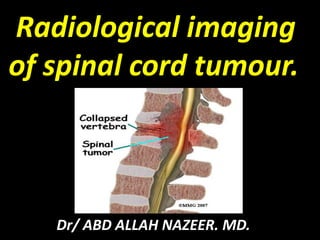
  • 1. Radiological imaging of spinal cord tumour. Dr/ ABD ALLAH NAZEER. MD.
  • 2.
  • 3. Spinal Cord Tumours: Extra-dural. Intra-dural extra-medullary Intra-medullary. Spinal Cord Tumors are classified into: Benign tumours. Cysts, and other benign tumour like masses. Malignant tumors. Metastasis.
  • 4. Extra-dural tumors: . Occurs outside the spinal dura. . Typically arises from the osseous spine, intervertebral discs and adjacent soft tissues. . Imaging criteria. Focal displacement of the theca away from the mass.
  • 5. Diagnosis: A thorough medical examination with emphasis on back pain and neurological deficits is the first step to diagnosing a spinal tumor. Radiological tests are required for an accurate and positive diagnosis. X-ray: Application of radiation to produce a film or picture of a part of the body can show the structure of the vertebrae and the outline of the joints. X-rays of the spine are obtained to search for other potential causes of pain, i.e. tumors, infections, fractures, etc. X-rays are not very reliable in diagnosing tumors. Computed tomography scan (CT or CAT scan): A diagnostic image created after a computer reads X-rays, a CT/CAT scan can show the shape and size of the spinal canal, its contents, and the structures around it. It also is very good at visualizing bony structures. Magnetic resonance imaging (MRI): A diagnostic test that produces three-dimensional images of body structures using powerful magnets and computer technology. An MRI can show the spinal cord, nerve roots and surrounding areas, as well as enlargement, degeneration, and tumors.
  • 6. Extra-dural benign tumors: -Hemangioma. -Osteoid osteoma. -Osteochondroma.
  • 7. Hemangioma. Slowly growing benign tumour of capillary, cavernous or venous origin. Most common benign spinal neoplasm and approximately 50% of osseous hemangiomas are found in the vertebral bodies (thoracic especially). Most epidural hemangiomas occur secondarily as extension of expanding intra-osseous lesions. Most hemangioma are asymptomatic and discovered accidentally.
  • 8. Radiological imaging of hemangiomas. Plain film: Findings include: prominent trabecular pattern, sclerotic vertebra with vertical trabeculae: Corduroy sign lytic calvarial lesions with spoke-wheel appearance irregular and lytic in long bones, with a honeycomb appearance. CT: Usually as an incidental finding, especially in the vertebrae. Better visualization of thickened vertical trabeculation (polka-dot appearance). MRI: Signal intensity is somewhat variable, depending largely on the amount of fat content. T1: high is more common (fat rich). intermediate to low signal intensity is seen in fat poor hemangioma. T2: high. T1 C+ (Gd): enhancement is often present.
  • 12. MRI: An epidural component of hemangioma of the LI vertebra with a lesion of its arch pedicle (А); hemangioma of ThXI vertebra, spreading paravertebrally and to the right pedicle of its arch (B).
  • 13. Sagittal T2-weighted image showing a T3 haemangioma with epidural extension compressing the spinal cord.
  • 14. Osteoid osteoma is a small, benign osteoblastic tumor consisting of a highly vascularized nidus of connective tissue surrounded by sclerotic bone. The nidus is usually < 1.5 cm. 10% occur in the spine, usually at the site of neural arch. The classical clinical presentation of spinal osteoid osteoma is that of painful scoliosis. Other clinical features include nerve root irritation and night pain. Osteoid osteoma has characteristic computed tomography (CT) findings( dense sclerosis surrounding a lytic lesion that has a central calcific nidus). Because magnetic resonance imaging (MRI) findings of the osteoid osteomas causing intense perinidal edema can be confusing, these patients should be evaluated with clinical findings and other imaging techniques. Nidus typically low to intermediate signal at T1 and T2WI. Bone Scan. Focal activity on both intermediate and delayed images.
  • 17. Osteochondromas are among the most frequent of benign bone tumors. They occur either as solitary lesions or as multiple osteochondromatosis, they arises through lateral displacement of the epiphyseal growth cartilage, results in a bony excrescence with cartilaginous –covered cortex and a medullary cavity. However, the spine is affected by these tumors in only 2% to 7%. Osteochondromas are commonly seen in the second or the third decade of life. Spinal osteochondromas present as asymptomatic palpable masses or more unusually, with neurological deficit, because the majority of the lesions grow out of the spinal canal and usually do not cause symptoms. But nerve root compression is an uncommon manifestation of exostosis developed within the intra-spinal canal. Neurological compromise is more common in the patient.
  • 18. Radiological imaging of osteochondroma. X-Ray films: A sessile or pedunculated bone like projection. CT Scan: The cortex of the parent bone flares into the cortex of the osteochondroma with which it is contiguous. The cartilaginous cap often contains calcification. MRI: Show mixed signal on both T1 and T2WI. Cartilage in the cap has high signal intensity on T2- weighted, spin-echo MRI scans. This characteristic allows measurement of the cap, which is an important consideration in malignant transformation.
  • 19. Osteochondroma at upper cervical cord in a patient with multiple exostoses.
  • 20. Osteochondroma of rib with neural foraminal extension and cord compression
  • 21. T2W MRI cervical spine showing the C3 osteochondroma compressing the cord, (b) CT axial section showing the pedunculated osteochondroma, (c) Post op MRI T2WI showing the cord contusion, (d) Histopathological examination showing cartilaginous cap with underlying irregular bony trabeculae.
  • 22. Uncommon extra-dural masses. Cysts, and other benign tumour like masses. -Eosinophilic granuloma. -Epidural lipomatosis. -Non-neoplastic cysts. Synovial and arachnoid cyst.
  • 23. Eosinophilic granuloma(EG) Langerhans Cell Histiocytosis. A benign non-neoplastic disorder, self limiting process of well demarcated bone resorption. 1st- 2nd decade, Male to female ratio 2:1 Spine involvement in 10-15% of EG. A classic cause of a single collapsed vertebral body as a lytic lesion without surrounding sclerosis. Associated with 2 systemic diseases: -Hand-Schuller-Christian disease. - Letterer-Siwe disease.
  • 24. Radiological imaging finding: X-Ray: Vertebral plana. CT Scan: Lytic lesions or collapse of the vertebral bodies(Vertebra plana). MRI: decreased signal intensity on T1- weighted images and high signal intensity on T2-weighted sequences. The lesion may enhance after the administration of a gadolinium-based contrast agent.
  • 28. Epidural lipomatosis. Epidural lipomatosis refers to excessive accumulation of fat within the epidural space, such that the thecal sac is compressed. Clinical presentation Patients may present with back pain resembling that of a disc herniation. In severe cases, symptoms of canal stenosis are produced. Radiographic features MRI spine There is a often generalized excess of fat seen in the extradural space. As a result the dural sac can appear narrowed or even resemble a "Y" shaped configuration. Signal characteristics follow fat on all sequences: T1 - high signal T1 (FS) - shows fat suppression T2 - high signal
  • 30. spinal epidural lipomatosis. The thecal sac has the typical "Y" configuration on axial section at the L5 level.
  • 32. SYNOVIAL CYST. A synovial cyst is a relatively uncommon cause of spinal stenosis in the spine. It is a benign (noncancerous) condition and the symptoms and level of pain and discomfort can remain stable for many years. A synovial cyst is a fluid-filled sac that develops as a result of degeneration of the joints in the spine. Since it develops as a result of the aging process, it is rarely seen in an individual less than 45 years of age and is most commonly seen in a person over the age of 65. The cyst causes compression of the nerves in the spinal canal and this causes the patient to experience symptoms of spinal stenosis. Spinal stenosis is defined as narrowing of the space for the spinal nerves.
  • 33. Radiological imaging finding: A synovial cyst is best visualized on an MRI scan of the spine. It shows up as a bright signal on the T2 portion of the scan. It often has the same appearance as the cerebrospinal fluid that surrounds the nerves in your spinal canal. Plain X-rays of the spine, including flexion/extension (bending) X-rays of the spine, are also performed to rule out any associated instability of the spine. It is very important to check for spinal instability as the involved joint often has an accompanying degenerative spondylolithesis. A spondylolithesis is defined as a slippage of one vertebrae over another and it indicates that the joint is unstable or incompetent. CT Scan: Show hypo to hyperdense area compared to the ligamentum flavum and these changes are associated with facet joint degeneration.
  • 35.
  • 36. Extradural arachnoid cyst. CSF-filled outpouching of arachnoid that protrude through a dural defect. Spinal extradural arachnoid cysts are uncommon expanding lesions. Idiopathic arachnoid cysts are not associated with trauma or other inflammatory insults. If they enlarge, they usually present with progressive signs and symptoms of neural compression. Spinal extradural arachnoid cysts are a rare cause of spinal cord compression. These cysts most commonly occur in the middle to lower thoracic spine (65%) but also have been reported in the lumbar and lumbosacral (13%), thoracolumbar (12%), sacral (7%), and cervical regions (3%). Cyst enlargement can result in symptomatic spinal cord compression.
  • 37. Radiological imaging finding: MRI is considered the imaging study of choice in identifying arachnoid cysts which appears CSF-equivalent extra-dural mass that cause spinal cord compression. MRI provides better resolution of tissue intensity, absence of bone interference, multiplanar capabilities, and is noninvasive. Plain films may show bony erosion of the spinal canal. If MRI made with a contrast medium: The signal in the cyst is the same as in the dural bag. CT scan is another examination method often used for the diagnosis of Tarlov cyst. Unenhanced CT scans may show widened interpedicular distance, scalloping of vertebral bodies or pedicle thinning and cystic masses that are have the same density with CSF. CT Myelogram is minimally invasive, and could be employed when MRI cannot be performed on patient.
  • 39.
  • 40.
  • 42. Primary Extradural Tumours and metastasis of the Spine. Primary malignant tumour: Chordoma. Lymphoma. Sarcoma. Osteogenic sarcoma. Chondrosarcoma. Ewing sarcoma. Plasmacytoma. Multiple Myeloma. Malignant Schwannoma. Metastasis.
  • 43. Chordoma Chordoma is a rare slow-growing neoplasm thought to arise from cellular remnants of the notochord. Grossly, locally invasive lobulated gelatinous –appearing masses. Typically, arise in the midline of the spinal column at any location from the clivus to the coccyx. Radiographic features: CT: Centrally located, well-circumscribed destructive lytic lesion, sometimes with marginal sclerosis expansile soft-tissue mass (usually hyper-attenuating relative to the adjacent brain, however inhomogeneous areas may be seen due to cystic necrosis or hemorrhage; the soft-tissue mass is often disproportionately large relative to the bony destruction) MRI: T1 intermediate to low signal intensity, small foci of hyperintensity (Intratumoral hemorrhage or a mucus pool). T2: most exhibit very high signal. T1 C+ (Gd): heterogeneous enhancement with a honeycomb appearance corresponding to low T1 signal areas within the tumour. GE (gradient echo): confirms hemorrhage if present with blooming.
  • 47.
  • 48. Lymphoma of the spinal cord: Lymphoma of the spinal cord is an uncommon manifestation of lymphoma. Although lymphoma more commonly involves the vertebral body (vertebral body tumours) or epidural compartment, intramedullary lymphoma does rarely occur. Radiographic features: The most common intramedullary location is the cervical cord, followed by the thoracic cord then the lumbar cord . Most are solitary lesions, however there may be multiple lesions throughout the spinal cord. MRI: Although spinal cord expansion is usually present, in some patients there is relatively minimal enlargement of the cord. The lesions are generally poorly defined and tumoural cysts are generally not a feature and secondary syringomyelia is rare. Lymphoma usually does not have a hemorrhagic component Reported signal characteristics include: T1: isointense to spinal cord T2: hyperintense (this is in contrast to the characteristic low T2 signal intensity that is seen in intracranial lesions) T1 C+ (Gd): usually solid and homogeneous enhancement. CT: Non specific bone destruction and hyperostosis.
  • 50. Arrows point to lymphoma causing epidural compression. Lymphoma of the spines.
  • 51. Osteosarcoma: Osteosarcomas are malignant bone forming tumours and the second most common primary bone tumour after multiple myeloma. They account for ~20% of all primary bone tumours. primary osteosarcoma: typically occurs in young patients (10- 20 years) with 75% occurring before the age of 20; which is logical because the growth centers of the bone are more active during the puberty to adolescence time period. Therefore the epidemiology can be easily understood. For less clear reasons, there is a slight male predominance. secondary osteosarcoma: occurs in the elderly, usually secondary to malignant degeneration of Paget's disease, extensive bone infarcts or post radiotherapy for other conditions.
  • 52. Radiographic features Plain film: Conventional radiography continues to play an important role in diagnosis. Typical appearances of conventional high grade osteosarcoma include: medullary and cortical bone destruction wide zone of transition, permeative or moth-eaten appearance aggressive periosteal reaction, sunburst type . CT: The role of CT is predominantly in assisting biopsy and staging, but adds little to plain x-ray films. MRI: T1: soft tissue non-mineralized component: intermediate signal intensity mineralized/ossified components: low signal intensity, peri-tumoural edema: intermediate signal intensity scattered regions of hemorrhage will have variable signal (see ageing blood on MRI) enhancement: solid components enhance T2: soft tissue non-mineralized component: high signal intensity mineralized/ossified components: low signal intensity peri-tumoural edema: high signal intensity.
  • 56. Chondrosarcoma. Chondrosarcoma is a malignant tumor that produces cartilage matrix. Primary chondrosarcoma is very uncommon, arises centrally in the bone, and is found in children. Secondary chondrosarcoma arises from benign cartilage defects such as osteochondroma or enchondroma. Imaging show lytic lesions with sclerotic and variable matrix calcification occurring in rings and arc. CT scanning may be useful for detecting subtle calcifications in the matrix when the diagnosis is in doubt. MRI: The investigation of choice for assessing the extent of a chondrosarcoma Helps delineate the extent of soft-tissue involvement Important for preoperative planning and for confirming or diagnosing recurrence at a surgically treated site
  • 57. Chondrosarcoma along the left posterior paraspinal location.
  • 59. Chondrosarcoma of the lower cervical spine.
  • 60. Plasmacytoma and multiple myeloma. Multiple myeloma is the most common primary malignant bone neoplasm in adults, and results in a wide range of radiographic abnormalities. Four main patterns are recognised: disseminated form: multiple defined lesions: predominantly affecting the axial skeleton disseminated form: diffuse skeletal osteopenia solitary plasmacytoma: single large/expansile lesion most commonly in a vertebral body or in the pelvis osteosclerosing myeloma. The spine is the most common location.
  • 61. Radiographic features: X-Ray Films: The vast majority of lesions are purely lytic, sharply defined/punched out with endosteal scalloping when abutting cortex. In only 3% of patients are the lesions sclerotic. CT: CT does not have a great role in the diagnosis of disseminated multiple myeloma, however it may be useful to determine the extent of extra-osseous soft tissue component in patients with a large disease burden. MRI: MRI is generally more sensitive in detecting multiple lesions compared to the standard plain film skeletal survey. Infiltration and replacement of bone marrow is exquisitely visualized, and newer scanners are able to perform whole body scans for this purpose which has been shown to be superior to both CT and skeletal surveys.
  • 63. Two cases of multiple myeloma.
  • 64. Spinal metastases: Spinal metastases is by far the most common extra-dural malignant neoplasm. Metastatic disease have any of the following: vertebral metastases (94%) may have epidural extension intradural extramedullary metastases(5%) intramedually metastases (1%). In adults, the initial site is the vertebral body, usually the posterior aspect. Most metastasis arises from the, breast: 22%, lung: 15%, prostate: 10%, lymphoma: 10%, kidney: 7%, gastrointestinal tract: 5%, melanoma: 4%, unknown: 4%, others: 24%.
  • 65. Radiographic features. Plain radiography is used to show erosion of the pedicles or the vertebral body. Owl-eye erosion of the pedicles in the anteroposterior (AP) view of lumbar spine is characteristic of metastatic disease and is observed in 90% of symptomatic patients. However, radiologic findings become apparent only when bone destruction reaches 30-50%. Osteoblastic or osteosclerotic changes are common in prostate cancer and Hodgkin disease; they are occasionally seen in breast cancer and lymphoma. CT scanning is useful in determining the integrity of the vertebral column. MRI- Is very sensitive in detecting vertebral metastasis. The most common pattern is multifocal lytic lesions characterized by low signal at the T1WI and of high signal at the T2WI. Scintigraphy- Very sensitive although non-specific in detecting bone metastasis.
  • 68. C2 metastasis in a 60-year-old male patient with renal cell carcinoma.
  • 69. Intradural extramedullary spinal tumours. Intradural extramedullary neoplasms are located outside the spinal cord but within the dural sheath. Schwannomas are the most common intradural extramedullary spinal lesions (30% cases), followed by meningiomas (25% cases). In the pediatric population, the most common intradural extramedullary neoplasms are leptomeningeal metastases resulting from primary brain tumours. Benign tumours: spinal schwannoma, spinal meningioma, spinal neurofibroma spinal paraganglioma and myxopapillary ependymoma Cysts and other benign tumourlike masses: spinal lipoma, intradural spinal lipoma, spinal epidermoid cyst, spinal dermoid cyst spinal neurenteric cyst and spinal arachnoid cyst. Malignant tumours: spinal leptomeningeal metastases.
  • 70. Spinal schwannoma and neurofibroma: Spinal schwannomas are schwannomas arising from nerves within the spinal canal. They are the most common intradural extramedullary spinal tumours, representing 30% of such lesions. They are most frequently seen in the cervical and lumbar regions, far more frequently than in the thoracic spine. In general schwannomas appear as rounded lesions, often with associated adjacent bony remodelling. when large they may either align themselves with the long axis of the cord, forming sausage shaped masses which can extend over several levels, or may protrude out of the neural exit foramen, forming a dumb-bell shaped mass. Plain X-Ray film: Show erosions of the pedicle with enlarged neural foramina. CT Scan: Shows bone erosion, and the density is hypo to slightly hyperdense. MRI: T1 - 75% are isointense, 25% are hypointense. T2 - more than 95% are hyperintense, often with mixed signal. T1 C+ (Gd) - virtually 100% enhance.
  • 75. Spinal meningioma. Meningiomas arising from the coverings of the spinal cord represent a minority of all meningiomas (approximately 12%5) but are the second most common intradural extramedullary spinal tumour representing 25% of all such tumours. Despite usually being small, due to the confines of the spinal canal they can result in significant neurologic dysfunction. spinal meningiomas are also not distributed evenly along the canal: cervical spine - 15% thoracic spine - 80% lumbosacral spine: uncommon. Meningiomas are often located posterolaterally in the thoracic region and anteriorly in the cervical region. Most meningiomas are solitary lesions (98%). Multiple meningiomas are most often associated with NF2.
  • 76. Radiographic features. Plain film: usually normal, rarely bone erosion or calcification CT (non-contrast) Isodense or moderately hyperdense mass hyperostosis may be seen but is not as common as in the intracranial forms. calcification may be present MRI: well-circumscribed, broad-based dural attachment dural tail signs They share similar signal characteristics to intracranial meningiomas: T1 isointense to slightly hypointense. may have a heterogenous texture. T2 - isointense to slightly hyperintense. T1 C+ (Gd) - moderate homogeneous enhancement. Occasionally, densely calcified meningiomas are hypointense on T1 and T2, and show only minimal contrast enhancement.
  • 78. Upper cervical spinal meningioma.
  • 82. Spinal epidermoid cyst. Spinal epidermoid cysts are cystic tumours lined by squamous epithelium. Unlike dermoid cysts, they do not contain skin appendages (hair follicles, sweat glands, sebaceous glands). They are usually extramedullary but rarely can be intramedullary . They may be congenital or acquired. Plain film: In advanced cases, there may be scalloping of the vertebral bodies or a scoliosis. CT: well circumscribed mass hypodense (similar to CSF), minimal to no enhancement calcification is rare MRI: well-defined lesion, generally no perilesional edema, Although variable, typical signal characteristics include: T1: hypointentse (similar to CSF) T2: hyperintense (similar to CSF) FLAIR: hyperintense compared to CSF T1 C+ (Gd): no enhancement or a thin rim of capsular enhancement DWI: bright (with corresponding low intensity on ADC map).
  • 83.
  • 84. Spinal dermoid cyst. Spinal dermoid cysts are uni or multilocular cystic tumours lined by squamous epithelium containing skin appendages (hair follicles, sweat glands, sebaceous glands) . They are congenital in origin. Forty percent are intramedullary, and 60% are extramedullary . Radiographic features: Spinal dermoid cysts are most often located in the lumbosacral region (60%) and cauda equina (20%) . They are rarely found in the cervical or thoracic spine. They have variable imaging appearances, but commonly appear as a mass of CSF density/intensity with fat density/intensity components . CT: well defined mass, isodense to CSF, often with hypodense components (fat) calcification may be present minimal enhancement may be demonstrated widening of the spinal canal, flattening of the pedicles and laminae and osseous erosions may be demonstrated MRI: Signal intensity may be homogeneous or heterogeneous. Typical signal characteristics include: T1: hypo or hyperintense hypointense: water content hyperintense: due to the presence of fatty secretions of sebaceous glands T2: hyperintense FLAIR: hyperintense compared to surrounding CSF T1 C+ (Gd): no enhancement or mild rim enhancement DWI: less likely to show diffusion restriction than epidermoid.
  • 86. Spinal arachnoid cyst: Spinal arachnoid cysts are relatively uncommon and may be either intra-dural (type III meningeal cyst) or extra-dural (type IA meningeal cyst). Radiographic features General: Most primary intradural spinal arachnoid cysts are dorsal to the cord. They occur at the following locations: thoracic: 80%, cervical: 15%, lumbar: 5% Secondary spinal arachnoid cysts can be located anywhere. MRI: As the cysts follow the intensity of CSF and their walls are generally not visible, they may not be identified unless the cord is displaced. T1: CSF intensity T2: CSF intensity, may even be brighter than CSF on account of no signal loss from pulsation/flow T1 C+ (Gd): no contrast enhancement phase-contrast imaging: decreased CSF flow within the cyst. DWI: No evidence of restricted diffusion
  • 90. Intradural extramedullary metastases: Intradural extramedullary metastases are rare and only accounts for approximately 5% of spinal metastases. Presentation is highly variable. As the most commonly affected site is the lumbosacral spine symptoms and signs include back or radicular pain, weakness, paresthesia, gait disturbance, cauda equina syndrome and symptoms/signs of meningeal irritation. Radiographic features: Plain films and CT are inadequate for the assessment of possible leptomeningeal metastatic disease, and in these cases, MRI is required. MRI MRI without contrast may be normal, and thus when suspected contrast should be administered. Typical signal characteristics include: T1: thickened nerve roots or nodular lesions that are isointense with the spinal cord may be seen. T2: cord edema may be seen with more extensive disease, especially if there is an intramedullary component. T1 C+ (Gd) enhancing tumour nodules on the spinal cord, nerve roots or cauda equina. “sugar coating” of the spinal cord and nerve roots.
  • 91. Multiple intramedullary extradural spinal cord metastases.
  • 92. Multiple intramedullary extramedullary spinal cord metastases.
  • 93. Intramedullary spinal tumours. Intramedullary spinal tumours are rare, representing 4-10% of all CNS tumours. They account for 20% of all intraspinal tumours in adults and 35% of all intraspinal tumours in children . Classification: They can be classified according to many ways: intramedullary neoplastic lesion: glial neoplasms: 90-95% of all intramedullary tumours spinal ependymoma: 60% of all glial spinal cord tumours spinal astrocytoma: 33% of all glial spinal cord tumours spinal ganglioglioma: 1% of all glial spinal cord tumours non-glial neoplasms: highly vascular lesions spinal hemangioblastoma and spinal paraganglioma other rare lesions intramedullary metastasis primary lymphoma of the spinal cord spinal primitive neuroectodermal tumour solitary fibrous tumour intramedullary benign masses: Syringohydromyelia, MS , transverse myelitis arachnoid and ependymal cyst.
  • 94. Syringohydromyelia. Syringohydromyelia refer to any pathologic CSF-containing cord cavity and can mimic spinal cord tumour. Imaging shows focal or diffuse cord expansion. CT Scan: Show distinct area of low density within the cord without enhancement at the post-contrast study. MRI: Cystic area within the cord which appears of low signal at the T1WI and of bright signal at the T2WI. No enhancement seen within the lesion at post-contrast study.
  • 95. Two cases of Syringohydromyelia.
  • 96. Multiple sclerosis. Spinal cord may be the earliest affected by MS plaques. Plaques occur preferentially in the dorsolateral cord and do not respect boundaries between grey and white matter. More predominant on the female. Early there is a predilection for the cervical cord. MRI: The most common finding on T2WI of one or more elongated, poorly marginated, hyperintense intramedullary lesions. The foci may has mass effect and enhance at the post-contrast study. Focal or generalized mild cord atrophy may be present.
  • 98. Multiple Sclerosis with disease activity.
  • 99. Transverse Myelitis (TM).) Focal inflammatory disorder of the spinal cord resulting in motor, sensory and autonomic dysfunction. Imaging findings: More than 2/3 of the cross sectional area is involved. Focal enlargement. T2WI hyperintensity Enhancement + / - Two forms of TM: Acute partial transverse myelitis - APTM Lesions extending less than two Segments. These patients are at risk of developing MS. Acute complete transverse myelitis - ACTM Lesions extending more than two Segments
  • 100. ATM on T2WI, CE-T1WI and STIR.
  • 103.
  • 104. Spinal ependymoma. Spinal ependymomas are the most common intramedullary neoplasm in adults, comprising 60% of all glial spinal cord tumours. They are the second most common intramedullary neoplasm in the pediatric population, representing 30% of pediatric intramedullary spinal neoplasms. Peak incidence is in the fourth decade, with 39 years being the mean age at presentation. Males are more commonly affected than females. There is an increased incidence with neurofibromatosis type 2. 6 histological subtypes are recognised: cellular (the most common intramedullary type). Papillary. clear cell. Tanycytic. Myxopapillary. virtually always located along the filum terminale with occasional extension into the conus medullaris melanotic (the least common type).
  • 105. Radiographic features: CT: CT may demonstrate non-specific canal widening isodense or slightly hyperdense compared with normal spinal cord intense enhancement with iodinated contrast. MRI: Typical signal characteristics: T1: most are isointense to hypointense; mixed signal lesions are seen if cyst formation, tumour necrosis or hemorrhage has occurred T2: hyperintense peritumoural edema is seen in 60% of cases associated hemorrhage leads to the “cap signs” (a hypointense hemosiderin rim on T2 weighed images) in 20-33% of cases. The cap sign is suggestive of but not pathognomonic for ependymoma as it may also be seen in hemangioblastomas & paragangliomas. T1 C+ (Gd): virtually all enhance strongly, somewhat inhomogeneously.
  • 106. An ependymoma of the cervical spine on MRI T2 image (left) and T1 with contrast (right). Note the associated syrinx seen on T2, contrast enhancing mass on T1, and overall expansion of the cord.
  • 107. Large filum terminalis ependymoma (arrows). Intramedullary cervical ependymoma (red arrows).
  • 108. Spinal astrocytoma. Spinal astrocytomas are the second most common spinal cord tumour overall, representing 40% of intramedullary tumours . They account for 60% of pediatric intramedullary tumours, making them the most common spinal cord tumour in children Spinal cord astrocytomas generally have a lower histologic grade than astrocytomas in the brain. In adults, 75% are low-grade neoplasms. Up to 25% are anaplastic astrocytomas. Glioblastoma multiforme represent only 0.2-1.5% of spinal cord astrocytomas. In children younger than 3, 80% are either grade I or II. All astrocytomas are characterised by hypercellularity and the absence of a surrounding capsule. In contrast to cord ependymomas, a cleavage plane is not present in most intramedullary spinal astrocytomas. Most common at cervical cord. High grade tumours are more likely to demonstrate extensive leptomeningeal spread seen in up to 60% of spinal cord glioblastomas.
  • 109. A fibrillary astrocytoma of the cervical spine on T2 (left) and T1 with contrast (right). Note the indistinct cord edema, expansion and partial contrast enhancement
  • 111. Spinal ganglioglioma are rare, comprising 1.1% of all spinal cord neoplasms . They are more frequent in children, representing 15% of intramedullary neoplasms in the pediatric age group.
  • 112. Spinal hemangioblastoma. Spinal hemangioblastomas are the third most common intramedullary spinal neoplasm, representing 2-6% of all intramedullary tumours. Angiography: A densely enhancing nidus with associated dilated arteries and prominent draining veins is characteristic for a hemangioblastoma. CT: On non-contrast CT They may be seen as a soft tissue nodule often with a prominent hypodense cyst-like component. Contrast administration results in vivid enhancement of the solid component. MRI: Although they usually appear as discrete nodules, there can be diffuse cord expansion. An associated tumour cyst or syrinx is common (50-100%) . T1: variable relative to normal spinal cord. hypo-isointense most common, and difficult to identify hyperintense (25%) T2: iso-hyperintense focal flow voids especially in larger lesions surrounding edema and associated syrinx are usually seen hemosiderin capping may be present T1 C+ (Gd): the tumour nodule enhances vividly.
  • 113. A hemangioblastoma of the thoracic spine on T1 with contrast in sagittal (left) and axial sections (right). Note the enhancing tumour nodule with associated cyst.
  • 114. Spinal cord hemangioblastoma is seen at L1-2 (arrowed).
  • 115. Lymphoma of the spinal cord. Lymphoma of the spinal cord is an uncommon manifestation of lymphoma. Radiographic features The most common intramedullary location is the cervical cord, followed by the thoracic cord then the lumbar cord . Most are solitary lesions, however there may be multiple lesions throughout the spinal cord. MRI: Although spinal cord expansion is usually present, in some patients there is relatively minimal enlargement of the cord. The lesions are generally poorly defined and tumoural cysts are generally not a feature and secondary syringomyelia is rare. Lymphoma usually does not have a hemorrhagic component . Reported signal characteristics include: T1: isointense to spinal cord T2: hyperintense (this is in contrast to the characteristic low T2 signal intensity that is seen in intracranial lesions) T1 C+ (Gd): usually solid and homogeneous enhancement
  • 116. Primary intramedullary spinal cord lymphoma.
  • 117. Intramedullary spinal metastasis. Intramedullary spinal metastases are rare, occurring in 0.9-2.1% of autopsied cancer patients. They represent 8.5% of central nervous system metastases and account for 5% of all intramedullary lesions. They are less common than leptomeningeal metastases. Radiographic features: CT (contrast enhanced) Hypervascular metastases may rarely be seen as enhancing intraspinal lesions. MRI Lesions are usually well-defined and typically produce cord expansion over several segments. In contrast to primary intramedullary neoplasms, associated cysts are rare. Typical MRI signal characteristics are: T1: hypointense T2: hyperintense prominent edema commonly surrounds the tumour nodule T1 C+ (Gd): avid homogeneous enhancement.