
Recommended
More Related Content
What's hot
What's hot (20)
Similar to Slit lamp
Similar to Slit lamp (20)
Recently uploaded
Recently uploaded (20)
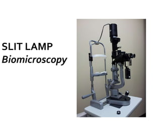
Slit lamp
- 2. Purkinje 1823 DeWecker 1863 Albert & Greenough 1891 Czapski 1897
- 3. Gullstrand 1911 Henker 1916
- 4. Observation System Illumination System Mechanical support System
- 5. Eyepiece +10D Objective lens +22D Prisms
- 7. Czapskiscope with rotating Objectives Haag Streit model, Bausch and Lomb, Thorpe model Littman-Galilean telescope principle Zeiss,Rodenstock and American optical slit lamps Zoom system Nikon photo SL, Zeiss 75 SL
- 14. Joy stick arrangement Patient support system Fixation target Mechanical coupling
- 15. Patient education Patient positioning Adjust Ocular eye pieces & IPD Fixation Magnification
- 16. Manipulating the Light source Focusing Examination & Special procedures Documentation
- 17. 1. Diffuse illumination 2. Direct focal illumination 3. Inirect illumination 4. Retroillumination 5. Specular reflection 6. Sclerotic scatter 7. Oscillating illumination of koeppe
- 18. General view of anterior eye and the palpebral conjunctiva. Contact lens fitting.
- 21. Optical section Parallelopiped of cornea Conical Beam Tangential Illumination
- 22. Narrow slit beam focused obliquely Knife like histological section
- 23. Tear layer – Bright anterior most Epithelium – Dark line Bowman’s membrane – Bright line Stroma –Wide granular and Grayish Descemet’s membrane & Endo – Bright zone
- 25. Corneal scars or infiltrates- bright Water clefts - black
- 26. To examine presence of aqueous flare and cells Magni- 25x
- 27. Observe surface structure Medium wide beam Magni 10, 16 or 25x Anterior and posterior cornea,anterior iris, anterior lens (pseudoexfoliation)
- 28. Uses: Corneal infiltrates Corneal microcysts Corneal vacuoles Epithelial cells
- 30. 16-25x
- 32. Obstructive – pigment, blood filled vessel. Repersive – epithelial oedema, precipitates Refractile - vacuole Vacuole I.D.R – Black area with bright surface D.R- illuminated area with black line
- 33. Solid / opaque precipitate Retroilluminaition from Fundus (Red Glow ) lamp & microscope coaxial 10- 16x Cornea,lens,vitreous pathologies D.R- Dark area I.D.R – Bright area away from illumination
- 34. Pupil- mid dilated Circular beam of light equal to size of pupil Microscope focused on iris Magni 10- 16x Use- Iris defects
- 35. Integrity of cornea or Lens Start from 10-16x Endothelial mosaic & Lens surface
- 38. Slit beam given oscillatory movement. Minute particles and filaments
- 39. SLIT LAMP EXAMINATION Suggested Power 6X or 10X external (lids,tear film,conjunctiva),contact lenses 16X angles,cornea,iris,lens,foreign bodies,corneal abrasions 25x aqueous flare,corneal endothelium 40X corneal endothelium Beam Width 1. narrowest angles,cornea,anterior chamber 2. a bit wider cornea,lens,etc 3. a bit wider yet external,contact lenses 4. full width external,applanation tension (with blue filter) Beam Height Full most areas and structures short checking anterior chamber for cells & flare Color/Filter White most areas and structures Blue (use fluorescein dye) applanation tensions,corneal staining,tear film,staining patterns of rigid contact lenses Green (red-free) evaluating blood vessels ,iron lines ,fleischer ring
- 40. Routine examination of ocular adnexa, anterior segment and anterior vitreous face Confirm fitting of contact lens Foreign body removal Suture removal Epilation Removal of membrane,crusts
- 41. Gonioscopy Fundus examination (fundus non contact lenses) Pachymetry Applanation tonometry Ophthalmodynamometry As delivery system for argon,diode andYAG laser Laser interferometry Potential acuity meter test Slitlamp photography Slitlamp videography