More Related Content
Similar to Ophthalmic & Physiological Optics ISSN 0275-5408 ABSTRACTS
Similar to Ophthalmic & Physiological Optics ISSN 0275-5408 ABSTRACTS (20)
Ophthalmic & Physiological Optics ISSN 0275-5408 ABSTRACTS
- 1. Ophthalmic & Physiological Optics ISSN 0275-5408
ABSTRACTS
The 2013 International Myopia Conference in California
August saw an influx of international researchers from
around the world to attend the 14th International Myopia
Conference, which took place at Asilomar in Northern California. The retreat style setting of Asilomar, which is
located in a Californian State park, made this conference
stand out from recent conferences in this series, and was
one of a number of strategies adopted to acknowledge the
passing early last year of Josh Wallman, one of the giants in
the myopia research field. Apart from his research accomplishments, Josh was known for his love of discussion and
debate, which the Asilomar environment fostered, and his
strong support for junior researchers. The weather also
behaved, providing fog cover sufficient to keep attendees
indoors for formal conference sessions and rolling back to
provide warm sunshine on the only afternoon break of the
conference.
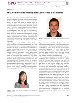
Continuing an already established tradition, the Chew
Sek-Jin Memorial Lecture, named in the memory of Dr. Sek
Jin Chew, another giant in this field, was given this year by
Seang Mei Saw (Figure 1), who is a Professor of Epidemiology and Ophthalmology in the Saw Swee Hock School of
Public Health and Yong Loo Lin School of Medicine at the
National University of Singapore. The title of her lecture
was ‘The Epidemic of Myopia in Asian and Beyond: Translations for Public Health and Clinical Practice’. This award
was sponsored by Cooper Vision. In recognition of the Josh
Wallman’s strong support of junior researchers, the 2013
International Myopia Conference featured the inaugural
Josh Wallman Memorial Lecture. Zeiss sponsored this young
investigator award, which went to Regan Ashby (Figure 2),
from the Research School of Biology, ARC Center of Excellence in Vision Science, Australian National University,
Canberra, Australia. He spoke on ‘The role of Light in the
Figure 1. Professor Seang Mei Saw, who gave the 2013 Chew Sek-Jin
Memorial Lecture.
Figure 2. Dr. Regan Asby, who gave the 2013 Josh Wallman Memorial
Lecture.
Regulation of Ocular Growth’, as the feature lecture in a
session otherwise featuring past students and collaborators
of Josh Wallman – Drs Debora Nickla, Chea-Su Kee, Frances Rucker and David Troilo, all world-renown myopia
researchers in academia. Five poster awards to junior
researchers, to Yijin Tao, China, Andrew Collins, New Zealand, and Baskar Arumugam, Ross Collery, and Mariana
Garcia, all USA-based, were a further acknowledgement of
Josh Wallman’s support of young researchers.
The conference opened with a keynote address by Professor Xingtao Zhou, Chief Physician and Director, Department of Optometry and Strabismus, Eye and ENT
Hospital, of Fudan University, Shanghai in China. His topic
was ‘Recent 15 years: Myopia Research and Associated
Translation Studies in Shanghai, China’. The program
included 10 other paper sessions, wide-ranging in topic –
covering fundamental research, mostly based on animal
models, including some new ones – squid and zebra fish, to
more clinically applied, including behavioral and optical
interventions studies involving children. Among the sessions attracting the most positive feedback were the two
panel discussion/debate sessions held on the last day, covering ‘Optical management of Myopia – How Many Options
and How Many Mechanisms’ and ‘Visual Environment versus Genetic Contributions to Myopia’. Both sessions served
to refocus attendees on the important unresolved issues in
this field. Posters were a significant feature of this conference, with a total of 91 presenters. They were also a major
attraction, with their physical location within the main
lecture hall providing a comfortable space for discussion
during coffee breaks, in addition to the formal poster
sessions.
© 2013 The Authors Ophthalmic & Physiological Optics © 2013 The College of Optometrists
Ophthalmic & Physiological Optics 33 (2013) 661–674
661
- 2. Abstracts
As host of this year’s International Myopia Conference, I
am also happy to pass over these duties to the myopia
researchers at Wenzhou, Zhejiang, China, where it was
agreed the 15th International Myopia Conference will be
held in 2015.
Christine Wildsoet
Center for Eye Disease & Development, School of Optometry,
University of California, Berkeley, USA
E-mail address: wildsoet@berkeley.edu
Abstracts of papers presentated at the 2013
International Myopia Conference in California
Recent 15 years: myopia research and associated
translational studies in Shanghai, China
Xingtao Zhou, Renyuan Chu, Zhiqiang Yu and Zhi Chen
Fudan University Eye and ENT Hospital, Shanghai,
China
Purpose: The prevalence of myopia is extremely high in China,
especially in urban areas like Shanghai. The recent 15 years saw a series
of impressive discoveries and constructive translational studies within
our laboratory, the Key Lab for Myopia Research of China’s Ministry
of Health, Fudan University Eye and ENT Hospital.
Methods: Basic researches and translational studies were carried
out.
Results: (1) ‘Medical Refraction’ as a term was developed to incorporate high myopia related medical implications, e.g. gene screening,
to regular refraction process in order to enhance myopia prevention in
clinical practice; (2) The first Chinese patented microkeratome for
refractive surgeries was developed, its safety and efficacy being adequately evaluated, then commercially available, and now in wide use in
China; (3) The effects of different monochromatic lights on refractive
development in guinea pigs and monkeys were discovered: animals
raised in long-wavelength lighting developed more myopia than in
short-wavelength lighting; the results were first applied to develop
color filters to accelerate emmetropization in hyperopic anisometropic
eyes and then used to manufacture chromatic reading materials, e.g.
text books, to interfere with myopia progression in children countrywide; (4) Effects of peripheral retina on myopic eye growth was
emphasized, and spectacle lenses incorporating concentric myopic
defocus were invented and patented for clinical prevention of myopia
in China; effects of pupil size on axial growth in orthokeratology was
discovered for the first time; (5) Slow stroboscopic lighting was found
to induce myopia in guinea pigs, especially at the flicker rate of 0.5 Hz,
as opposed to the myopia-preventing effect with higher flicker rate
stroboscopic lightings.
Conclusion: These basic and associated translational studies have
yielded significant progress in China’s myopia research and have
provided constructive strategies in myopia intervention.
The epidemic of myopia in Asia and beyond:
translations for public health and clinical practice
Seang-Mei Saw
Saw Swee Hock School of Public Health, Singapore Eye
Research Institute, Singapore, Singapore
Purpose: To determine the current and future preventive and treatment options for myopia.
662
Methods and results: For primary prevention, epidemiologic studies and community-based randomized trials have evaluated time outdoors as a modifiable preventive measure for myopia. Other
environmental factors such as reading/writing/education as well as
novel genes from recent GWAS studies also contribute to the onset of
myopia. For children with early onset myopia and a risk of high myopia in adulthood, interventions such as eye drops may decrease the
progression of myopia and lead to a less severe final refractive error.
0.01% atropine eye drops have recently been found to be effective in
randomized clinical trials with very few adverse effects. As several new
genes have and will be identified from GWAS and exome sequencing
studies, genetic tests could be developed in the future to identify highrisk children who will benefit most from eye drops. Cross-sectional
and longitudinal studies have demonstrated that elderly adults with
high myopia are at risk of visually blinding pathologic myopia complications such as chorioretinal atrophy and thus older individuals should
be screened regularly.
Conclusion: From a clinical and public health perspective, there are
many important strategies that could be adopted by eye care practitioners and governments at a national level. The major primary preventive measure should be to encourage increased amount of time
outdoors in very young children to prevent myopia onset through
public education as well as school and family based programs. For children with early onset myopia who are at risk of high myopia, 0.01%
atropine eye drops could be considered on a case-by-case basis. As we
predict an epidemic of pathologic myopia in certain Asian countries
because of the recent aging populations and cohort effect, it would be
prudent to screen elderly highly myopic adults at regular intervals and
provide low vision programs or surgical/medical options.
Large differences in myopia prevalence among
primary school children in adjoining provinces of
rural western China
Zhongqiang Zhou1, Xiaochen Ma2, Hongmei Yi3,
Xiaopeng Pang4, Yaojiang Shi5, Qianyun Chen1, Mirjam
Meltzer1, Mingguang He1, Scott Rozelle6 and Nathan
Congdon1,7
1
State Key Laboratory of Ophthalmology and Division
of Preventive Ophthalmology, Zhongshan Ophthalmic
Center, Sun Yat-sen University, Guangzhou, China,
2
Department of Agriculture and Resource Economics,
University of California Davis, Davis, USA, 3Center for
Chinese Agricultural Policy, Institute of Geographical
Sciences and Natural Resources Research, Chinese
Academy of Sciences, Beijing, China, 4School of
Agricultural Economics and Rural Development,
Renmin University of China, Beijing, China, 5School of
Economic Management, Xibei University, Xi’an, China,
6
Freeman Spogli Institute of International Studies,
Stanford University, Stanford, USA, and 7ORBIS
International, New York, USA
Purpose: To study myopia prevalence and spectacle use in middleincome (Shaanxi) and poor (Gansu) provinces in western China.
Methods: In September, 2012, a randomly-selected populationbased sample of primary school children in the 4th and 5th grade (age
7–17 years) from 120 schools in Tianshui Prefecture (Gansu province)
and 133 schools in Yulin Prefecture (Shaanxi province) underwent
screening of their distance visual acuities (VA). Those with uncorrected visual acuity (VA) ≤ 6/12 in either eye underwent cycloplegic
automated refraction with subjective refinement. Spectacle ownership
and subject and family characteristics were assessed by questionnaire.
Myopia was defined as spherical equivalent refractive error
© 2013 The Authors Ophthalmic & Physiological Optics © 2013 The College of Optometrists
Ophthalmic & Physiological Optics 33 (2013) 661–674
- 3. Abstracts
(SE) ≤ À0.5D in both eyes and uncorrected VA ≤ 6/12 in at least one
eye.
Results: Myopia prevalence among 9667 children in Shaanxi (mean
age 10.4 Æ 1.0 years, 53.6% male) was 23.1%, nearly twice that among
10 308 children (mean age 10.7 Æ 1.2 years, 50.6% boys) in Gansu at
13.4% (p < 0.0001). Myopia prevalence increased with age (20.7%
among those 7–9 years to 25.6% 12–17 years) in Shaanxi, but not
Gansu. Spectacle ownership was low among children with refractive
error in both Shaanxi (464/2362 = 19.6%) and Gansu (250/
1472 = 17.0%). Among children with 3.5–4.5 diopters of myopia,
fewer than half had glasses (Shaanxi 45.2%, Gansu 45.8%). In regression models, ownership of spectacles was associated with greater absolute SE in the better-seeing eye [Relative risk (RR) for 1.0 Diopter
increase: Gansu 1.39, p < 0.001; Shaanxi 1.24, p < 0.001], greater family wealth in the combined population (RR 1.31, p = 0.03) though not
for Shaanxi or Gansu separately, and one or both parents with
>12 years of education in Shaanxi (RR 1.48, p < 0.001). Age, sex,
province, boarding at school and parental out-migration were un-associated with spectacle ownership. Three quarters of children owning
glasses had them at school in both Shaanxi (343/464 = 74.1%) and
Gansu (200/250 = 80.0%).
Conclusion: Population prevalence of myopia in China’s poor provinces, previously unreported, appears far lower than in better-studied
wealthier areas. Spectacle ownership is not common in western China,
even among children with significant refractive errors. Studies are
needed to determine if low myopia prevalence in poor areas is due to
modifiable factors such as outdoor activity.
High myopia registry in Guangzhou
Mingguang He1,2, Linxing Chen1,2, Ian Morgan1,2 and
Brien Holden1,2
1
Zhongshan Ophthalmic Center, Sun Yat-sen
University, Guangzhou, China, and 2Brien Holden
Vision Institute, Australia
Purpose: To establish a high myopia registry in Guangzhou that
aims to identify the clinical features and natural history of high myopia
and to identify risk factors for the development of visual damage and
complications.
Methods: Subjects with sphere <À6.00 Diopter (D) in both eyes and
aged 7 years or older were recruited from both optometric clinic and
community screening. Baseline data were collected on cycloplegic
refraction, visual acuity, ocular biometry, strabismus, lens opacity,
intraocular pressure, visual field, B scan ultrasound, fundus photography, autofluorescence and optical coherence tomography. Fundus
fluorescein angiography (FFA) and magnetic resonance imaging
(MRI) were performed on the participants (25% for FFA and 10% for
MRI) who are selected randomly and stratified by age and refraction
status. Blood samples were collected for DNA extraction and genotyping. A questionnaire was administrated to collect information on medical history of myopia, family history and habitual lifestyle. Follow-up
examinations were scheduled to carry out every 1 year for 5 years.
Results: Study recruitment was accomplished on October 2012 and
a total of 917 eligible subjects were enrolled. The mean age at baseline
was 22.5 Æ 12.5 years old (range 7–72), and 438 (47.8%) were male.
Baseline sphere of the right eye ranged from À30.00 to À6.00 D, with
a mean of À9.23 Æ 3.36 D. The mean axial length of the right eye was
27.3 Æ 1.46 mm (range: 23.9–32.0 mm). One hundred and seventyeight out of 914 subjects (19.5%) had best corrected visual acuity <20/
40 in either eye. Other baseline characteristics were under analysis.
Conclusion: This study will help us to have a better understanding
on the clinical features, nature history, progression and gene environment interaction in high myopia people. It will also help understand
the risk factors and determinants for the development of myopiarelated complications and vision impairment.
Prevalence of refractive error in Western Europe
Katie Williams and on behalf of the European Eye
Epidemiology (E3) Consortium
Departments of Ophthalmology and Twin Research,
King’s College London
Purpose: (1) To report the distribution and prevalence of refractive
error in Western Europe, within the European Eye Epidemiology (E3)
consortium. (2) To investigate if there is evidence of a cohort effect for
increasing myopia prevalence, as identified elsewhere. (3) To examine
the effect of educational level on the risk of myopia.
Methods: A meta-analysis of refractive data was performed from
members of the European Eye Epidemiology Consortium including
cohort data from TwinsUK (n = 6095), 1958 British Birth Cohort
(n = 2495), EPIC-Norfolk (n = 7444), Tromso Eye Study (n = 5792),
Gutenberg Health Study (n = 14 069), Thessaloniki Eye Study
(n = 1952), EUREYE (n = 2882), Rotterdam Study (n = 6506), ERF
(n = 2662), POLA (n = 2285), and Alienor (n = 511) Studies. The
mean spherical equivalent of the two eyes was considered for each
individual and those who reported cataract surgery, retinal detachment
or laser refractive surgery were excluded. In total 52 653 participants
were included in a meta-analysis of the prevalence of myopia (defined
as ≤À0.75 D), hyperopia (≥1.00 D) and astigmatism (≥Æ1.00 D).
Subjects with myopia were divided into low (≤À0.75 to >À3.00 D),
moderate (≤À3.00 to >6.00 D) or high (≤À6.00 D) categories, and
subjects with hyperopia were classified as low (≥1.00–<3.00 D) or high
(≥3.00 D) hyperopes. The prevalence of myopia in different birth
cohorts was examined and the effect of education on myopia risk was
assessed by stratifying participants into those completing primary education (<16 years of age of completing full-time education), secondary
(16–19 years old) and higher education (≥20 years old).
Results: Median ages of the included cohorts ranged from 44 to
78 years old. There was a slight female predominance in the combined
cohort (57.9% females, 42.1% male) and minimal variation in ethnicity (99% European ancestry). Refractions were performed between
1990 and 2012 using either autorefraction or subjective refraction.
Mean spherical equivalent of the 52 653 participants was 0.13 diopters
(D) [95% confidence interval (CI) 0.11–0.15 D]. The overall myopia
prevalence was 23.5% (95% CI 23.13–23.84); 14.6% had low myopia,
6.1% moderate and 2.0% high myopia. The prevalence of hyperopia
was 31.4% (95% CI 31.0–31.8), 7.2% had high hyperopia. Astigmatism
prevalence was 22.0% (95% CI 21.7–22.3). There was a cohort effect of
increasing myopia prevalence: participants born between 1950 and
1970 had approximately 10% higher prevalence of myopia at the same
ages than those born between 1930 and 1950. Educational level was
significantly associated with myopia: 16% participants with only primary education were myopic compared to a prevalence of 35% in
those who completed higher education.
Conclusion: A quarter of over 50 000 subjects in epidemiological
studies of age-related eye diseases in Europe are myopic. The sharing
of data within the E3 Consortium finds a cohort effect of rising prevalence, and will allow projections of the rising burden of sight-threatening complications. The clear association of myopia with higher levels
education will be explored as a possible reason for the rise in myopia
prevalence.
Nearwork and outdoor activities in monozygotic
twins in relation to discordance in refraction
Xiaohu Ding, Xinxing Guo, Fan Xiang, XiaoboGuo,
Ian Morgan and Mingguang He
State Key Laboratory of Ophthalmology, Zhongshan
Ophthalmic Center, Sun Yat-sen University,
Guangzhou, China
Purpose: To evaluate the effect of nearwork and outdoor activity
differences on discordant refractions in monozygotic (MZ) twins.
© 2013 The Authors Ophthalmic & Physiological Optics © 2013 The College of Optometrists
Ophthalmic & Physiological Optics 33 (2013) 661–674
663
- 4. Abstracts
Methods: A longitudinal twins study was launched in 2009, Guangzhou City, China. A standard questionnaire was administered by interview to obtain estimates of daily activities, including time spent on
near work and outdoor activities. Refraction was measured by autorefraction under cycloplegia. Cross-sectional analyses of associations of
refractive discordance with environmental factors were conducted
among monozygotic (MZ) twins.
Results: A total of 490 MZ twin pairs were eligible, the refraction
was À1.50 Æ 2.14 D (Mean Æ SD), nearwork time was 4.2 Æ 1.5 h,
and outdoor activity time was 1.4 Æ 0.9 h. In the mixed model, we
found that nearwork activities conferred increased risk of myopic
spherical equivalent (SE) whereas outdoor activity had a marginal protective effect to myopic SE. The variation in nearwork activities
explained about 2.0% of total phenotypic discordance and outdoor
activity explained about 0.5%.
Conclusions: Our results confirm that nearwork is a risk factor of
myopia, while outdoor activity exerts protective effects. Given the very
marked genetic similarity of MZ twins, and the small effects of known
risk factors on discordance, we suggest that the discordance between
MZ twins largely results from stochastic variations, at the genomic or
epigenetic levels.
Short-term changes in axial length during simulations
of typical far, intermediate and near tasks
Atanu Ghosh, Michael J. Collins, Scott A. Read,
Brett A. Davis and Payel Chatterjee
School Of Optometry and Vision Science, Queensland
University of Technology, Brisbane, Australia
Purpose: To investigate the changes in axial length with the combined effect of accommodation and angle of gaze (convergence and
downward gaze) over 5 minutes in groups of myopes and emmetropes.
Methods: A total of 31 subjects (nine emmetropes, 10 low myopes,
and 12 moderate to high myopes) aged from 18 to 31 years were
recruited. To measure ocular biometrics in infero-nasal gaze with
accommodation, an optical biometer (Lenstar LS900) was inclined on
a tilt and height adjustable stage, with the subject’s chinrest mounted
on a rotary stage to induce various levels of convergence by rotation of
the subject’s head in primary or downward gaze. Initially, the subjects
performed a distance viewing task in primary gaze for 10 minutes to
provide a ‘wash-out’ period for prior visual tasks, and then the subject’s axial length and ocular biometrics were measured in nine different combinations of gaze/accommodation over 5 minutes. These nine
sessions for all gaze measurements (i.e. three levels of accommodation 9 three levels of convergence) were completed across 3 days of
testing (one accommodation condition on each day).The nine combinations of gaze/accommodation were based on those required to view
the centre, right and left edges of a distant TV at 6 m in primary gaze,
an intermediate task (i.e. computer at 50 cm in 10° downward gaze)
and a near task (i.e. reading A4 page at 20 cm in 20° downward gaze).
Subjects were wearing a custom built three-axes head tracker throughout the experiment that monitored subjects’ relative head movements
(roll, pitch and yaw) during measurements.
Results: A significant increase in axial length occurred with the
combined effect of accommodation, convergence and downward gaze
(repeated measures ANOVA, p < 0.001), with the greatest axial elongation during the near task in downward gaze with convergence (i.e.
downward 20°/inward 33°, with 5 D accommodation) (mean change
33 Æ 13 lm, after 5 minutes task) followed by the intermediate task
(i.e. downward 10°/inward 25°, with 2 D accommodation) (mean
change 14 Æ 11 lm, after 5 minutes task).Changes in axial length for
the distance task (i.e. primary gaze/9° convergence, with 0.16 D
accommodation) were not statistically significant (mean change
4 Æ 8 lm, after 5 minutes task, p > 0.05). Moderate to high myopes
had a greater change in the axial length (mean change 40 Æ 11 lm
after 5 minutes of near task) than that of emmetropes (mean change
29 Æ 15 lm after 5 minutes of near task) and low myopes (mean
664
change 29 Æ 16 lm after 5 minutes of near task) associated with time
(p = 0.02) and accommodation by time (p = 0.03).
Conclusions: The combination of accommodation, convergence
and downward angle has a significant short term effect on axial length
over time. The near task in downward gaze with convergence caused a
greater change in axial length than the intermediate and distant visual
tasks. The greater axial elongation measured in the infero-nasal direction with accommodation is most likely associated with a combination
of biomechanical factors such as, extraocular muscle forces and ciliary
muscle contraction.
The role of light in the regulation of ocular growth
Regan Ashby
Discipline of Biomedical Sciences, Faculty of ESTeM,
University of Canberra, Australia and Research School
of Biology, Australian National University, Australia
Experimental evidence has shown that lighting conditions affect ocular
growth in laboratory studies of experimental myopia. For instance,
extended rearing of chicks under either constant light or constant dark
leads to excessive vitreous chamber elongation, but, because of severe
corneal flattening, an overall hyperopic shift in refraction. In terms of
spectral composition, studies in both chicks and guinea pigs have indicated that rearing animals under red light enhances the development
of myopia, while blue light retards it, presumably due to differences in
the focal point of these wavelengths within the eye. More recently, the
role of light intensity has become a major research focus due to evidence that time spent outdoors is protective against the development
of myopia. It has been postulated that such a protective effect might be
mediated by the light-stimulated release of dopamine from the retina.
In agreement with this hypothesis, rearing animals under high illumination levels (15 000–25 000 lux) significantly retards the development of deprivation-myopia in chicks, rhesus monkeys and tree
shrews. In chicks, this protective effect is abolished by the administration of the D2 receptor antagonist spiperone, implicating lightinduced dopamine release and D2 dopamine receptors as critical in
this pathway. In both chicks and tree shrews, high light also reduces
the rate of compensation for negative lenses, although full compensation (target refraction) is still achieved. In rhesus monkeys, however,
high light levels were found not to alter the rate of compensation for
negative lenses. In chicks, high light also enhances the rate of compensation for plus lenses, suggesting that in all conditions so far tested, elevated illumination levels reduces the rate of ocular growth. Consistent
with this hypothesis, normal refractive development in diurnally reared
chicks also appears to depend, to some degree, on illumination levels,
as chicks reared under low illumination levels (50 lux), for a period
of90 days, develop significant amounts of myopia (approximately
À2.41 D), as compared to animals reared under medium (500 lux,
approximately +0.03 D) or high light levels (10 000 lux, approximately
+1.1 D). Furthermore, contralateral control eyes of both tree shrews
and rhesus monkeys kept under high light show a small but significant
hyperopic shift. A number of possibilities could explain the ability of
light to retard the development of experimental myopia, including;
pupil constriction, vitamin D levels, spectral composition, changes in
optic flow rates, or increased physical activity. However, at present, the
data supports a role for light-induced increases in retinal dopamine
release.Time outdoors has been identified as a protective factor against
the development of myopia. Currently, the mechanism of this effect is
not completely established, although some of the animal work supports a role for light intensity. Irrespective of whether or not light levels are the critical mechanism, evidence from experimental studies
demonstrates that rearing animals under high illumination levels
retards the development of experimental myopia, for the most part,
and induces a hyperopic shift in normal eyes. Although caution must
be taken in translating the findings from animal studies to preventive
regimes in humans, the implications of these findings are worthy of
further investigation.
© 2013 The Authors Ophthalmic & Physiological Optics © 2013 The College of Optometrists
Ophthalmic & Physiological Optics 33 (2013) 661–674
- 5. Abstracts
Muscarinic agonists thin chick choroids and stimulate
scleral extracellular matrix synthesis in eyecups
without the RPE
Debora L. Nickla1, Xiaoying Zhu1 and Josh Wallman2
1
Bioscience Department, The New England College of
Optometry, Boston, USA, and 2The City College of New
York, CUNY, New York, USA
Purpose: We have shown that intravitreal injections of the muscarinic agonists carbachol, oxotremorine, and arecaidine result in choroidal thinning within 24 h in intact eyes, but only oxotremorine
resulted in ocular growth stimulation (Nickla, Zhu and Wallman,
OPO 2013). These same agonists, and also pilocarpine, caused thinning of choroids in eyecups containing RPE, choroid and sclera. The
antagonist pirenzepine caused choroidal thickening both in vivo and in
vitro; no other tested antagonist had an effect in vitro. We ask whether
the RPE is required for the thinning effect on the choroid.
Methods: Paired eyecups of choroid and sclera were made from
untreated 1-week old chicks. Eyecups were paired with choroids of
matching thicknesses. All drugs were tested on one eyecup, and its pair
in plain medium. Drugs used were the antagonists pirenzepine, oxyphenonium and dicyclomine, and the agonists carbachol, oxotremorine, arecaidine and dicyclomine, Choroidal thickness was measured at
0, 1, 3 and 24 h using high frequency A-scan ultrasonography. For carbachol and oxotremorine, after 24 h of culture with drugs, eyecups
were put into plain medium radiolabeled with sulfur-35 for another
24 h. Scleral glycosaminoglycan (GAG) synthesis was determined by
precipitation of radiolabeled GAGs and scintillation counting.
Results: There was no effect on choroidal thickness for any of the
three antagonists at 1 or 3 h, but at 24 h, dicyclomine, which had no
effect on eyecups with RPE, caused choroidal thinning (mean X vs C:
103 vs 169 lm; p < 0.05). Neither of the other two antagonists had a
significant effect at 24 h. For the agonists, arecaidine and oxotremorine caused choroidal thinning by 1 h (X vs C: arecaidine: À37 vs
38 lm; oxotremorine: À17 vs 31 lm; p < 0.05 for both); choroids cultured in oxotremorine were still thinner than controls at 24 h (115 vs
183 lm; p = 0.07) and choroids in carbachol became thinner at this
time point (23 vs 110 lm; p < 0.05). Pilocarpine had no significant
effect. Both carbachol and oxotremorine resulted in significant
increases in scleral GAG synthesis (X/C log ratio: carbachol: 1.15; oxotremorine: 1.63; p < 0.05 for both).
Conclusion: For three of the four agonists, thinning was similar in
eyecups without the RPE, suggesting that the RPE is not required as a
signal relay for acetylcholine in the compensatory responses to hyperopic defocus. The stimulation of scleral GAG synthesis by the agonists
is consistent with a role for the choroid in ocular growth control, or a
direct effect on the sclera.
Characteristics of astigmatism induced by altered
visual experience in chicks
Chea-Su Kee
School of Optometry, The Hong Kong Polytechnic
University, Hong Kong, China
Purpose: Astigmatism frequently coexists with myopia and hyperopia in humans. While numerous studies have focused on the mechanism underlying myopia, the etiology of astigmatism has received little
attention. This study aimed to summarize the characteristics of astigmatism in chicks exposed to a variety of visual manipulations.
Method: White Leghorn chicks (Gallus gallus domesticus) were
reared with form deprivation, spherical defocus (negative and positive), constant light or astigmatic defocus from P5 for 1–3 weeks. Biometric data, including ocular optical and axial changes, as well as
mRNA expression data for genes related to corneal and scleral structural remodeling were collected from both the treated and control eyes
for analysis.
Results: While the majority of visual manipulations led to astigmatism of a particular orientation, exposing the chicks to lower magnitudes of astigmatic defocus (À8D) induced orientation-specific
astigmatism. In chicks that developed high myopia and astigmatism in
response to form deprivation, mRNA levels of three genes (MMP-2,
TIMP-2, TGF-b2) involved in structural remodeling were significantly
upregulated in specific scleral regions. Interestingly, compared to other
regions, superior scleral region had more correlations between mRNA
expressions and several ocular parameters including astigmatic components.
Conclusions: Visual experience can modulate the characteristics of
astigmatism. Further studies are in need to understand the structural
and molecular mechanism underlying these characteristics.
Visual signals for defocus
Frances Rucker
Department of Biomedical Science and Disease, New
England College of Optometry, Boston, USA
Purpose: There are three visual pathways in the human eye. Information carried by these visual pathways can be used to determine the
sign of defocus in three ways: (1) Changes in luminance contrast with
defocus (2) Comparison of relative cone contrast or color contrast (3)
Relative changes in luminance and color contrast with defocus. How
does the eye assimilate the color and luminance information and focus
the retinal image?
Methods: Three experiments were performed. Chicks were exposed
for 3 days to (1) red (620 nm) or blue (460 nm) monochromatic light
(0.67 or 0.2 chick lux) wearing monocular Æ 6 or 8 D lenses, (2) white
light with alternate, monocular viewing of a 2 or 5 c/d sinusoidal
printed simulations of hyperopic or myopic defocus and (3)2 Hz sinusoidally modulated white light or red/green light (mean 680 lux).
Results: Firstly, the eye can use changes in luminance contrast to
provide information on defocus. In monochromatic light, defocus
causes an equal reduction in luminance contrast for all three cone
types, but the eye can bring the image into focus by changing the defocus level to maximize luminance contrast. However, in white light,
there is a conundrum, because all three cones will have maximum contrast at different focal planes. The second method resolves this issue,
since the relative difference in cone contrast with defocus produces a
color signal that indicates the sign of defocus. Hyperopic defocus produces higher contrast in short-wavelength sensitive cones, while myopic defocus produces higher contrast in long-wavelength sensitive
cones. However, in white light, there is another dilemma. In white light
the focal plane that maximizes luminance contrast and red/green color
contrast lies close to the peak of the Vk function, while the focal plane
for blue/yellow color contrast lies approximately 0.50 D more myopic.
This dilemma can be resolved with the inclusion of a third method for
determining focus that involves comparison of the relative contrast in
the color and luminance visual pathways.
Conclusion: In summary, the focusing mechanism is able to compare both color and luminance contrast information to determine the
optimal plane of focus in a variety of different visual situations.
Manipulation of eye growth and refractive state using
contact lenses
David Troilo, Ann Nour and Alexandra Benavente-Perez
SUNY College of Optometry, New York, USA
Purpose: Experimental studies with animal models show that
imposing retinal defocus results in predictable compensatory changes
in eye growth and refractive state. In this presentation we summarize
our results from several recent experimental studies with contact lenses
that show this, and how combining positive and negative defocus, or
restricting defocus to the peripheral retina, may be effective for controlling the development of refractive errors, particularly myopia.
Methods: We treated marmosets with either single vision contact
lenses or one of several different bifocal designs. We used a monocular
© 2013 The Authors Ophthalmic & Physiological Optics © 2013 The College of Optometrists
Ophthalmic & Physiological Optics 33 (2013) 661–674
665
- 6. Abstracts
treatment paradigm and examined the effects of experimental lenses
on eye growth (VC) and refractive state (Rx) relative to contralateral
plano-lens-treated control eyes. Treatments began at 70 days and continued for 10–12 weeks. We used one concentric design that had alternating powers of Æ5 D (N = 10), and three annular bifocal contact
lens designs with central plano zones of 1.5 or 3 mm, and either +5D
or À5D in the periphery (N = 10 per group). Age-matched untreated
marmosets (N = 25) and marmosets treated using single vision negative contact lenses (SVN, À5D, N = 16) and single vision positive contact lenses (SVP, +5D, N = 19) were also used as controls.
Results: As in other species examined, negative power lenses
increased axial growth and produced compensatory myopia while
positive power lenses reduced growth and produced compensatory hyperopia (mean Æ SE exp-con; SVN VC: +0.123 Æ 0.064 mm,
Rx: À2.13 Æ 1.11 D; SVP VC: À0.045 Æ 0.026 mm; Rx:
+1.62 Æ 0.46 D). The concentric multizone design imposed simultaneous hyperopic and myopic defocus across the retina, and resulted in
shorter and more hyperopic treated eyes compared to controls (VC:
À0.017 Æ 0.030 mm, Rx: +0.38 Æ 0.46 D), which was a similar but
attenuated response compared to imposing myopic defocus using single vision positive lenses. The annular bifocal contact lenses imposed
relative myopia or hyperopia on the peripheral retina while providing
clear vision on-axis. Positive power annular lenses resulted in shorter
and more hyperopic eyes (1.5 mm annular +5 VC:
À0.054 Æ 0.015 mm, Rx: +1.08 Æ 0.47 D). Negative power annular
lenses resulted in larger and more myopic eyes (3 mm annular À5 VC:
+0.076 Æ 0.051 mm, Rx: À0.03 Æ 0.56 D). The effects increased with
increasing peripheral treatment zones (VC R2 = 0.83 p = 0.011; Rx
R2 = 0.78 p = 0.019), but were variable and not as strong as those seen
in animals treated with single vision lenses.
Conclusion: These results, together with several recent studies in
myopic children, support the hypothesis that multifocal contact lens
designs that add positive defocus to the myopia correction may be an
effective treatment for myopia control. Our experimental studies suggest that restricting positive addition to the periphery may be effective
in reducing eye growth, but not as well as when it is provided simultaneously across the entire retina.
Emmetropisation in an invertebrate
Philip Turnbull, John R Phillips and Simon Backhouse
Department of Optometry and Vision Science, The
University of Auckland, New Zealand
Purpose: Previous animal emmetropisation research has focused on
vertebrate eyes because they are most similar to our own eye. However,
the vertebrate retina is complex and little is known of the retinal mechanisms underlying emmetropisation. Squid also possess a high acuity,
camera-type eye, but with a simple photoreceptor-only retina. To
determine whether squid eyes display optically guided emmetropisation, we investigated squid eye growth under different chromatic conditions.
Methods: Experiment 1: Two cohorts of laboratory hatched squid
(Sepioteuthis australis) were raised in separate salt-water tanks, one
covered with a blue filter and one with an orange filter. At 60 days
post-hatch ocular biometry measurements were made in vivo on infrared transilluminated eyes from both groups using infrared photography. The squid were then replaced in the same tank. At day 90, the
squid were switched between tanks, and ocular biometry measures
were repeated daily for five consecutive days. Experiment 2: Two
cohorts of squid were independently raised until day 30 in white or
orange tanks. At 30, 45 and 60 days post-hatching each group was successively switched between orange and blue tanks in a multiple crossover design. A third cohort remained in the blue tank throughout.
Ocular biometry measurements were made on day 30, 45, and 60.
Results: Experiment 1: At day 60, there was no significant difference
in absolute ocular dimensions between animals in either tank. However, squid from the orange tank possessed a significantly higher Matthiessen’s Ratio (MR) than squid from the blue tank (2.48 Æ 0.07 vs
2.35 Æ 0.07, p = 0.002; n = 20). Following crossover, MR decreased
666
in animals crossed from orange to blue, and MR slightly increased in
animals crossed from blue to orange, approaching significance at
5 days post-crossover (p = 0.07). There was no significant effect of
time for either tank, but a significant contrast of tank*time
(p = 0.014). Experiment 2: At day 30, MR of eyes from the orange tank
(2.77 Æ 0.19; n = 5) was significantly higher than of eyes from both
the blue (2.38 Æ 0.12, p = 0.002; n = 4) and white (2.41 Æ 0.06,
p = 0.006, n = 5) tanks. At day 45 crossover, MR in eyes crossed to
the orange tank increased significantly (2.74 Æ 0.17) from those
crossed to the blue tank and from the third cohort which remained in
blue.(2.31 Æ 0.17, p = 0.010; 2.35 Æ 0.16, p = 0.013). At day 60
crossover
again
reversed
MR
(orange = 2.49 Æ 0.24,
blue = 2.18 Æ 0.066, p = 0.078) All MR decreased over time in all
three groups (p = 0.03).
Conclusion: Squid appear able to regulate their eye growth by
changing the relative growth of the lens and retina in response to
altered focal planes. The last common ancestor of vertebrates and
invertebrates likely did not possess an optical eye, suggesting that emmetropisation has independently evolved in invertebrate camera-type
eyes. The simple retina of cephalopods may allow new approaches in
the understanding of emmetropisation signaling.
Altered retinoid homeostasis contributes to the
myopia of lrp2 mutant zebrafish
Kerry N. Veth, Ross F. Collery and Brian A. Link
Medical College of Wisconsin, Milwaukee, USA
Purpose: Zebrafish with mutations in lrp2 result in pathological
myopia. Lrp2 is a large transmembrane protein involved in receptormediated endocytosis and trans-cellular trafficking. Within the eye,
Lrp2 protein is expressed exclusively in the retinal pigment epithelium
and associated ciliary epithelia. Lrp2 has many identified ligands,
including the plasma retinol carrier, Rbp4. Because altered retinoid
signaling has been implicated in experimental myopia, we explored the
role of this pathway in the lrp2 mutant phenotype.
Methods: Optical Coherence Tomography (OCT) was used to evaluate the relative eye size and refractive errors or lrp2 mutants when
combined with additional genetic mutations or following pharmacological manipulations. In addition, transgenic fish were constructed to
evaluate the role of Lrp on trafficking of retinol complexed with its
serum carrier protein, Rbp4.
Results: Descriptive studies indicated that lrp2 mutant fish have
altered retinoid homeostasis and signaling. High-performance liquid
chromatography (HPLC) analysis indicated serum retinol levels were
significantly reduced in mutants compared to wild-type fish (0.65 vs
0.21 lg/mL; p < 0.01, T-test). Transcript profiling showed altered levels of retinoic acid (RA) target genes in lrp2 mutant eyes, including
rlbp1, crabp2a and crabp2b. In addition, mutant fish exposed to RAtreated water from 1 to 2 months of age were more sensitive to its
effects as compared to wild-type sibling controls. Importantly, mutant
fish exposed to the lowest dose of RA (1 nM) showed ocular enlargement greater than DMSO treated mutants, as well as RA or DMSO
treated wild-type fish. Specifically, the relative eye sizes as measured by
the eye area to body length ratio were: lrp2À/À with RA, 0.66; lrp2À/
À with DMSO, 0.49; wild-type with RA, 0.36; and wild-type with
DMSO, 0.30 (p < 0.001, ANOVA). Furthermore, lrp2 mutants showed
genetic interaction with mutants for cyp26a1. The cyp26a1 gene
encodes a cytochrome P450-type enzyme that degrades RA. In humans
the cyp26a1 locus has recently been shown to be associated with myopia. Our genetic experiments showed that lrp2À/À;cyp26a1+/À animals exhibited exacerbated myopia compared to lrp2À/À mutant fish
alone (0.73 vs 0.52, relative eye size, p < 0.01, t-test). Finally, we constructed transgenic animals in which GFP-Rbp4 was secreted into the
serum from liver hepatocytes, the endogenous source of Rbp4. GFPRbp4 accumulated 3.4-fold within the sclera and choroid layers of
lrp2À1/À1 mutant eyes as compared to wild-type sibling fish (relative
pixel intensity; p < 0.001 t-test).
Conclusion: These and additional results suggest that Lrp2 associated with RPE cells controls homeostasis of periocular retinoids, and
© 2013 The Authors Ophthalmic & Physiological Optics © 2013 The College of Optometrists
Ophthalmic & Physiological Optics 33 (2013) 661–674
- 7. Abstracts
dysregulation of this process contributes to the observed high myopia
in lrp2 mutant fish.
Involvement of GABA transporters in atropinetreated myopic retina as revealed by iTRAQ
quantitative proteomics
Veluchamy A. Barathi1,2,3, Michael Poidinger4, Siew
Kwan Koh1, Candice E. H. Ho1, Roger W. Beuerman1,2,3
and Lei Zhou1,2,3
1
Singapore Eye Research Institute, Singapore,
Singapore, 2Department of Ophthalmology, Yong Loo
Lin School of Medicine, National University of
Singapore, Singapore, Singapore, 3SRP Neuroscience
and Behavioral Disorder, DUKE-NUS Graduate Medical
School, Singapore, Singapore, and 4Singapore
Immunology Network, A*Star, Singapore, Singapore
Purpose: Atropine, a muscarinic antagonist, is known to inhibit
myopia progression in several animal models and humans. However,
the mode of action is not established yet. The purpose of present study
is to establish quantitative mouse retinal proteome and study the differences between the lens-induced myopia (LIM) and atropine-treated
LIM retinal proteomes.
Methods: Myopic group received a (À15 D) spectacle lens over the
right eye on post-natal day 10 with or without atropine eye drops starting on post-natal day 24. Axial length was measured by OLCI, ACMaster and refraction was measured by automated infrared photorefractor at post-natal 24, 38, 52 days. Retinal tissue samples were pooled
from six eyes for each group. The experiments were repeated twice and
technical replicates were also performed for LC-MS/MS analysis. Metacore was used to perform gene profiling for pathway analysis. In this
study, we compared quantitative iTRAQ proteomic analysis in the retinae collected from control and LIM mouse eyes treated with atropine.
Results: The GABAergic transmission in the neural retina plays a
pivotal role in the maintenance of axial eye growth in mammals. We
identified a total of 3883 unique proteins with <1% FDR by analyzing
the samples in replicates for two independent experiments. This is the
largest number of mouse retina proteome reported to date. Forty-eight
proteins were found to be up-regulated (ratio for myopia/control > 1.5) and 50 proteins were down-regulated (ratio for myopia/
control < 0.67) in myopic eyes as compared to control retinas. Pathway analysis using MetaCore revealed regulation of c-aminobutyric
acid (GABA) levels in the myopic eyes. Detailed analysis of the quantitative proteomics data showed that the levels of GABA transporters
(GATs)- GAT1 and GAT3 were elevated in myopic retina and significantly reduced after atropine treatment. These results were further validated with immunohistochemistry and western blot analysis.
Conclusion: In conclusion, this study provides a comprehensive
quantitative proteomic analysis of atropine-treated mouse retinal tissue while indicating that GATs could be a potential target for regulating the anti-myopic effects of atropine in mouse eyes. Identification of
myopia susceptible proteins will provide valuable insight into the
molecular basis of this eye disorder and help identify pathways that are
involved in eye growth and development. These efforts may lead to the
development of potential therapeutic strategy to tackle this preventable
blinding disorder.
Differential gene expression in tree shrew retinal
pigment epithelium (RPE) in response to 6 h of
minus-lens wear
Michael R. Frost, Li He and Thomas T. Norton
Department of Vision Sciences, The University of
Alabama at Birmingham, Birmingham, USA
Purpose: There is extensive evidence that implies local regulation of
ocular growth and that the retina itself must be the source of the signals that modulate this growth. The retinal pigment epithelium (RPE)
occupies a strategic location between the retina and choroid and is
therefore likely to play a critical role in receiving retinally-generated
signals and transmitting (or translating) them to the choroid which, in
turn, produces scleral remodeling and regulation of the ocular growth
rate. In juvenile tree shrews, we examined early gene expression patterns in the RPE from eyes responding to minus-lens wear, a stimulus
for increased axial elongation.
Methods: Starting 24 days after normal eye opening (days of visual
experience), a group of five tree shrews wore a monocular À5 D lens
for 6 h to initiate increased axial elongation and the development of
lens-induced myopia. The untreated contralateral eyes served as controls. Quantitative real-time PCR was used to measure the relative difference (treated vs control) in mRNA expression for 22 genes of
interest. These genes were chosen from an Ingenuity Pathway Analysis,
seeded in part with RPE expression data from the Wildsoet lab, to
identify a plausible interaction network that may be involved in the
transmission/translation of retinal signals.
Results: After 6 h of minus-lens wear, seven genes showed significant differential expression (APOE, DRD1, HIF1A, SLC18A2, SST,
SSTR2, and VIP); all were slightly down-regulated. These relatively
small changes in gene expression are induced prior to measurable
changes in refraction or axial length. The following genes were not significantly regulated: AQP4, BMP2, BMP4, BMPR1B, BMPR2, DRD2,
FGF1, FGFR2, IGF1, KDR, LRP2, RARB, SERPINF1, TYR, and VIPR1.
This RPE expression profile is dissimilar, where the same genes have
been examined, to those in both retina after 1 day of minus-lens wear
and choroid after 2 days. Therefore, it is unlikely that this RPE expression profile results from either retinal or choroidal tissue contamination.
Conclusion: Six hours of minus-lens wear is sufficient to produce
differential gene expression in tree shrew RPE. The pattern of gene
expression differs from that in adjacent structures, suggesting that emmetropization-related signaling is transformed as it moves from retina,
through RPE, and on into choroid. Some of the differentiallyexpressed genes in tree shrew RPE may also behave similarly in chick.
As a monolayer of very similar cells, changes in gene expression in RPE
may be particularly useful in examining the signals involved in the
control of axial elongation and refractive error.
Efficacy of Chinese eye exercise on accommodation
and visual acuity in school-aged children: a
randomized controlled trial
Meng-Tian Kang1, Shi-Ming Li1, Xiao-Xia Peng2,
Si-Yuan Li1, Yang Wang1, Jing Yu1, Luo-Ru Liu3 and
Ning-Li Wang1
1
Beijing Tongren Eye Center, Beijing Tongren Hospital,
Capital Medical University, Beijing, China, 2Capital
Medical University, Beijing, China, and 3Anyang Eye
Hospital, Henan Province, China
Purpose: To investigate the instant effect of Chinese eye exercise,
which has been popularized in China as a daily routine to alleviate eyestrain and prevent myopia in school-aged children for 50 years, on
accommodative lag as well as visual acuity at distant and near with
only one time of performance of the exercise.
Methods: We conducted a randomized, controlled, double-blind
trial on Chinese eye exercises. Children were randomly allocated into
three groups: standard Chinese eye exercises (SCEE) group, where the
children were trained by traditional Chinese medicine doctor for standard operation of exercises; sham point eye exercises (SPEE) group,
where the children were instructed to massage on non-acupuncture
points; and eye closed (EC) group, where the children were asked to
only close their eyes without any massage. All children were asked to
perform respective exercises for one time which lasted 5 minutes, then
© 2013 The Authors Ophthalmic & Physiological Optics © 2013 The College of Optometrists
Ophthalmic & Physiological Optics 33 (2013) 661–674
667
- 8. Abstracts
the ocular examinations were taken. The primary measurement was
change in accommodative lag. The secondary measurements included
corrected near visual acuity, corrected distant visual acuity, pupil
diameters and visual discomfort score. All of the measurements were
taken under 10 minutes.
Results: Of 190 participants aged 10–14 years with emmetropia to
moderate myopia (0.5 D to À6.0D), 63 were randomly assigned to
SCEE group, 64 to SCEE group and 63 to EC group. The baseline characteristics were similar in three groups. The SCEE group had significant alleviation in accommodative lag (À0.10D) than that of SPEE
group (À0.03D) and EC group (0.07D; p = 0.04). There were no significant differences in corrected near and distant visual acuity, pupil
diameters and visual discomfort score between three groups. The proportion of children with alleviation in accommodation lag were significantly higher in SCEE group (54.0%) than in SPEE group (32.8%) and
EC group (34.9%) (v2 = 11.591, p = 0.03). There were no significant
differences in the proportions of children for other outcomes. The alleviation of accommodation lag was associated with age (OR = 0.448,
95% CI = 0.247–0.815) and interventions (OR = 0.019, 95%
CI = 0.444–0.929). No side effects were observed.
Conclusion: Chinese eye exercises can alleviate accommodative lag
in school-aged children after only one time of performance, with
greater effect in younger children and standard performance. Chinese
eye exercise may be a beneficial intervention to prevent myopia in
school-aged children which deserves long-term study.
Protective effect of time outdoors on the development
of juvenile-onset myopia: the Guangzhou outdoor
activity longitudinal (GOAL) study: a 3-year cluster
randomized trial
Fan Xiang, Yangfa Zeng, Jian Zhang, Ian Morgan,
Kathy Rose and Mingguang He
Zhongshan Ophthalmic Center, Sun Yat-sen University,
Guangzhou, China
Purpose: To assess the effectiveness of increasing time spent outdoors in preventing the development of myopia in urban Chinese children.
Methods: Grade 1 children aged 6.6 years (Æ0.34) from 12 primary
schools in Guangzhou were enrolled, with six allocated to the control
group and six to the intervention group. Schools in the control and
intervention groups were matched in pairs on the basis of the change
in visual acuity in the schools measured in previous years. For intervention schools, one additional class (40 minutes) of scheduled outdoor activities each day was added to the school curriculum, and
information campaigns encouraged parents to engage their children in
outdoor pursuits. Background demographic data was collected prior
to the baseline visit, and data on cycloplegic refraction and ocular
biometry, academic performance, near-work and outdoor activities
were collected annually over the 3-year follow-up period. Myopia was
defined as À0.50 D or greater. Primary outcomes were spherical equivalent refraction (SER). Secondary outcome measures were axial length
(AL) in the current preliminary analysis. Data were analyzed by intention to treat using available data and mixed-model analysis of variance.
Cluster effect due to randomization by school was treated as random
effects and was adjusted in the mixed model. Cumulative incidence
rates of myopia were calculated based on the principle of last observation carried forward.
Results: Of the 1903 children enrolled, 951 were in the intervention
group and 952 were in control group. The 3-year retention rate was
89.6%. At baseline, there was no significant difference for SER, AL and
myopia prevalence between intervention and control groups. During
the 3-year follow up, cumulative incidence rates of myopia were 28.8%
(95% CI: 25.7–31.8%) in the intervention and 38.2% (34.4–42.0%) in
the control group. Change in SER from baseline to 3 years in the intervention group was À1.43 D (95% CI, À1.50 to À1.35) and À1.68 D
(95% CI, À1.78 to À1.58) in the control group; the mean difference
668
was À0.25 D (95% CI, À0.38 to À0.13; t test; p < 0.0001). Mixed
model analysis of all time points also showed significant difference in
SER (mean of difference, À0.24 D; 95% CI, À0.40 to À0.07;
p = 0.0042). Intervention did not reduce axial length elongation based
on single variable analysis [intervention: mean, 0.96 (95% CI, 0.93–
0.99); control: mean, 1.00; (95% CI, 0.96–1.04); mean difference, 0.04
(95% CI, À0.01 to 0.09); t test, p = 0.09]. However, in a mixed-model
analysis, there was a statistically significant difference in AL at 3 years,
[intervention: mean, 23.56 (95% CI, 23.50–23.62), vs control: mean,
23.67; (95% CI, 23.61–23.73); mean difference, 0.11; (95% CI, 0.02–
0.19); p = 0.04].
Conclusions: This school-based trial showed statistically significant
reductions in myopic progression, axial elongation and myopia incidence, which suggested that the intervention is having effect. However,
the differences in terms of absolute myopic progression between two
groups were not clinically significant. The small effect size may be due
to the small increase in time outdoors achieved. These data therefore
provide ‘proof-of-principle’ for the effectiveness of the intervention,
but suggest that greater exposures will be required to obtain clinically
significant effects in future studies.
Intermittent episodes of bright light enhance the
protective effect against myopia
Weizhong Lan1,2,3, Marita Feldkaemper3 and
Frank Schaeffel3
1
Zhongshan Ophthalmic Center, State Key Laboratory
of Ophthalmology, Sun Yat-sen University, China,
2
Graduate School of Cellular and Molecular
Neuroscience, University of Tuebingen, Germany, and
3
Section of Neurobiology of the Eye, Ophthalmic
Research Institute, University of Tuebingen, Germany
Purpose: It was previously found that exposure to continuous bright
light for 6 h inhibits the development of deprivation myopia in chickens. In the present study, we tested whether the inhibitory effect can be
further enhanced by modulating the temporal features of bright light
exposure.
Methods: Six groups of eight-day-old chickens wore translucent diffusers over their right eyes, while the left eyes remained open. The reference group (n = 4) was kept under 500 human lux ambient
illuminance at a 10:14 light:dark cycle (light on at 8 AM and off at
6 PM). Paradigm I: exposure to continuous bright light for 5 h (15 000
human lux, from 10 AM to 3 PM; n = 4) and extended exposure for
10 h (15 000 human lux over the entire light phase; n = 4). Paradigm
II: exposure to intermittent episodes of bright light, either 30:30 minutes 15 000 lux:500 lux (n = 4), or 15:15 minutes (n = 6), or 1:1 minutes (n = 7). The total daily exposure to bright light was always the
same (5 h). Refraction and axial length were measured prior to and
immediately after the 5-day experiment. Changes were analyzed by
paired t-tests, and differences among groups were tested by one-way
ANOVA.
Results: At baseline, no difference existed among the groups in their
refractive errors and axial lengths. In paradigm I, exposure to bright
light for 5 h significantly inhibited the deprivation myopia, compared
with the reference group (À3.24 Æ 2.39 vs À10.88 Æ 1.08 D,
p = 0.014) but this effect was not enhanced when the exposure time
was doubled to 10 h (p = 0.79). It also made no difference whether
bright light was provided continuously for 5 h or in episodes of
30:30 minutes for 10 h (À3.24 Æ 2.39 vs À5.42 Æ 1.43 D,
p = 0.463). However, when provided at shorter pulses (Paradigm II),
the protective effect of light was significantly enhanced. It increased
with decreasing durations of the episodes (À5.42 Æ 1.43,
À3.08 Æ 0.66, À0.47 Æ 0.38 D for the 30:30, 15:15 and 1:1 minutes
exposure, respectively; F = 10.445, p = 0.002).
Conclusion: Increasing exposure time to bright light from 5 to 10 h
does not enhance the protective effect against deprivation myopia in
© 2013 The Authors Ophthalmic & Physiological Optics © 2013 The College of Optometrists
Ophthalmic & Physiological Optics 33 (2013) 661–674
- 9. Abstracts
chickens. However, shorter periods of intermittent exposure increase
the benefit, in positive correlation with the frequency of change.
Controlling myopia progression with soft contact
lenses
Xu Cheng, Jing Xu, Khaled Chehab and Noel Brennan
Johnson & Johnson Vision Care
Purpose: The goal of the study was to evaluate the efficacy and
visual performance of a novel soft contact lens design for controlling
myopia progression in children.
Methods: A soft contact lens (D) with positive spherical aberration
(SA) in the optical design was evaluated in a randomized, double
masked, controlled clinical trial between 2008 and 2011. A soft contact
lens (A) with a conventional spherical optical design and the same lens
material and parameters as the investigational lens served as a control.
A total of 127 eligible subjects (primarily Asian) between 8 and
12 years of age were enrolled in the study. Subjects were followed for
up to 2 years and their myopia progression (cycloplegic auto refraction and axial length) was monitored every 6-month. Over-the-lens
high contrast logMAR visual acuity, near Snellen visual acuity, wavefront aberrations and off-axis refraction were also measured throughout the study. After ceasing treatment, a subset of subjects continued
to be followed for additional 1.5 years after ceasing treatment.
Results: One hundred and nine subjects completed 1-year lens wear
follow up. During the first year, axial elongation from baseline was less
in D group than in A group by 0.11 mm (65%) and 0.14 mm (38%)
at 6- and 12-months, respectively (p < 0.05). After 6-months, refraction change from baseline was less in D group than in A group by
0.21 D (54%, p < 0.05). However, the difference in refraction change
at 1-year was not statistically significant between the two groups. Due
to an early termination of the study, only 42 and 25 subjects completed
the 18-month and 2-year follow up visits, respectively. Despite significant reduction in sample sizes, the differences in axial elongation from
baseline between the two groups were still statistically significant during the second year (i.e. axial elongation from baseline in D group was
less than A group by 0.19 mm (40%) and 0.13 mm (21%) at 18- and
24-months, respectively). Refraction change was found to be statistically less in D group than A group at 18-months only (0.36D, 39%).
Distance visual acuity with lens D was approximately 20/25 + 2, which
was about half a line worse than the control. Near visual acuity with
the study lenses was 20/20 for both two groups. With lens D on eye,
SA at the 5 mm pupil margin was 1.84 D, which was significantly
higher than lens A. Compare to A, lens D also caused a myopic shift of
relative peripheral refraction in the temporal retinal field (25°). Subjects followed after ceasing treatment showed no sign of regression,
and best-corrected visual acuity was similar between the two groups.
Conclusion: SA has potential to control myopia progression by
slowing the axial growth of the eye. However, achieving designs that
achieve a substantial and increasing separation between treated and
non-treated groups without interfering with vision remains a substantial challenge.
Effect of low-additional soft contact lens with
decentered optical design on myopia progression in
children
Takashi Fujikado1, Sayuri Ninomiya2, Takuma
Kobayashi2, Asaki Suzaki1,3, Mitsuhiko Nakada3 and
Kohji Nishida4
1
Department of Applied Visual Science, Osaka
University Graduate School of Medicine, Suita, Japan,
2
Itami Central Eye Clinic, Itami, Japan, 3Clinical
Department, Menicon Co., Ltd., Nagoya, Japan, and
4
Department of Ophthalmology, Osaka University
Graduate School of Medicine, Suita, Japan
Purpose: To investigate the effect of low-additional soft contact lens
(SCL) with decentered optical design on myopia progression in children by randomized control study with cross-over design.
Methods: Forty-seven Japanese children, aged 10–16 years, with
baseline myopia from sphere À0.75 to À3.50D and cylinder ≤1.00 D
were recruited. Test SCL was designed to have nasal decentration so as
to fit the optical center with line of sight and had a progressive additional power peripherally with +0.5 D. Mono-focal SCLs were used as
control lenses. A pair of test lenses or control lenses were randomly
assigned and children wore the lenses for 12 months (1st phase). Then
the type of lenses were changed and children were observed another
12 months (2nd phase). The age and the baseline spherical equivalent
values were not statistically different between the test -lens group and
the control group. The primary endpoint was the axial length and the
secondary endpoint was the objective refraction under cycloplegia.
Results: In the first phase, the change of the axial length in the testlens group (0.09 Æ 0.08 mm) was significantly smaller (47%) than
that in the control group (0.17 Æ 0.09 mm; p < 0.01, Mann–Whitney
U-test). The change of the objective refraction in the test-lens group
(À0.33 Æ 0.33 D) was significantly smaller (32%) than that in the
control group (À0.49 Æ 0.26 D; p < 0.05). In the second phase, neither the change of objective refraction nor the axial length in the testlens group were significantly different from that in the control group.
Conclusion: The low-additional SCL with decentered optical design
could reduce the progression of myopia in children.
Multifocal orthokeratology (MOK) shortens vitreous
chamber depth in children with progressive myopia
Martin Loertscher, John R. Phillips and Simon Backhouse
Department of Optometry and Vision Science, The
University of Auckland, New Zealand
Purpose: To investigate the efficacy of a novel multifocal orthokeratology (MOK) lens in slowing axial eye elongation and myopia progression in children. Overnight MOK lens wear molds a concentric,
multifocal surface on the cornea.
Methods: Thirty children with mean spherical refraction of
À2.71 Æ 0.76 D and mean age of 12.2 Æ 1.3 years were enrolled in an
18 month, prospective, paired-eye comparison, investigator-masked
study. Overnight they wore an MOK lens in one eye (randomly
assigned) and a conventional OK lens in the fellow (control) eye. Lowcoherence reflectometry (Haag Streit Lenstar LS900) was used to monitor axial eye dimensions including vitreous chamber depth (VCD),
choroidal thickness and inner axial eye length (IAL: posterior cornea
to choroid/sclera boundary) in both eyes at baseline, then immediately
after successful lens fitting (post-fit) and then every 6 months. We also
measured peripheral refractions (PR) in both eyes in the horizontal
meridian (every 5° from 35° nasal to 35° temporal) with an open field
autorefractor (Shin Nippon NVision-K 5001). Here we report data
after 1 year.
Results: Eyes fitted with MOK lenses underwent an immediate postfit shortening of À0.057 Æ 0.02 mm relative to baseline (p = 0.004).
At 1 year, VCD in these eyes was still shorter (À0.046 Æ 0.18 mm relative to baseline but no longer significant: p = 0.08) whereas VCD had
significantly elongated (+0.082 Æ 0.13 mm: p = 0.0003) in eyes fitted
with conventional OK lenses. For each repeated measure, VCD was
significantly different between the eyes: immediate post-fit
(p = 0.016), 6 months (p = 0.003) and 1 year (p = 0.003). With
MOK, IAL decreased by À0.022 Æ 0.17 mm, but with OK lens wear
IAL increased by +0.086 Æ 0.16 mm relative to baseline after 1 year.
Each repeated measure for IAL was significantly different between eyes:
immediate post-fit (p = 0.043), 6 months (p = 0.018) and 1 year
(p = 0.029). Change in choroidal thickness at 1 year (vs baseline) was
significantly different (p = 0.021) between the eyes wearing MOK and
OK lenses, with an increase in thickness with MOK lenses and a
© 2013 The Authors Ophthalmic & Physiological Optics © 2013 The College of Optometrists
Ophthalmic & Physiological Optics 33 (2013) 661–674
669
- 10. Abstracts
decrease in thickness with OK lenses.The pattern of PR was not different between the eyes (p = 0.119).
Conclusion: Eyes fitted with MOK lenses responded with an immediate (post-fit vs baseline) shortening of VDC and IAL. The shortening
was sustained after 1 year of MOK lens wear, whereas significant eye
elongation occurred in eyes wearing conventional OK lenses. We conclude that the on-axis simultaneous myopic defocus induced by the
multifocal optics and the myopic peripheral refraction associated with
conventional OK have an additive effect in slowing myopia progression.
A statistical modeling framework for optically
manipulating retinal images to design strategies to
modulate cone activity and control myopia
Brian Schmidt, Maureen Neitz and Jay Neitz
Graduate Program in Neurobiology and Behavior and
Department of Ophthalmology, University of
Washington, Seattle, USA
Purpose: Myopia results when the eye grows too long for its optics.
Our group has argued that relative activity of cone photoreceptors
produces the most relevant signal during eye development. Therefore,
to construct treatment plans to ameliorate a myopically active cone
mosaic it is important to have a rigorous account of the expected activity of photoreceptors in response to retinal images during eye development and emmetropization. The response properties of a cone will be
largely determined by three factors: the statistical structure of the scene
falling on the eye, the optics of the eye, and the receptive field of the
photoreceptor. By modeling each of these components, the present
work develops a statistical framework to exhaustively describe cone
responses that can be used in efforts to design the most effective treatment strategy for modulating cone activity and controlling myopia.
Methods: The frequency spectrum of a group of calibrated natural
images (Tkaik et al. 2011 PLoS One 6:e20409) were computed with a
c
fast Fourier transform and fit with a 1/fa power law. Custom written
ray tracing software based on the C++ GNU optical design library,
Goptical, was developed to iteratively derive a family of modulation
transfer functions of a schematic eye (Escudero-Sanz Navarro
1999 J. Opt. Soc. Am. A. 16:1881–91) varying field angle, accommodation state, wavelength of the traced rays and object distance. The receptive field of a photoreceptor was modeled as a difference of Gaussians,
with an excitatory center and an inhibitory surround arising through
feedback from nearby cones connected via horizontal cells. Finally, for
each set of parameters, the response of a cone to a typical natural scene
was estimated through multiplication of the image power spectrum,
transfer function of the eye and the contrast sensitivity function of the
photoreceptor.
Results: All four factors included in the current analyses produced
robust effects on the predicted activity of cone photoreceptors. As
expected, deviations from on-axis infinity optics reduce the quality of
transfer functions and degrade image quality. This loss of predominantly medium and high spatial frequencies under degraded optical
conditions greatly reduces the excitation of photoreceptors at all
eccentricities.
Conclusion: We have developed a mathematical model of photoreceptor activity that takes into account the most relevant features of the
statistical properties of natural scenes, the optics of the human eye and
the receptive field of cone photoreceptors. Our model attempts to provide a comprehensive framework with which to predict the activity of
a photoreceptor mosaic during development. As emmetropization is a
visually guided process, we take this to be an important step in understanding the precise role photoreceptors play in modulating the signal
used by the eye to control axial growth. Through a more complete
understanding of the visual and optical parameters that drive photoreceptor activity, we can refine treatments that specifically modulate the
visual parameters most associated with myopic progression.
670
Profiles of GWAS-identified genetic variants on
childhood refractive error trajectories, and tests for
gene-environment interaction
Jeremy A. Guggenheim1, Cathy Williams2, Kate
Northstone2, George McMahon2, Beat St Pourcain2 and
e
The CREAM Consortium
1
Centre for Myopia Research, Hong Kong Polytechnic
University, Kowloon, Hong Kong SAR, China, and
2
School of Social and Community Medicine, University
of Bristol, Bristol, UK
Purpose: Genome-wide association studies have identified dozens
of genetic variants associated with refractive error in adulthood
(CREAM Consortium) or age-at-diagnosis of myopia (23andMe company). In at least one species (the chicken) genetic variants are also
known to be the major determinants of myopia susceptibility induced
by cues from the visual environment, suggesting that gene-environment (G 9 E) interactions may also be important in human myopia.
We sought to examine how the newly-identified genetic variants influenced refractive error over childhood, and whether they showed evidence of G 9 E interactions.
Methods: Participants in the Avon Longitudinal Study of Parents
and Children (ALSPAC) birth cohort were invited to research clinics
when they were aged approximately 7½, 10½, 11½, 12½ and 15½ years
old. At each visit, refractive error was assessed using non-cycloplegic
autorefraction. For unrelated subjects of White European ancestry,
genotype dosage at the 39 known GWAS loci were imputed from genome-wide SNP array data. Time spent reading for pleasure and time
spent outdoors were assessed by parental questionnaire completed at
age 8–9 years. Refractive error ‘growth’ trajectories were modeled
using the lme package of R, for subjects autorefracted at three or more
research clinic visits (N = 5200).
Results: Ten of the 39 GWAS SNPs (26%) showed significant main
effects, suggesting they had an early-onset influence that persisted
through childhood. Eleven SNPs (28%) showed significant genotype 9 age interactions, consistent with an increasing impact on
refractive error as children got older. Five SNPs (13%) showed both
main effects and an interaction with age. These results would not be
expected to occur by chance (p 0.0001). There were 23 SNP (59%)
that displayed no association (p 0.05) with refractive error, which
may be due to lack of power or due to these SNPs having effects only
in adulthood. Only three of 78 G 9 E interactions involving either
time reading or time outdoors showed an association (p = 0.01 to
p = 0.04). Further data will be needed to determine if these G 9 E
interaction results are true positive or chance findings.
Conclusion: For those SNPs found to influence refractive development of ALSPAC participants, a quarter exhibited a progressively
increasing impact with age, a quarter showed a consistent effect with
age (i.e. early-onset), and the remainder demonstrated both earlyonset and age-related effects. Further clarification of the mechanisms
involved in these SNP effects may lead ultimately to public health
interventions for children that would reduce myopia prevalence and/
or severity.
The change in relative peripheral refraction caused by
lens thinning could play an important role in myopia
development
Ji C. He
New England College of Optometry, Boston, USA
Purpose: Peripheral hyperopia is believed to be a risk factor for
myopia development, but the sources responsible for the hyperopic
peripheral refraction have not been well explored. In a previous theoretical model, deepening of the anterior chamber depth (ACD) was
found to make the relative peripheral refraction (RPR) to be more
hyperopic. Thinning of the crystalline lens, combined with flattening
© 2013 The Authors Ophthalmic Physiological Optics © 2013 The College of Optometrists
Ophthalmic Physiological Optics 33 (2013) 661–674
- 11. Abstracts
in its surfaces, is a process of eye development in children, and it could
be accompanied with ACD deepening. The purpose of this study was
to theoretically model the effect of ACD deepening and lens surface
flattening on RPR.
Method: Ray-tracing was performed on Navarro et al’s model eye
by using a self-developed MatLab program to calculated Zernike aberrations up to 5th order at eccentricities of visual field across À60 to
+60° along the horizontal meridian. The lens thickness was assumed to
thin from 4.0 to 3.5 mm while the ACD is deepened in three ways: (1)
without any ACD deepening; (2) with ACD deepening only and no
posterior lens surface(PLS) forward movement; (3) with ACD deepening and PLS forward movement together in equal amount. The surface
radius was assumed to increase 4.0 mm in anterior lens surface (ALS)
and 1.0 mm in PLS in three ways: (1) flattening in ALS only; (2) flattening in PLS only; and (3) flattening in both lens surfaces. Spherical
equivalent refractive error was derived from 2nd order Zernike aberrations and spherical aberration.
Results: Lens thinning without any ACD deepening or with flattening in PLS only causes RPR to be more myopic. But, more hyperopic
change in RPR is resulted if the lens thinning was coupled with ACD
deepening only or with ALS flattening only. Combination of the ACD
deepening and lens surface flattening produces different RPR shifts,
depending on the amounts of ACD deepening and lens surface flattening. A lens thinning of 0.2 mm, for example, causes RPR at 50°
become À0.51D more myopic if the changes were 0.0, 2.0 and 0.5 mm
in ACD, ALS radius and PLS radius respectively. But the same amount
of lens thinning produces 0.58 D more hyperopic RPR at the same
eccentricity if the changes were 0.2, 0.5 and 0.5 mm in ACD, ALS
radius and PLS radius respectively. Differences in peripheral refraction
between the two different types of lens thinning are 0.20, 0.76, and
1.46 D at 20, 40 and 60° eccentricities respectively.
Conclusion: The results suggest that lens thinning is a developmental process responsible not only for the reduction in lens power at the
visual axis but also for the relative change of refraction at the peripheral field. More flattening in the posterior lens surface and less deepening in ACD produce more myopic shift in RPR and therefore could be
a protective factor for maintaining emmetropia. But, lens thinning
with more ACD deepening and/or more flattening in anterior lens surface could be risky for myopia development because it causes RPR to
be more hyperopic.
The role of peripheral refraction in the temporal
integration of induced eye growth in marmosets
Alexandra Benavente-Perez, Ann Nour, Luying Yan,
Keisha Roden, Kathleen Abarr and David Troilo
SUNY College of Optometry, New York, USA
Purpose: To evaluate the role of peripheral refraction as a predictor
of induced eye growth in marmosets where À5D lens wear was interrupted early or late during treatment.
Methods: A total of 13 marmosets were treated with À5D contact
lenses on their right eyes for 8 weeks starting at 10 weeks of age (left
eyes wore plano controls). Six marmosets had both contact lenses
removed for 30 minutes twice per day (mid-morning and mid-afternoon) during the first 4 weeks of treatment. Seven had them removed
during the second four weeks of treatment. Peripheral refraction and
on-axis vitreous chamber depth were measured at baseline (T0), and
after 4 (T4) and 8 (T8) weeks of treatment. During the daily interruptions, marmosets watched videos of natural scenes from the center of a
cylinder with a 1 m radius, and their on-axis refractive state was measured every 5 minutes using an infrared video photorefractor.
Results: At T4, the interocular difference in ocular growth in marmosets with interrupted lens wear was smaller than those with continuous lens wear (rate Æ SE exp-con: À0.2 Æ 1.1 vs +3.2 Æ 1.1 lm/day
p = 0.056), and baseline peripheral refraction alone did not predict the
induced ocular growth changes. When peripheral refraction was analyzed in a multiregression model that included the on-axis refraction
during the interruption, animals that at baseline had relative peripheral
myopia and experienced clear vision axially during the interruption
periods in the drum grew less (R2 = 0.94, p = 0.01). At T8, after interrupting lens wear in the group that wore lenses continuously during
the first 4 weeks and had completely compensated for the imposed
À5D, both groups had the same amount of ocular growth (+2.9 Æ 0.6
vs +3.2 Æ 1.5 lm/day p = 0.89). In these animals, relative peripheral
hyperopia alone predicted greater changes in eye growth (R2 = 0.97,
p = 0.007).
Conclusion: Interrupting negative defocus with periods of clear
vision is more effective at slowing induced eye growth when it occurs
early during treatment than when it occurs later after treated eyes have
begun to compensate for the imposed defocus and become myopic.
Peripheral refraction can predict the induced changes in ocular growth
only when it is considered along with the central vision experienced,
or after treated eyes have begun to compensate for the imposed hyperopic defocus. These results suggest that, in the marmoset, peripheral
refraction might change as a consequence of myopia development and
can predict myopia progression when treated eyes have already begun
to develop myopia.
New method to derive posterior retinal contour
combining optical biometry and ray tracing
Faria-Ribeiro Miguel1, Lpez-Gil Norberto2, Navarro
o
Rafael3, Lopes-Ferreira Daniela1, Jorge Jorge1 and
Gonzlez-Mijome Jos Manuel1
a
e
e
1
Clinical Experimental Optometry Research Lab,
School of Sciences (Optometry), University of Minho,
Braga, Portugal, 2CiViUM. Facultad de Optica y
Optometr Universidad de Murcia, Murcia, Spain, and
ıa,
3
Instituto de Ciencia de Materiales de Aragn, Consejo
o
Superior de Investigaciones Cient
ıficas – Universidad de
Zaragoza, Spain
Purpose: To describe a new methodology that derives two-dimensional posterior retinal contours using measures of eye length obtained
with corrected partial coherence interference (PCI) biometry that
accounts for the effect of refraction inside the eye using numerical ray
tracing.
Methods: Optical simulations were produced for theoretical eyes
using Zemax (Zemax Development Corporation) software. Chief rays
were traced for seven horizontal visual field eccentricities correspondent to the central 60°. The position of the peripheral retina was interactively changed until the theoretical optical path length value
matched the one obtained by the biometers infrared beam at each of
the seven eccentricities. The chief ray point of ‘impact’ at the retina,
for each of the eccentricities under evaluation, was determined. A Matlab (matrix laboratory, MathWorks) software was developed to fit the
best conic curve to the set of points previous obtained. The technique
was applied to a sample of 55 myopic eyes (À2.59 Æ 1.29 D) with an
average AL of 24.48 Æ 0.81 mm.
Results: Conic sections were fitted to the retinal contour data of
the 55 myopes. The average values obtained for retinal apical radius
and conic constant where À11.78 Æ 2.67 mm and À0.29 Æ 1.24,
respectively. Conic sections apex coordinates and orientation were
also calculated; the average values (x0 = À0.34 Æ 0.60 mm;
z0 = 0.03 Æ 0.09 mm; a = 0.09 Æ 16.68°) seem to reveal a nasal-temporal asymmetry.
Conclusions: Optical modelization based on this approach might
help to improve the predictability of the local optical behavior of
devices attempting to customize the ocular refraction for a wide field.
© 2013 The Authors Ophthalmic Physiological Optics © 2013 The College of Optometrists
Ophthalmic Physiological Optics 33 (2013) 661–674
671
- 12. Abstracts
The internal visual coding for blur matches the ocular
optics
Susana Marcos, Lucie Sawides, Carlos Dorronsoro,
Maria Vinas and Aiswaryah Radhakrishnan
Institiuto de Optica, CSIC, Spain
Purpose: There is increasing evidence that subjects can adapt to the
blur produced by defocus, astigmatism, or higher order aberrations.
Short-term adaptation is evidenced by shifts in the perceived best focus
(after-effects) to images with various forms of optical degradation, and
demonstrates that the visual system recalibrates constantly upon
changes in the environment or within the observer (such as a new
spectacle prescription). Furthermore, images with similar degradation
are perceived differently across observers, suggesting individual internal visual coding for blur. We estimated this internal visual coding for
blur, and its relationship with the ocular optics of the subjects.
Methods: A custom-developed Adaptive Optics visual simulator
was used to present images to the observers upon identical optics, by
correcting defocus (with a Badal optometer) and astigmatism and
higher order aberrations (HOA) by an electromagnetic deformable
mirror. Images were convolved with real Point Spread Functions (PSF)
to generate the desired blur on the retina. In a first experiment, the
best perceived focus under natural adaptation was measured. Images
were degraded by PSFs from 128 real eyes, with Strehl Ratio (SR, optical quality) from 0.097 to 0.1932 (5-mm pupils). In a QUEST psychophysical paradigm, the subjects responded whether the presented
images were blurred or sharp. The experiment was also performed
using images blurred by pure defocus and same SR range. In a second
experiment, the PSF template which best matched the orientation of
the best perceived focus under natural adaptation was estimated.
Images were degraded by real PSFs, scaled to produce subject’s own
SR, but with different orientations. In an image-classification inspired
paradigm, the subject responded which of two random images from
the series was best perceived, providing a ranking score. The template
PSF was obtained by a weighted average of the positive responses.
Experiments were performed on a total of 15 young subjects. Both eyes
were tested to study potential interocular transfer of the natural state
of adaptation.
Results: There was a high correlation (slope = 0.94; r = 0.89;
p 0.0001) between the natural SR (ocular optical degeneration) and
the SR perceived as best focused. This good correspondence was found
both for images degraded by real PSFs (with HOA) or by equivalent
defocus, and indicates that subjects are naturally adapted to the
amount of blur produced by their HOA. The best perceived focus was
similar for right and left eye of same subject, and generally matched
the sharpest (lower SR) eye. The classification-images method showed
that for a constant amount of blur (matched to the subject’s SR), there
is a bias towards the subject’s own HOA orientation. The orientation
of classified map of positive responses (PSF template) lied within
21 Æ 12° of the orientation of the natural PSF, while the classified
map of negative responses was rotated 76 Æ 10° from the natural PSF.
Conclusion: The internal visual coding for blur is driven by the
overall amount of blur produced by the ocular optics. In addition, it is
biased by the specific orientation pattern of higher-order aberrations.
672
The huge number of posters unfortunately means that
we were unable to provide the abstracts of all of them
in the hard copy of the journal, but they are all
available online as supplementary material.
The abstracts of the five award winning posters are
provided below
cAMP regulates visual development and myopia
occurrence in Guinea pigs
Yijin Tao1, Miaozhen Pan1, Shufeng Liu1, Fang Fang1,
Jiang-Fan Chen2, Jia Qu1 and Xiangtian Zhou1
1
School of Ophthalmology and Optometry, Wenzhou
Medical College, Wenzhou, Zhejiang, China, and
2
Department of Neurology, Boston University School of
Medicine, Boston, Massachusetts, USA
Purpose: Scleral extracellular matrix may play a important role in
development of myopia. Based on the altered scleral remodeling with
reduced collagen synthesis in myopia and the demonstrated cAMP
control of collagen synthesis in lung and myocardial fibroblasts, we
hypothesized that the cAMP level is a critical regulator of visual development and myopia occurrence.
Methods: We investigated the associations between cAMP and myopia development in guinea pigs. Form-deprived myopia (FDM) was
induced by unilateral masking of guinea pig eyes. We examined the
cAMP levels in the retina and sclera in normal and FDM (n = 61) guinea pigs with a 125I-cAMP radioimmunoassay (RIA) kit. The effects
of cAMP level on normal visual development and form deprivationinduced myopia in guinea pigs (n = 172) were evaluated by subconjunctival injection of adenylyl cyclase (AC) activator forskolin and adenylyl cyclase inhibitor SQ22536. The refractive error, ocular biometric
parameters, and intraocular pressure were measured in drug- or vehicle-treated eyes during normal postnatal development and following
form-deprivation-induced myopia before and after treatment for 2 or
4 weeks. Scotopic and photopic electroretinograms (ERGs) were
recorded after 4 weeks of forskolin or vehicle treatment.
Results: Two weeks of form deprivation induced significant myopia
in guinea pigs. Scleral cAMP levels increased selectively in the FDM
eyes (FDM eye vs Fellow eye: 2597.8 Æ 181.9 vs 2004.4 Æ 164.1 fM,
p = 0.004) and returned to normal levels after unmasking and recovery (Recovery eye vs Fellow eye: 1388.8 Æ 171.5 vs
1328.1 Æ 133.9 fM). The cAMP levels in the retinas of the FDM eyes
were not significantly different from the fellow eyes or normal eyes,
and also displayed no significant change after recovery. Treatment of
forskolin for four weeks induced myopia (À2.42 Æ 1.07 D), accompanied by significantly increased vitreous chamber depth (VCD) and
axial length (AL) of eye in normal guinea pigs. However, increasing
cAMP levels had no effect on form-deprived eyes. Unilateral subconjunctival treatment with the AC activator forskolin resulted in a myopic shift without affecting intraocular pressure and retinal functions.
After daily subconjunctival injection on form-deprived eyes for
4 weeks, SQ22536 reduced form deprivation-induced myopic refraction (À6.21 Æ 1.55 D) in comparison with the vehicle-treated animals
(À8.67 Æ 1.81 D). The elongation of VCD and AL by form deprivation was also reduced compared to the vehicle-treated animals. However reducing cAMP levels had no effect on normal eyes.
Conclusions: The alteration of cAMP levels in sclera during myopia
development, effects of pharmacological altering cAMP levels on myopia development strongly argue for the critical role of abnormal high
level of cAMP level in development of myopia. Targeting cAMP level
in the sclera may selectively counteract myopic pathogenesis without
affecting normal visual development. It could be a novel therapeutic
strategy for preventing and treating myopia.
© 2013 The Authors Ophthalmic Physiological Optics © 2013 The College of Optometrists
Ophthalmic Physiological Optics 33 (2013) 661–674
- 13. Abstracts
Effect of high light levels on form-deprivation myopia
and retinal dopamine levels in the Guinea pig
Andrew V. Collins, Simon Backhouse and John R. Phillips
Department of Optometry and Vision Science, The
University of Auckland, New Zealand
Purpose: Epidemiological evidence suggests that time spent outdoors reduces myopia prevalence in children. This effect is thought to
be mediated by the significantly higher light levels experienced outdoors. The aim of this study was to investigate the influence of high
light levels on Form Deprivation Myopia (FDM) development by rearing monocularly-occluded guinea pigs under both indoor and simulated outdoor illumination conditions.
Methods: Guinea pigs (1–2 weeks old) were raised for 2 weeks
under a 12:12 h light:dark cycle with one eye occluded by a translucent
diffuser to induce FDM. During the light phase the animals were
exposed to either: (1) 300 lux standard indoor fluorescent illumination
(General Electric T5/f28W) (n = 10); or (2) 10 000 lux daylight halogen lamps (SoluxTM, 50W, 4700 K) at a 50% duty cycle and 1 h period,
with 300 lux background illumination (n = 10). Outcome measures of
refractive error by auto-refraction and axial component dimensions by
high frequency A-scan ultrasound were taken at the end of the 2 week
exposure period. Retinal tissue samples were collected and analyzed for
dopamine content by HPLC/MS.
Results: Guinea pigs raised under the high illumination conditions
developed significantly less myopia (relative to the non-occluded eye)
than those raised under the standard indoor illumination conditions
(À2.38 Æ 1.29 vs À4.01 Æ 1.91 DS, p = 0.038). This was primarily
due to a reduction in myopia induced in the form-deprived eye (high
illum FD eye: +0.34 Æ 1.69 DS vs std illum FD eye:
À0.343 Æ 2.52 DS) although this difference by eye did not achieved
statistical significance. While no statistically significant differences in
corneal curvature, axial length or vitreous chamber depth were found
between the illumination groups, the trends within the data suggested
that a difference in corneal curvature may be the primary contributor
to the difference in refraction. Retinal dopamine concentration
appeared to be increased in the non-occluded eyes of the high illumination group (6.58 Æ 1.97 ng/mL) compared to the FD eyes
(5.17 Æ 2.32 ng/mL) of the same group, and to both the nonoccluded (5.17 Æ 2.32 ng/mL) and FD eyes (4.97 Æ 1.51 ng/mL) of
the standard illumination group, although these differences were not
statistically significant.
Conclusions: Exposure to periodic simulated outdoor high illumination levels of 10 000 lux (at a 50% duty cycle and 1 h period) is sufficient to significantly reduce FDM in guinea pigs. However, we were
unable to demonstrate a significant correlation between retinal dopamine level and refractive outcome following form-deprivation in the
Guinea Pig.
Effects of simultaneous, dual-focus lenses on
refractive development in rhesus monkeys
Baskar Arumugam1,2, Li-Fang Hung1,2, Juan Huang1,2
and Earl L. Smith III1,2
1
College of Optometry, University of Houston,
Houston, USA, and 2Vision CRC, Sydney, Australia
Purpose: The eye often experiences many different focal planes
simultaneously, particularly in in-door environments. How these different signals are integrated by the mechanisms that regulate emmetropization influences the overall growth of the eye and refractive
development. The aim of this study was to investigate the effects of
simultaneous dual-focus lenses on refractive errors in monkeys.
Methods: The subjects were infant rhesus monkeys that were
obtained at 3 weeks of age and reared with dual-focus, multizone spectacle lenses over both eyes. The treatment lenses had central 2 mm
zones of zero power and concentric annular zones (Fresnel design) that
had alternating powers of +3.0 and 0 D (n = 7; +3D/pl) or À3.0 D
and 0 D (n = 7; À3D/pl). The monkeys wore the treatment lenses over
both eyes continuously until 150 Æ 4.4 days; subsequently, the lenses
were removed and the animals allowed unrestricted vision. Comparison data were obtained from monkeys reared with either full field (FF)
+3 D (n = 4) or À3 D (n = 5) lenses over both eyes. Refractive status,
corneal power and axial dimensions were assessed every 2 weeks
throughout the lens rearing period and every 2–4 weeks during the
recovery period. Control data were obtained from 33 monkeys reared
with unrestricted vision.
Results: At the end of treatment period, the median refractive error
and average vitreous chamber depth for the +3D/pl monkeys were similar to the those for animals reared with FF+3 D lenses (right eye:
+5.25 D vs +4.63 D, p = 0.32 and 9.31 vs 9.62 mm, p = 0.10) and significantly more hyperopic than normal controls (+2.41 D,
p = 0.0002). On the other hand, at the end of treatment period, only
one of the À3D/pl monkeys had developed a relative axial myopia. For
the other six À3D/pl monkeys, refractive development was dominated
by the zero-powered portions of the lenses. The median refractive error
for the À3D/pl monkeys was more hyperopic than that found in the
FF-3D monkeys (right eyes: +3.41 vs À1.72 D, p = 0.005), but similar
to that in the control animals (p = 0.15). After the removal of the
treatment lenses, most of the lens-reared monkeys exhibited systematic
reductions in the magnitude of any treatment-induced refractive
errors.
Conclusions: The results demonstrate that under simultaneous dual
defocus conditions, refractive development is dominated by the least
hyperopic/more myopic focal plane. This study supports the idea that
imposing relative myopic defocus over a large extent of the retina
would be an effective means for slowing ocular growth.
An in vitro assay to quantify 3D cell migration from
intact monolayers into hydrogels
Mariana Garcia1, Amit Jha2, Kevin Healy2 and Christine
Wildsoet1
1
Vision Science Graduate Group, Berkeley, USA, and
2
Bioengineering Department – UC Berkeley, Berkeley,
USA
Purpose: Injectable hyaluronic acid (HyA)-based hydrogels grafted
with cell-binding peptides (bsp-RGD(15) and heparin groups for sustained release of growth factors are being developed for myopia control
applications, specifically for rehabilitation of the myopic sclera. Here,
we describe a 3D migration assay used to optimize the polymer design
for this application.
Methods: A stable cell monolayer was established by seeding primary guinea pig scleral fibroblasts onto gelatin-coated 8 mm glass coverslips. The cells were allowed to attach overnight, at which point they
were placed, cell-side down, on polymerized HyA-based hydrogels
with an elastic modulus of 200 Pa. Hydrogels incorporated three different concentrations of transforming growth factor-beta cocktail
(TGF-b) 1, 2, and 3 in the following ratio 2 TGF-b1: 33 TGF-b2: 1
TGF-b3, as determined by Jobling et al, 2004. Three hydrogel samples
were made for each of the four total TGF-b concentrations (0, 0.055,
0.55, 5.5 ng/mL). The coverslip-hydrogel systems were kept in cell
growth media for 48 h, at 5% CO2 and 37°C. The systems were then
fixed with 4% paraformaldehyde and the cell nuclei were stained with
DAPI (4′,6-diamidino-2-phenylindole). Z-stacks (with 10 µm slices)
were acquired from four different areas in the hydrogels using a swept
field confocal microscope, the areas imaged being approximately the
same in all gels. Cell migration was quantified by first assembling the
Z-stacks in ImageJ, and then counting the cells in each slice; data are
reported as% of cells that migrated into the hydrogel as well as average
and maximum migration distances.
Results: The percentage of migrated cells was greatest for hydrogel
containing 0.55 ng/ml TGF-b cocktail (72.7 Æ 15.1%). Interestingly,
the cell migration in the control (no TGF -b) and other TGF -b
concentrations were not significantly different from each other (no
TGF-b: 18.4 Æ 15.1%, 0.055 ng/mL: 22.8 Æ 12.3%, 5.5 ng/mL:
© 2013 The Authors Ophthalmic Physiological Optics © 2013 The College of Optometrists
Ophthalmic Physiological Optics 33 (2013) 661–674
673